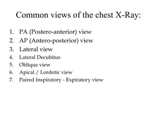
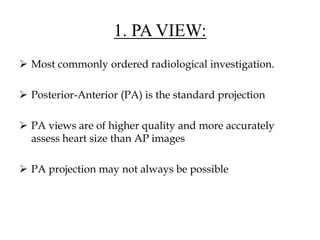
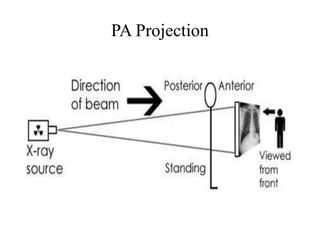
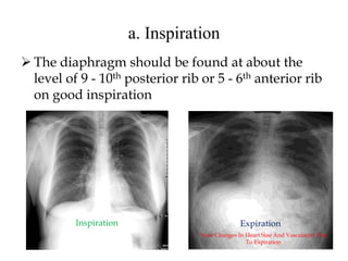
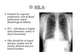
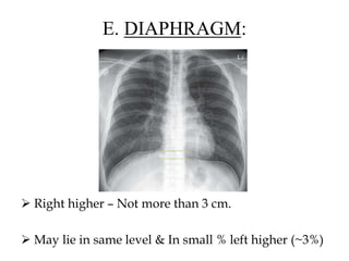
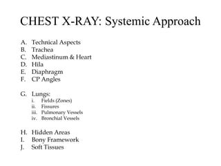
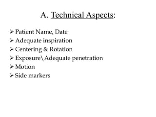
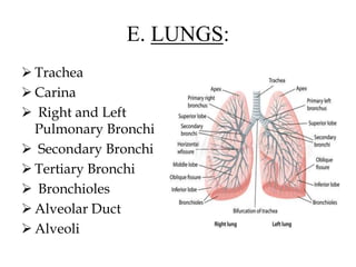
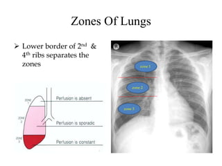
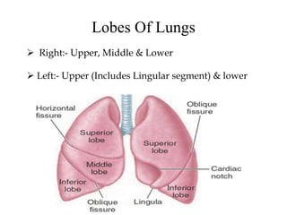
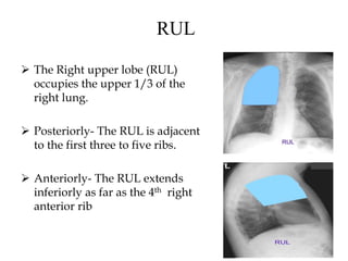
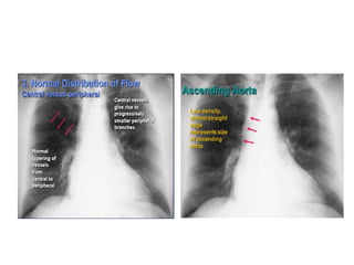
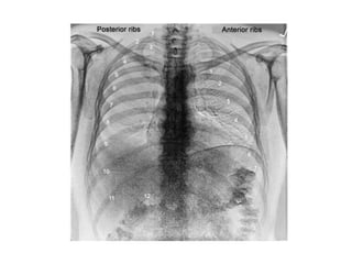
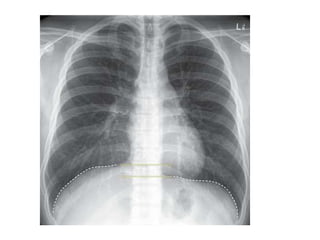
NORMAL CHEST X-RAY
Dr. Bijay Kumar Yadav
(1st Year Radiology Resident)
I.K KSMA, Bishkek
x-ray
Tube
Table
Film
cassette
Wilhelm Conrad Roentgen - Father of Radiology
 Nov 8, 1895 – Discovered
unknown radiations with
photographic effect which he
named ‘axa rays’
 He got the Nobel prize in 1901.
Common views of the chest X-Ray:
1. PA (Postero-anterior) view
2. AP (Antero-posterior) view
3. Lateral view
4. Lateral Decubitus
5. Oblique view
6. Apical / Lordotic view
7. Paired Inspiratory - Expiratory view
1. PA VIEW:
 Most commonly ordered radiological investigation.
 Posterior-Anterior (PA) is the standard projection
 PA views are of higher quality and more accurately
assess heart size than AP images
 PA projection may not always be possible
PA Projection
2. AP VIEW:
Lower quality
 Heart magnification (farther from film)
 Clavicles are projected more cranially above
lung apex
 Scapulae overlie lung fields
 Ribs appear horizontal
 Gastric bubble is absent
AP Projection
AP view CXR
PA view v/s AP View
IN PA VIEW:-
 Clavicles don’t project too high into the apices or
thrown above the apices (more horizontal)
 Heart wont be magnified over the mediastinum
therefore preventing the appearance of
cardiomegaly
 Scapula are away from the lung fields
 Ribs are obliquely oriented in PA view
 Spine and posterior ends of ribs are clearly seen
3. LATERAL VIEW
 Routinely left lateral film
obtained
 In specific lesion, the
side of the interest is
positioned adjacent to
the film
 Should be viewed along
with the PA film
CHEST X-RAY: Systemic Approach
A. Technical Aspects
B. Trachea
C. Mediastinum & Heart
D. Hila
E. Diaphragm
F. CP Angles
G. Lungs:
i. Fields (Zones)
ii. Fissures
iii. Pulmonary Vessels
iv. Bronchial Vessels
H. Hidden Areas
I. Bony Framework
J. Soft Tissues
A. Technical Aspects:
 Patient Name, Date
 Adequate inspiration
 Centering & Rotation
 ExposureAdequate penetration
 Motion
 Side markers
a. Inspiration
 The diaphragm should be found at about the
level of 9 - 10th posterior rib or 5 - 6th anterior rib
on good inspiration
Inspiration Expiration
Note Changes In Heart Size And Vascularity Due
To Expiration.
Inspiration - Expiration
 Films taken after maximal inspiration &
expiration
 Advantage
• Helps in detection of focal or diffuse air
trapping – advantage in suspecting FB & small
pneumothorax
• Demonstration of diaphragmatic movement
b. Penetration
 On a good PA film, the
thoracic spine disc spaces
should be barely visible
through the heart but bony
details of the spine are not
usually seen.
 On the other hand
penetration is sufficient that
bronchovascular structures
can usually be seen through
the heart.
UNDERPENETRATION: Likelihood
of missing an abnormality
overlying by another structure
OVERPENETRATION: Results in loss
of visibility of low density lesion
e.g. Early Consolidation
c. Centering & Rotation
 Can be assessed by observing the clavicular
heads and determining whether they are equal
distance from the spinous process of the thoracic
vertebral bodies.
 Good centering: 1/3 of heart is to right & 2/3 to
left of midline.
c. Motion:
 Cardiac margin, diaphragm and pulmonary
vessels should be sharply marginated in a
completely still patient.
B. TRACHEA:
 Should be examined for:
• Narrowing
• Displacement
• Intraluminal lesions
 Position: Central, slightly deviated towards
Right around the aortic knuckle
 Calibre:
• Even
• Max. Coronal: 25mm (M), 21mm (F)
 Carina angle: 60 - 750 widening
C. MEDIASTINUM & HEART:
 Central dense shadow is formed by:
• Heart
• Mediastinum
• Sternum
• Spine
Cardiac Shadow
 Good centering:
– Heart:
• 2/3 left
• 1/3 right
 In chest x-ray heart examined for size, shape,
position, silhouette.
 Size measurement:
• CT ratio: < 50%
• Transverse cardiac
diameter:
< 15.5 cm (M)
< 14.5 cm (F)
 Heart size appears
enlarged on expiration,
supine film, AP film &
when diaphragms are
elevated
Borders
Silhouette sign:
 The silhouette sign is the absence of depiction
of an anatomic soft-tissue border resulting
from the juxtaposition of structures of similar
radiographic attenuation.
 Density difference  delineation of the outline.
 There are four basic densities in x-ray images:
a. Gas
b. Fat
c. Water / soft tissue
d. Bone / calcium
 Loss of density difference of the adjacent
structures  loss of silhouette.
D. HILA:
 Formed by superior
pulmonary vein & Basal
pulmonary artery
(Radiological Hilum)
 97% - left hilum is higher
(left pulmonary artery is
above bronchus)
 Hila should be of equal
density, similar size &
clearly defined concave
lateral borders
Structures in the Hilum
1. Pulmonary arteries & upper lobe veins-
significant contribution to hilar shadow
2. Normal LN- Not seen in plain radiography
3. Bronchi- walls seen end on
E. DIAPHRAGM:
 Right higher – Not more than 3 cm.
 May lie in same level & In small % left higher (~3%)
 On inspiration – Anterior 6th rib , Posterior 10th
rib (Erect film)
 Both domes – gentle curves- steepen towards
posterior angles
 Clearly defined upper borders except area where
heart rests & anterior cardio-phrenic angles (fat
pad)
 Loss of outline – adjacent tissue no air-
consolidation or pleural effusion
 Free intra peritoneal gas-under surface of
diaphragm: 2-3mm thick
Diaphragm (Normal Variants)
 Diaphragmatic Hump: Rt side anteriorly
 Eventration: Part of the muscle is absent- Left
side
 Scalopping: Rt side- short curves convex
upwards
 Muscle Slips: Rt side- short curves concave
upwards
Left v/s Right Dome Of Diaphragm:
 Anterior left hemidiaphragm is obliterated by the
cardiac contact; right is seen in entirity
 By identifying the fissures: left oblique fissure is contacts
diaphragm ~5 cm behind the anterior costophrenic angle
 On left lateral film, the right anterior and posterior
costophrenic sulci should project beyond the
corresponding left sided sulci as a result of x-ray beam
divergence
 By seeing air in stomach and splenic flexure below the
left hemidiaphragm
CP Angles
 Acute and well
defined
 Obliterated when
diaphragms are flat
E. LUNGS:
 Trachea
 Carina
 Right and Left
Pulmonary Bronchi
 Secondary Bronchi
 Tertiary Bronchi
 Bronchioles
 Alveolar Duct
 Alveoli
Zones Of Lungs
 Lower border of 2nd &
4th ribs separates the
zones zone 1
zone 2
zone 3
Lobes Of Lungs
 Right:- Upper, Middle & Lower
 Left:- Upper (Includes Lingular segment) & lower
RUL
 The Right upper lobe (RUL)
occupies the upper 1/3 of the
right lung.
 Posteriorly- The RUL is adjacent
to the first three to five ribs.
 Anteriorly- The RUL extends
inferiorly as far as the 4th right
anterior rib
RML
 The right middle lobe is
typically the smallest of the
three, and appears
triangular in shape, being
narrowest near the hilum
RLL
 The right lower lobe is the
largest of all three lobes,
separated from the others
by the major fissure.
 Posteriorly, the RLL extend
as far superiorly as the 6th
thoracic vertebral body, and
extends inferiorly to the
diaphragm.
 Review of the lateral plain
film surprisingly shows the
superior extent of the RLL.
Right Lobe Fissures
 These lobes can be separated
from one another by two
fissures.
 The minor fissure separates
the RUL from the RML, and
thus represents the visceral
pleural surfaces of both of
these lobes.
 The major fissure oriented
obliquely extends posteriorly
and superiorly
approximately to the level of
the 4th vertebral body.
LUL
 The lobar architecture of
the left lung is slightly
different than the right.
 Because there is no
defined left minor fissure,
there are only two lobes
on the left; the left upper
LLL
 Left lower lobes
 These two lobes are separated by
a major fissure, identical to that
seen on the right side, although
often slightly more inferior in
location.
 The portion of the left lung that
corresponds anatomically to the
right middle lobe is incorporated
into the left upper lobe.
Fissures Of Lungs
 Major: Right & Left oblique Fissure
 Minor: Right Horizontal Fissure
Pulmonary Vessels
1. Measure the right descending Pulmonary artery
Diameter- (16mm in Male & 15mm in Female)
2. Distribution of flow from apex to base
• At first intercostal space– normal vessels not more than 3
mm in diameter
• Erect- Lower lobe vessels prominent
• Supine- Equalize
3. Distribution of flow from central to peripheral -
tapering
• Vascular lung markings- Central 2/3rd
F. HIDDEN AREAS:
Apices: Partially obscured by ribs, costal cartilage,
clavicles & soft tissues
 Central lesions obscured by mediastinum and
hila
 Posterior & lateral basal segments of lower lobes
& posterior sulcus obscured by the downward
curve of the posterior diaphragm
 Hidden areas due to bones
G. BONY STRUCTURES:
• RIBS
• SCAPULA
• CLAVICLES
• SPINE
• STERNUM
H. SOFT TISSUE:
 General survey in chest wall, shoulders &
lower neck.
 Breast Shadows
 Supraclavicular areas
 Axillae
 Tissues along sides of Breasts
THANK YOU!!

Normal Chest X-Rays & Its Systemic Approach- Anatomy

  • 1.
    NORMAL CHEST X-RAY Dr.Bijay Kumar Yadav (1st Year Radiology Resident) I.K KSMA, Bishkek x-ray Tube Table Film cassette
  • 2.
    Wilhelm Conrad Roentgen- Father of Radiology  Nov 8, 1895 – Discovered unknown radiations with photographic effect which he named ‘axa rays’  He got the Nobel prize in 1901.
  • 3.
    Common views ofthe chest X-Ray: 1. PA (Postero-anterior) view 2. AP (Antero-posterior) view 3. Lateral view 4. Lateral Decubitus 5. Oblique view 6. Apical / Lordotic view 7. Paired Inspiratory - Expiratory view
  • 4.
    1. PA VIEW: Most commonly ordered radiological investigation.  Posterior-Anterior (PA) is the standard projection  PA views are of higher quality and more accurately assess heart size than AP images  PA projection may not always be possible
  • 5.
  • 7.
    2. AP VIEW: Lowerquality  Heart magnification (farther from film)  Clavicles are projected more cranially above lung apex  Scapulae overlie lung fields  Ribs appear horizontal  Gastric bubble is absent
  • 8.
  • 9.
  • 10.
    PA view v/sAP View IN PA VIEW:-  Clavicles don’t project too high into the apices or thrown above the apices (more horizontal)  Heart wont be magnified over the mediastinum therefore preventing the appearance of cardiomegaly  Scapula are away from the lung fields  Ribs are obliquely oriented in PA view  Spine and posterior ends of ribs are clearly seen
  • 11.
    3. LATERAL VIEW Routinely left lateral film obtained  In specific lesion, the side of the interest is positioned adjacent to the film  Should be viewed along with the PA film
  • 12.
    CHEST X-RAY: SystemicApproach A. Technical Aspects B. Trachea C. Mediastinum & Heart D. Hila E. Diaphragm F. CP Angles G. Lungs: i. Fields (Zones) ii. Fissures iii. Pulmonary Vessels iv. Bronchial Vessels H. Hidden Areas I. Bony Framework J. Soft Tissues
  • 13.
    A. Technical Aspects: Patient Name, Date  Adequate inspiration  Centering & Rotation  ExposureAdequate penetration  Motion  Side markers
  • 14.
    a. Inspiration  Thediaphragm should be found at about the level of 9 - 10th posterior rib or 5 - 6th anterior rib on good inspiration Inspiration Expiration Note Changes In Heart Size And Vascularity Due To Expiration.
  • 15.
    Inspiration - Expiration Films taken after maximal inspiration & expiration  Advantage • Helps in detection of focal or diffuse air trapping – advantage in suspecting FB & small pneumothorax • Demonstration of diaphragmatic movement
  • 16.
    b. Penetration  Ona good PA film, the thoracic spine disc spaces should be barely visible through the heart but bony details of the spine are not usually seen.  On the other hand penetration is sufficient that bronchovascular structures can usually be seen through the heart.
  • 17.
    UNDERPENETRATION: Likelihood of missingan abnormality overlying by another structure OVERPENETRATION: Results in loss of visibility of low density lesion e.g. Early Consolidation
  • 18.
    c. Centering &Rotation  Can be assessed by observing the clavicular heads and determining whether they are equal distance from the spinous process of the thoracic vertebral bodies.  Good centering: 1/3 of heart is to right & 2/3 to left of midline.
  • 20.
    c. Motion:  Cardiacmargin, diaphragm and pulmonary vessels should be sharply marginated in a completely still patient.
  • 21.
    B. TRACHEA:  Shouldbe examined for: • Narrowing • Displacement • Intraluminal lesions  Position: Central, slightly deviated towards Right around the aortic knuckle  Calibre: • Even • Max. Coronal: 25mm (M), 21mm (F)
  • 22.
     Carina angle:60 - 750 widening
  • 23.
    C. MEDIASTINUM &HEART:  Central dense shadow is formed by: • Heart • Mediastinum • Sternum • Spine
  • 24.
    Cardiac Shadow  Goodcentering: – Heart: • 2/3 left • 1/3 right  In chest x-ray heart examined for size, shape, position, silhouette.
  • 25.
     Size measurement: •CT ratio: < 50% • Transverse cardiac diameter: < 15.5 cm (M) < 14.5 cm (F)  Heart size appears enlarged on expiration, supine film, AP film & when diaphragms are elevated
  • 26.
  • 27.
    Silhouette sign:  Thesilhouette sign is the absence of depiction of an anatomic soft-tissue border resulting from the juxtaposition of structures of similar radiographic attenuation.  Density difference  delineation of the outline.  There are four basic densities in x-ray images: a. Gas b. Fat c. Water / soft tissue d. Bone / calcium  Loss of density difference of the adjacent structures  loss of silhouette.
  • 28.
    D. HILA:  Formedby superior pulmonary vein & Basal pulmonary artery (Radiological Hilum)  97% - left hilum is higher (left pulmonary artery is above bronchus)  Hila should be of equal density, similar size & clearly defined concave lateral borders
  • 29.
    Structures in theHilum 1. Pulmonary arteries & upper lobe veins- significant contribution to hilar shadow 2. Normal LN- Not seen in plain radiography 3. Bronchi- walls seen end on
  • 30.
    E. DIAPHRAGM:  Righthigher – Not more than 3 cm.  May lie in same level & In small % left higher (~3%)
  • 31.
     On inspiration– Anterior 6th rib , Posterior 10th rib (Erect film)  Both domes – gentle curves- steepen towards posterior angles  Clearly defined upper borders except area where heart rests & anterior cardio-phrenic angles (fat pad)  Loss of outline – adjacent tissue no air- consolidation or pleural effusion  Free intra peritoneal gas-under surface of diaphragm: 2-3mm thick
  • 32.
    Diaphragm (Normal Variants) Diaphragmatic Hump: Rt side anteriorly  Eventration: Part of the muscle is absent- Left side  Scalopping: Rt side- short curves convex upwards  Muscle Slips: Rt side- short curves concave upwards
  • 33.
    Left v/s RightDome Of Diaphragm:  Anterior left hemidiaphragm is obliterated by the cardiac contact; right is seen in entirity  By identifying the fissures: left oblique fissure is contacts diaphragm ~5 cm behind the anterior costophrenic angle  On left lateral film, the right anterior and posterior costophrenic sulci should project beyond the corresponding left sided sulci as a result of x-ray beam divergence  By seeing air in stomach and splenic flexure below the left hemidiaphragm
  • 34.
    CP Angles  Acuteand well defined  Obliterated when diaphragms are flat
  • 35.
    E. LUNGS:  Trachea Carina  Right and Left Pulmonary Bronchi  Secondary Bronchi  Tertiary Bronchi  Bronchioles  Alveolar Duct  Alveoli
  • 36.
    Zones Of Lungs Lower border of 2nd & 4th ribs separates the zones zone 1 zone 2 zone 3
  • 37.
    Lobes Of Lungs Right:- Upper, Middle & Lower  Left:- Upper (Includes Lingular segment) & lower
  • 38.
    RUL  The Rightupper lobe (RUL) occupies the upper 1/3 of the right lung.  Posteriorly- The RUL is adjacent to the first three to five ribs.  Anteriorly- The RUL extends inferiorly as far as the 4th right anterior rib
  • 39.
    RML  The rightmiddle lobe is typically the smallest of the three, and appears triangular in shape, being narrowest near the hilum
  • 40.
    RLL  The rightlower lobe is the largest of all three lobes, separated from the others by the major fissure.  Posteriorly, the RLL extend as far superiorly as the 6th thoracic vertebral body, and extends inferiorly to the diaphragm.  Review of the lateral plain film surprisingly shows the superior extent of the RLL.
  • 41.
    Right Lobe Fissures These lobes can be separated from one another by two fissures.  The minor fissure separates the RUL from the RML, and thus represents the visceral pleural surfaces of both of these lobes.  The major fissure oriented obliquely extends posteriorly and superiorly approximately to the level of the 4th vertebral body.
  • 42.
    LUL  The lobararchitecture of the left lung is slightly different than the right.  Because there is no defined left minor fissure, there are only two lobes on the left; the left upper
  • 43.
    LLL  Left lowerlobes  These two lobes are separated by a major fissure, identical to that seen on the right side, although often slightly more inferior in location.  The portion of the left lung that corresponds anatomically to the right middle lobe is incorporated into the left upper lobe.
  • 44.
    Fissures Of Lungs Major: Right & Left oblique Fissure  Minor: Right Horizontal Fissure
  • 45.
    Pulmonary Vessels 1. Measurethe right descending Pulmonary artery Diameter- (16mm in Male & 15mm in Female) 2. Distribution of flow from apex to base • At first intercostal space– normal vessels not more than 3 mm in diameter • Erect- Lower lobe vessels prominent • Supine- Equalize 3. Distribution of flow from central to peripheral - tapering • Vascular lung markings- Central 2/3rd
  • 48.
    F. HIDDEN AREAS: Apices:Partially obscured by ribs, costal cartilage, clavicles & soft tissues  Central lesions obscured by mediastinum and hila  Posterior & lateral basal segments of lower lobes & posterior sulcus obscured by the downward curve of the posterior diaphragm  Hidden areas due to bones
  • 49.
    G. BONY STRUCTURES: •RIBS • SCAPULA • CLAVICLES • SPINE • STERNUM
  • 52.
    H. SOFT TISSUE: General survey in chest wall, shoulders & lower neck.  Breast Shadows  Supraclavicular areas  Axillae  Tissues along sides of Breasts
  • 54.