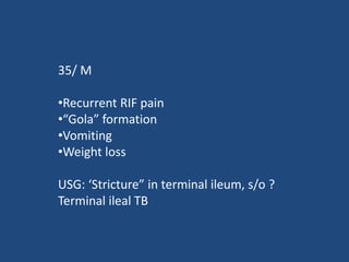
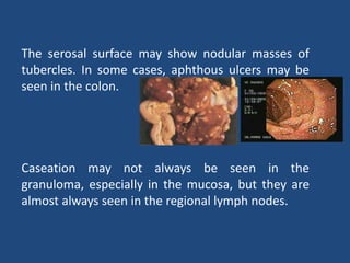
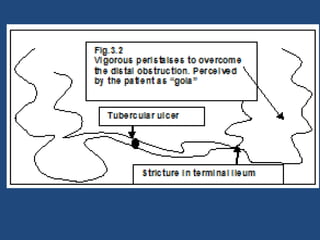
Abdominal Tuberculosis
Dr Manoj K Ghoda M.D., M.R.C.P.
Consultant Gastroenterologist
mkghoda@yahoo.com
35/ M
•Recurrent RIF pain
•“Gola” formation
•Vomiting
•Weight loss
USG: ‘Stricture” in terminal ileum, s/o ?
Terminal ileal TB
Abdominal TB fact file
•Tuberculosis (TB) is very common in the developing
world. Its reappearance has increased in association with
the AIDS.
•TB in its various forms remains an important cause of
morbidity and mortality in developing countries and in
patients with AIDS.
•TB can occur in persons of any age, although it is more
common in children and in older persons whose immune
systems are weak.
•TB can be seen in any age group that is
immunocompromised
•The occurrence of abdominal TB is independent of
pulmonary disease in most patients, with an incidence
of coexisting disease varying from 5 to 36%.
•In patients with abdominal TB, the highest incidence
of disease was noted in the GI tract and in the
peritoneum, followed by the mesenteric lymph nodes.
•Within the GI tract, the ileocecal area is the most
common site of involvement. A third of patients will
report a family history of tuberculosis.
The mode of spread:
•The majority of abdominal disease is either through
hematogenous spread from active pulmonary or
miliary tuberculosis, swallowing of infected sputum
or ingestion of contaminated milk or food, and
contiguous spread from adjacent organs.
•Associated active pulmonary tuberculosis is only
seen in 5-36% of cases.
Pathology:
Three types of intestinal lesion are
seen;
•Ulcerative,
•Stricturous,
•Hypertrophic,
though the three may co-exist.
(1) The ulcerative form of
TB is seen in approximately
60% of patients. Multiple
superficial ulcers largely
confined to the epithelial
surface. This is considered a
highly active form of the
disease with the long axis of
the ulcers perpendicular to
the long axis of the bowel.
(2) The stricturous form shows multiple
or single stricture, often very tight.
(3) The hypertrophic form is seen in
approximately 10% of patients and
consists of thickening of bowel wall with
scarring, fibrosis, and a rigid, mass-like
appearance that mimics carcinoma.
The ulcerohypertrophic form is a
subtype seen in 30% of patients.
These patients have a combination of
features of the ulcerative and
hypertrophic forms.
The serosal surface may show nodular masses of
tubercles. In some cases, aphthous ulcers may be
seen in the colon.
Caseation may not always be seen in the
granuloma, especially in the mucosa, but they are
almost always seen in the regional lymph nodes.
Clinical presentations of abdominal TB:
•The commonest presentation is non-specific abdominal pain,
associated with anorexia, weight loss and low grade fever.
•Systemic manifestations including low grade fever, lethargy,
malaise, night sweats, and anorexia and weight loss are present
in approximately a third of patients with abdominal
tuberculosis.
•Alteration in bowel habit, diarrhea, constipation or together,
malabsorption, rectal bleeding etc.
•Ascites.
Complications of abdominal TB:
•Subacute or acute intestinal obstruction due to stricture or
adhesions is the commonest complication.
•Hemorrhage and perforation are recognized complications of
intestinal TB, although free perforation is less frequent than in
Crohn’s disease.
•Malabsorption may be caused by obstruction that leads to
bacterial overgrowth, a variant of stagnant loop syndrome.
Involvement of the mesenteric lymphatic system, known as
tabes mesenterica, may retard chylomicron removal because
of lymphatic obstruction and result in malabsorption.
•TB is a well recognized cause of rectal stricture. Isolated rectal
involvement is rare and may be mistaken for rectal malignancy
Investigations:
Endoscopy may show,
•Ulcers,
•Nodules,
•Deformed cecum and ileocecal valve,
•Strictures,
•Multiple fibrous bands, and
•Polypoid lesions
Barium examination:
A small-bowel barium study may show dilated loops,
thickened folds or even a stricture.
USG:
•Intra-abdominal fluid, free or loculated; clear or
complex with septae or debris
•Inter loop ascites.
•Lymphadenopathy, discrete or conglomerated,
•Bowel wall thickening, and
•Pseudo kidney sign.
CT :
•The CT features suggestive of abdominal TB include,
•Irregular soft-tissue densities in the omentum,
•Low-attenuating masses surrounded by thick solid
rims,
•Low-attenuating necrotic nodes,
•High-attenuating ascitic fluid and bowel loops forming
poorly defined masses.
•Splenomegaly and hepatomegaly with nodules,
•Pleural effusion,
•Intrahepatic, intrasplenic, and intrapancreatic masses.
In clinical practice, typical GI symptoms mentioned
above with USG evidence of either terminal ileal
involvement or ascites are reasonable evidence in a
proper set up.
Further confirmation is by diagnostic ascitic tapping
and endoscopic biopsy.
Differential Diagnosis:
•Crohn’s disease,
•Gastrointestinal malignancy,
•Sarcoma,
•Amebiasis,
•Yersinia infection, and
•Periappendiceal abscess
Treatment:
•Due to the difficulty and in the case of culture the delay
associated with making a diagnosis of abdominal tuberculosis,
empirical treatment of suspected cases may be warranted.
•Even lesions causing partial bowel obstruction often respond to
medical treatment.
•
•In the HIV co-infected patient as in this case the decision as to
whether to start both quadruple therapy and HAART depends on
the CD4 count. If CD4 > 200 HAART is deferred until completion of
TB treatment, if 100-200 then HAART should start 2 months after
initiation of TB treatment and soon after TB treatment if CD4
<100.
Surgery in TB:
Surgery is required if there is a tight fibrotic stricture
which is symptomatic. Resection anastomosis or
stricturoplasty are the standard approaches. Results
are excellent.
Entrance test
The following are true about abdominal tuberculosis:
1. Abdominal tuberculosis (TB) is very common in the
developing world but it is rarely seen in western countries.
2. Its reappearance has increased in association with the
acquired immunodeficiency syndrome (AIDS).
3. The occurrence of abdominal TB is dependent of pulmonary
disease in most patients.
4. In patients with abdominal TB, the highest incidence of
disease was noted in the gastrointestinal tract and in the
peritoneum, followed by the mesenteric lymph nodes.
5. Within the gastrointestinal tract, the ileocecal area is the most
common site of involvement.
6. A third of patients will report a family history of tuberculosis.
7. TB can occur in persons of any age, although it is uncommon in
children and in older persons whose immune systems are
weak.
8. The mode of spread is either through hematogenous spread
from active pulmonary or miliary tuberculosis, swallowing of
infected sputum or ingestion of contaminated milk or food,
and contiguous spread from adjacent organs.
9. Most cases of abdominal tuberculosis involve the intestine
with the commonest site being the ileocecal region due to
abundance of lymphoid tissue (Payer’s patches).
10. Ileocecal junction is involved in 80-90% of the patients.
11. Proximal small intestinal disease is seen more commonly with
Mycobacterium avium-intracellulare (MAI) complex infection,
predominantly one involving the jejunum.
12. Three types of intestinal lesion are seen; Ulcerative,
Stricturous and hypertrophic.
13. Patients may present with non-specific abdominal pain,
with ascites, alteration in bowel habit, diarrhea,
constipation or together, malabsorption, rectal bleeding
etc.
14. Colonic TB rarely is associated with ileal TB.
15. Subacute or acute intestinal obstruction due to stricture is
the commonest complication.
16. Hemorrhage and perforation are recognized
complications of intestinal TB, and free perforation is
more frequent than in Crohn’s disease.
17. TB never involve rectum and isolated rectal involvement
suggests rectal malignancy.
18. A small-bowel barium study is the main radiographic
method for the evaluation of intestinal TB in regions of the
world where the disease is endemic.
19. However, because peritonitis is common in GI TB, abdominal
CT may be performed as a preferred examination, which
nearly always suggest the diagnosis in the presence of
necrotic lymph nodes or changes suggestive of TB
peritonitis.
20. Early changes on barium examinations reveal nodular
thickening of mucosal folds with loss of symmetry in fold
pattern.
21. Definitive diagnosis is by showing AFB microbiologically by
culture or by both smear and PCR positivity.
22. If the diagnosis of TB is not possible using both of these
methods, a clinical diagnosis of TB is ruled out.

Abdominal tuberculosis

  • 1.
    Abdominal Tuberculosis Dr ManojK Ghoda M.D., M.R.C.P. Consultant Gastroenterologist mkghoda@yahoo.com
  • 2.
    35/ M •Recurrent RIFpain •“Gola” formation •Vomiting •Weight loss USG: ‘Stricture” in terminal ileum, s/o ? Terminal ileal TB
  • 3.
  • 4.
    •Tuberculosis (TB) isvery common in the developing world. Its reappearance has increased in association with the AIDS. •TB in its various forms remains an important cause of morbidity and mortality in developing countries and in patients with AIDS. •TB can occur in persons of any age, although it is more common in children and in older persons whose immune systems are weak. •TB can be seen in any age group that is immunocompromised
  • 5.
    •The occurrence ofabdominal TB is independent of pulmonary disease in most patients, with an incidence of coexisting disease varying from 5 to 36%. •In patients with abdominal TB, the highest incidence of disease was noted in the GI tract and in the peritoneum, followed by the mesenteric lymph nodes. •Within the GI tract, the ileocecal area is the most common site of involvement. A third of patients will report a family history of tuberculosis.
  • 6.
    The mode ofspread: •The majority of abdominal disease is either through hematogenous spread from active pulmonary or miliary tuberculosis, swallowing of infected sputum or ingestion of contaminated milk or food, and contiguous spread from adjacent organs. •Associated active pulmonary tuberculosis is only seen in 5-36% of cases.
  • 8.
    Pathology: Three types ofintestinal lesion are seen; •Ulcerative, •Stricturous, •Hypertrophic, though the three may co-exist.
  • 9.
    (1) The ulcerativeform of TB is seen in approximately 60% of patients. Multiple superficial ulcers largely confined to the epithelial surface. This is considered a highly active form of the disease with the long axis of the ulcers perpendicular to the long axis of the bowel.
  • 10.
    (2) The stricturousform shows multiple or single stricture, often very tight. (3) The hypertrophic form is seen in approximately 10% of patients and consists of thickening of bowel wall with scarring, fibrosis, and a rigid, mass-like appearance that mimics carcinoma. The ulcerohypertrophic form is a subtype seen in 30% of patients. These patients have a combination of features of the ulcerative and hypertrophic forms.
  • 11.
    The serosal surfacemay show nodular masses of tubercles. In some cases, aphthous ulcers may be seen in the colon. Caseation may not always be seen in the granuloma, especially in the mucosa, but they are almost always seen in the regional lymph nodes.
  • 12.
    Clinical presentations ofabdominal TB: •The commonest presentation is non-specific abdominal pain, associated with anorexia, weight loss and low grade fever. •Systemic manifestations including low grade fever, lethargy, malaise, night sweats, and anorexia and weight loss are present in approximately a third of patients with abdominal tuberculosis. •Alteration in bowel habit, diarrhea, constipation or together, malabsorption, rectal bleeding etc. •Ascites.
  • 13.
    Complications of abdominalTB: •Subacute or acute intestinal obstruction due to stricture or adhesions is the commonest complication. •Hemorrhage and perforation are recognized complications of intestinal TB, although free perforation is less frequent than in Crohn’s disease. •Malabsorption may be caused by obstruction that leads to bacterial overgrowth, a variant of stagnant loop syndrome. Involvement of the mesenteric lymphatic system, known as tabes mesenterica, may retard chylomicron removal because of lymphatic obstruction and result in malabsorption. •TB is a well recognized cause of rectal stricture. Isolated rectal involvement is rare and may be mistaken for rectal malignancy
  • 15.
    Investigations: Endoscopy may show, •Ulcers, •Nodules, •Deformedcecum and ileocecal valve, •Strictures, •Multiple fibrous bands, and •Polypoid lesions
  • 16.
    Barium examination: A small-bowelbarium study may show dilated loops, thickened folds or even a stricture.
  • 17.
    USG: •Intra-abdominal fluid, freeor loculated; clear or complex with septae or debris •Inter loop ascites. •Lymphadenopathy, discrete or conglomerated, •Bowel wall thickening, and •Pseudo kidney sign.
  • 18.
    CT : •The CTfeatures suggestive of abdominal TB include, •Irregular soft-tissue densities in the omentum, •Low-attenuating masses surrounded by thick solid rims, •Low-attenuating necrotic nodes, •High-attenuating ascitic fluid and bowel loops forming poorly defined masses. •Splenomegaly and hepatomegaly with nodules, •Pleural effusion, •Intrahepatic, intrasplenic, and intrapancreatic masses.
  • 19.
    In clinical practice,typical GI symptoms mentioned above with USG evidence of either terminal ileal involvement or ascites are reasonable evidence in a proper set up. Further confirmation is by diagnostic ascitic tapping and endoscopic biopsy.
  • 21.
    Differential Diagnosis: •Crohn’s disease, •Gastrointestinalmalignancy, •Sarcoma, •Amebiasis, •Yersinia infection, and •Periappendiceal abscess
  • 22.
    Treatment: •Due to thedifficulty and in the case of culture the delay associated with making a diagnosis of abdominal tuberculosis, empirical treatment of suspected cases may be warranted. •Even lesions causing partial bowel obstruction often respond to medical treatment. • •In the HIV co-infected patient as in this case the decision as to whether to start both quadruple therapy and HAART depends on the CD4 count. If CD4 > 200 HAART is deferred until completion of TB treatment, if 100-200 then HAART should start 2 months after initiation of TB treatment and soon after TB treatment if CD4 <100.
  • 23.
    Surgery in TB: Surgeryis required if there is a tight fibrotic stricture which is symptomatic. Resection anastomosis or stricturoplasty are the standard approaches. Results are excellent.
  • 24.
  • 25.
    The following aretrue about abdominal tuberculosis: 1. Abdominal tuberculosis (TB) is very common in the developing world but it is rarely seen in western countries. 2. Its reappearance has increased in association with the acquired immunodeficiency syndrome (AIDS). 3. The occurrence of abdominal TB is dependent of pulmonary disease in most patients. 4. In patients with abdominal TB, the highest incidence of disease was noted in the gastrointestinal tract and in the peritoneum, followed by the mesenteric lymph nodes. 5. Within the gastrointestinal tract, the ileocecal area is the most common site of involvement.
  • 26.
    6. A thirdof patients will report a family history of tuberculosis. 7. TB can occur in persons of any age, although it is uncommon in children and in older persons whose immune systems are weak. 8. The mode of spread is either through hematogenous spread from active pulmonary or miliary tuberculosis, swallowing of infected sputum or ingestion of contaminated milk or food, and contiguous spread from adjacent organs. 9. Most cases of abdominal tuberculosis involve the intestine with the commonest site being the ileocecal region due to abundance of lymphoid tissue (Payer’s patches). 10. Ileocecal junction is involved in 80-90% of the patients. 11. Proximal small intestinal disease is seen more commonly with Mycobacterium avium-intracellulare (MAI) complex infection, predominantly one involving the jejunum.
  • 27.
    12. Three typesof intestinal lesion are seen; Ulcerative, Stricturous and hypertrophic. 13. Patients may present with non-specific abdominal pain, with ascites, alteration in bowel habit, diarrhea, constipation or together, malabsorption, rectal bleeding etc. 14. Colonic TB rarely is associated with ileal TB. 15. Subacute or acute intestinal obstruction due to stricture is the commonest complication. 16. Hemorrhage and perforation are recognized complications of intestinal TB, and free perforation is more frequent than in Crohn’s disease.
  • 28.
    17. TB neverinvolve rectum and isolated rectal involvement suggests rectal malignancy. 18. A small-bowel barium study is the main radiographic method for the evaluation of intestinal TB in regions of the world where the disease is endemic. 19. However, because peritonitis is common in GI TB, abdominal CT may be performed as a preferred examination, which nearly always suggest the diagnosis in the presence of necrotic lymph nodes or changes suggestive of TB peritonitis. 20. Early changes on barium examinations reveal nodular thickening of mucosal folds with loss of symmetry in fold pattern. 21. Definitive diagnosis is by showing AFB microbiologically by culture or by both smear and PCR positivity. 22. If the diagnosis of TB is not possible using both of these methods, a clinical diagnosis of TB is ruled out.