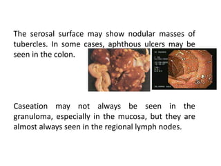
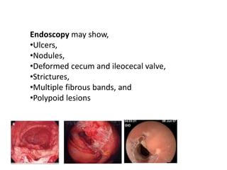
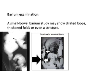
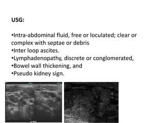
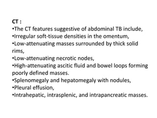
2. Introduction
⢠TB can involve any part of GIT from mouth to
anus, peritoneum & pancreatobiliary system.
⢠Very varied presentation possible ď
TB of GIT- 6th most frequent extrapulmonary
site.
3.
4. Pathogenesis
⢠Mechanisms by which M. tuberculosis reach the
GIT:
â Hematogenous spread from primary lung focus
â Ingestion of bacilli in sputum from active pulmonary
focus.
â Direct spread from adjacent organs.
â Via lymph channels from infected LN
5. ⢠Most common site - ileocaecal region
â Increased physiological stasis
â Increased rate of fluid and electrolyte absorption
â Minimal digestive activity
â Abundance of lymphoid tissue at this site.
6. Distribution of tuberculous lesions
Ileum > caecum > ascending colon > jejunum
>appendix > sigmoid > rectum > duodenum
> stomach > oesophagus
⢠More than one site may be involved
7. ⢠Peritoneal involvement occurs from :
â Spread from LN
â Intestinal lesions or
â Tubercular salpingitis
⢠Abdominal LN and peritoneal TB may occur
without GIT involvement in ~ 1/3 cases.
8. Peritoneal tuberculosis occurs in 3 forms.
⢠Wet type - ascitis.
⢠Encysted (loculated) type - localized swelling.
⢠Fibrotic type - masses composed of mesenteric &
omental thickening, with matted bowel loops.
9. Pathology:
Three types of intestinal lesion are
seen;
â˘Ulcerative,
â˘Stricturous,
â˘Hypertrophic,
though the three may co-exist.
10. (1) The ulcerative form of
TB is seen in approximately
60% of patients. Multiple
superficial ulcers largely
confined to the epithelial
surface. This is considered a
highly active form of the
disease with the long axis of
the ulcers perpendicular to
the long axis of the bowel.
11. (2) The stricturous form shows multiple
or single stricture, often very tight.
(3) The hypertrophic form is seen in
approximately 10% of patients and
consists of thickening of bowel wall with
scarring, fibrosis, and a rigid, mass-like
appearance that mimics carcinoma.
The ulcerohypertrophic form is a
subtype seen in 30% of patients.
These patients have a combination of
features of the ulcerative and
hypertrophic forms.
12. The serosal surface may show nodular masses of
tubercles. In some cases, aphthous ulcers may be
seen in the colon.
Caseation may not always be seen in the
granuloma, especially in the mucosa, but they are
almost always seen in the regional lymph nodes.
13. HIV & TB
⢠Before era of HIV infection > 80% TB
confined to lung
⢠Extrapulmonary TB increases with HIV
⢠40 â60% TB in HIV+ pt - extrapulmonary
⢠Globally, propotion of coinfected pt > 8 %
⢠~ 0.4 million people in India coinfected.
⢠16.6% abdominal TB pt in Bombay HIV +.
15. Clinical Features
⢠Mainly disease of young adults
⢠~ 2/3 of pt. are 21-40 yr old
⢠Sex incidence equal.
⢠Clinical presentation ďŽ Acute / Chronic / Acute
on Chronic.
17. Tuberculosis of esophagus
⢠Rare ~ 0.2% of total cases
⢠By extension from adjacent LN
⢠Low grade fever / Dysphagia / Odynophagia /
Midesophageal ulcer
⢠Mimics esophageal Ca
18. Gastroduodenal TB
⢠Stomach and duodenum each ~ 1% of total cases
⢠Mimics PUD - shorter history, non response to t/t
⢠Mimics gastric Ca.
⢠Duodenal obstruction - extrinsic compression by tuberculous
LN
⢠Hematemesis / Perforation / Fistulae / Obstructive jaundice
⢠Cx-Ray usually normal
⢠Endoscopic picture - non specific
19. Ileocaecal tuberculosis
⢠Colicky abdominal pain
⢠âBall of windâ rolling in abdomen
⢠Borborygmi
⢠Right iliac fossa lump - ileocaecal region,
mesenteric fat and LN
20. Obstruction
⢠Most common complication
Pathogenesis
â Hyperplastic caecal TB
â Strictures of the small intestine--- commonly multiple
â Adhesions
â Adjacent LN involvement ďŽ traction, narrowing and fixation
of bowel loops.
21. Perforation
⢠2nd commonest cause after typhoid
⢠Usually single and proximal to a stricture
⢠Clue - TB Chest x-ray, h/o SAIO
⢠Pneumoperitoneum in ~ 50% cases
22. Malabsorption
⢠Pathogenesis
â bacterial overgrowth in stagnant loop
â bile salt deconjugation
â diminished absorptive surface due to ulceration
â involvement of lymphatics and LN
23. Segmental / Isolated colonic tuberculosis
⢠Involvement of the colon without involvement of the
ileocaecal region
⢠9.2% of all cases
⢠Multifocal involvement in ~ 1/3 (28% to 44%)
⢠Median symptom duration <1 year
24. Colonic tuberculosis
⢠Pain --- predominant symptom ( 78%-90% )
⢠Hematochezia in < 1/3 - usually minor
Overall, TB accounts for ~ 4% of LGI bleeding
⢠Other features--- fever / anorexia / weight loss /
change in bowel habits
25. Rectal and Anal Tuberculosis
⢠Hematochezia - most common symp. Due to mucosal
trauma by stool
⢠Constitutional symptoms
⢠Constipation
⢠Rectal stricture
⢠Anal fistula â usually multiple
26. Diagnosis and Investigations
⢠Non specific findings---
â Raised ESR
â Positive Mantoux test
â Anemia
â Hypoalbuminaemia
28. Ascitic fluid examination
⢠Straw coloured
⢠Protein >3g/dL
⢠TLC of 150-4000/¾l, Lymphocytes >70%
⢠SAAG < 1.1 g/dL
⢠ZN stain + in < 3% cases
⢠+ culture in < 20% cases
29. Adenosine Deaminase (ADA)
Aminohydrolase that converts adenosine ď inosine
⢠ADA increased due to stimulation of T-cells by
mycobacterial Ag
â Serum ADA > 54 U/L
â Ascitic fluid ADA > 36 U/L
â Ascitic fluid to serum ADA ratio > 0.985
⢠Coinfection with HIV ďŽ normal or low ADA
32. USG:
â˘Intra-abdominal fluid, free or loculated; clear or
complex with septae or debris
â˘Inter loop ascites.
â˘Lymphadenopathy, discrete or conglomerated,
â˘Bowel wall thickening, and
â˘Pseudo kidney sign.
33. CT :
â˘The CT features suggestive of abdominal TB include,
â˘Irregular soft-tissue densities in the omentum,
â˘Low-attenuating masses surrounded by thick solid
rims,
â˘Low-attenuating necrotic nodes,
â˘High-attenuating ascitic fluid and bowel loops forming
poorly defined masses.
â˘Splenomegaly and hepatomegaly with nodules,
â˘Pleural effusion,
â˘Intrahepatic, intrasplenic, and intrapancreatic masses.
34. In clinical practice, typical GI symptoms mentioned
above with USG evidence of either terminal ileal
involvement or ascites are reasonable evidence in a
proper set up.
Further confirmation is by diagnostic ascitic tapping
and endoscopic biopsy.
38. Management
⢠ATT for at least 6 months including 2 months of Rif, INH,
Pzide and Etham
⢠However in practice t/t often given for 12 to 18 months
⢠2 recent reports ďŽ obstructing lesions may relieve with ATT
alone
However most will need surgery
39. Surgery in TB:
Surgery is required if there is a tight fibrotic stricture
which is symptomatic. Resection anastomosis or
stricturoplasty are the standard approaches. Results
are excellent.
40. Clinical Pearls
⢠Abdominal tuberculosis (TB) is very common in the
developing world but it is rarely seen in western countries.
⢠Its reappearance has increased in association with the
acquired immunodeficiency syndrome (AIDS).
⢠The occurrence of abdominal TB is dependent of pulmonary
disease in most patients.
⢠In patients with abdominal TB, the highest incidence of
disease was noted in the gastrointestinal tract and in the
peritoneum, followed by the mesenteric lymph nodes.
⢠Within the gastrointestinal tract, the ileocecal area is the most
common site of involvement.
41. ⢠A third of patients will report a family history of tuberculosis.
⢠TB can occur in persons of any age, although it is uncommon in
children and in older persons whose immune systems are
weak.
⢠The mode of spread is either through hematogenous spread
from active pulmonary or miliary tuberculosis, swallowing of
infected sputum or ingestion of contaminated milk or food,
and contiguous spread from adjacent organs.
⢠Most cases of abdominal tuberculosis involve the intestine
with the commonest site being the ileocecal region due to
abundance of lymphoid tissue (Payerâs patches).
⢠Ileocecal junction is involved in 80-90% of the patients.
⢠Proximal small intestinal disease is seen more commonly with
Mycobacterium avium-intracellulare (MAI) complex infection,
predominantly one involving the jejunum.
42. ⢠Three types of intestinal lesion are seen; Ulcerative,
Stricturous and hypertrophic.
⢠Patients may present with non-specific abdominal pain,
with ascites, alteration in bowel habit, diarrhea,
constipation or together, malabsorption, rectal bleeding
etc.
⢠Colonic TB rarely is associated with ileal TB.
⢠Subacute or acute intestinal obstruction due to stricture is
the commonest complication.
⢠Hemorrhage and perforation are recognized
complications of intestinal TB, and free perforation is
more frequent than in Crohnâs disease.
43. ⢠TB never involve rectum and isolated rectal involvement
suggests rectal malignancy.
⢠A small-bowel barium study is the main radiographic
method for the evaluation of intestinal TB in regions of the
world where the disease is endemic.
⢠However, because peritonitis is common in GI TB, abdominal
CT may be performed as a preferred examination, which
nearly always suggest the diagnosis in the presence of
necrotic lymph nodes or changes suggestive of TB
peritonitis.
⢠Early changes on barium examinations reveal nodular
thickening of mucosal folds with loss of symmetry in fold
pattern.
⢠Definitive diagnosis is by showing AFB microbiologically by
culture or by both smear and PCR positivity.
⢠If the diagnosis of TB is not possible using both of these
methods, a clinical diagnosis of TB is ruled out.