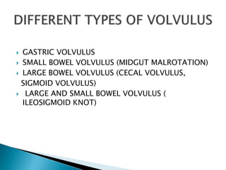
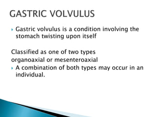
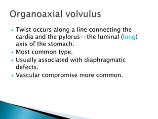
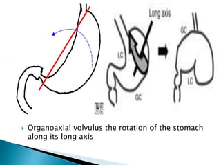
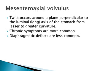
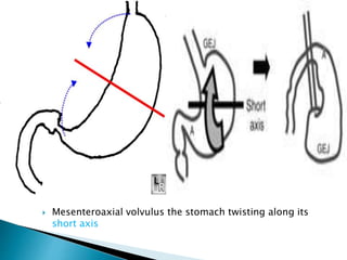
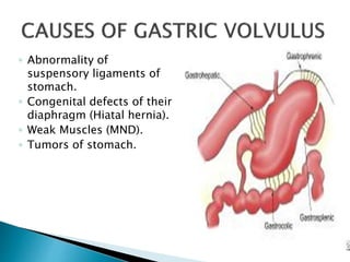
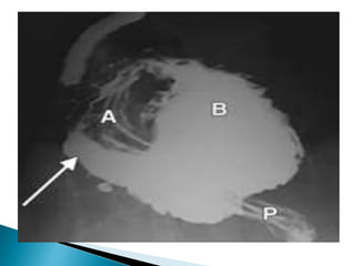
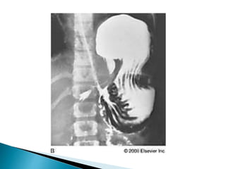
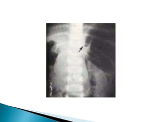
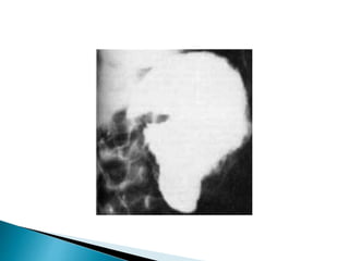
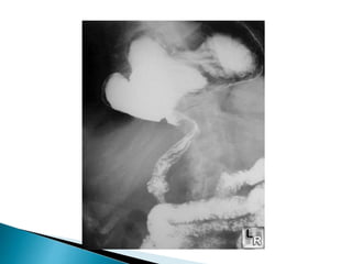
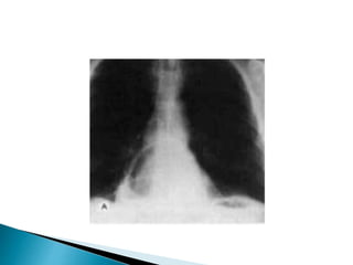
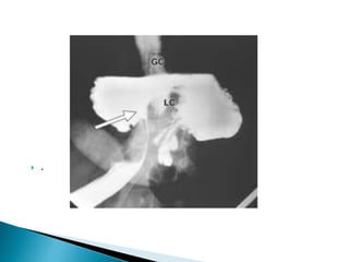
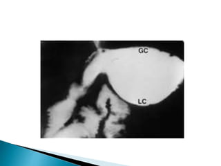
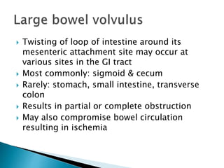
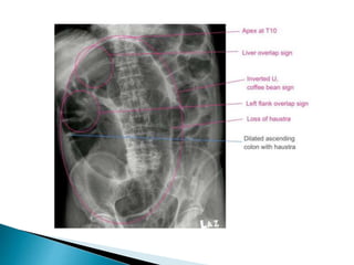
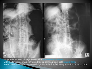
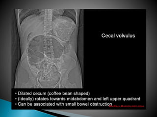
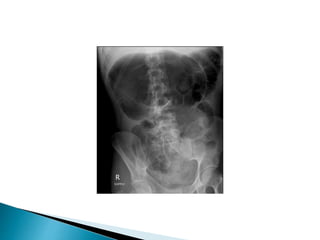
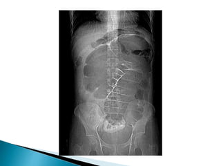
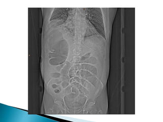
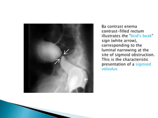
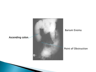
 GASTRIC VOLVULUS
 SMALL BOWEL VOLVULUS (MIDGUT MALROTATION)
 LARGE BOWEL VOLVULUS (CECAL VOLVULUS,
SIGMOID VOLVULUS)
 LARGE AND SMALL BOWEL VOLVULUS (
ILEOSIGMOID KNOT)
 Gastric volvulus is a condition involving the
stomach twisting upon itself
Classified as one of two types
organoaxial or mesenteroaxial
 A combination of both types may occur in an
individual.
 Twist occurs along a line connecting the
cardia and the pylorus--the luminal (long)
axis of the stomach.
 Most common type.
 Usually associated with diaphragmatic
defects.
 Vascular compromise more common.
 Organoaxial volvulus the rotation of the stomach
along its long axis
 Twist occurs around a plane perpendicular to
the luminal (long) axis of the stomach from
lesser to greater curvature.
 Chronic symptoms are more common.
 Diaphragmatic defects are less common.
 Mesenteroaxial volvulus the stomach twisting along its
short axis
◦ Abnormality of
suspensory ligaments of
stomach.
◦ Congenital defects of their
diaphragm (Hiatal hernia).
◦ Weak Muscles (MND).
◦ Tumors of stomach.
 Those with defects of the diaphragm
commonly suffer with the common type
(organoaxial volvulus), and it is the most
serious form, needing urgent surgical
intervention.
 The mesenteroaxial type does not often lead
to compromise of blood supply to the
stomach speedily, and may run a chronic
course.
◦ Unless acute, patients are frequently
asymptomatic.
◦ When acute and obstructing
 Abdominal pain
 Attempts to vomit without results
 Inability to pass an NG tube
 Together, these three findings comprise
the Borchardt triad which is diagnostic of
acute volvulus .
 In mesenteroaxial volvulus, distended
stomach appears spherical on supine images.
 Two air-fluid levels visible on upright film: in
fundus and in antrum.
 Upright image often demonstrates a beak
where the esophagogastric junction is seen
on normal images.
 peanut sign- in a case of chronic gastric
volvulus.
 The ultrasonographic features consist of a
constricted segment of stomach, with 2
dilated segments located above and below
the constricted part, akin to a peanut.
 .
 Gastric ischemia
◦ Gastric emphysema
◦ Twisting of stomach may tear spleen from its
normal attachments
◦ Perforation is rare
 Torsion of the entire gut around superior
mesenteric artery (SMA) due to a short
mesenteric attachment of small intestine in
malrotation.
 AGE
o Usually neonate or young infant
o Occasionally older child and adult
 ASSOCIATED WITH (IN 20%)
o Duodenal atresia
o Duodenal diaphragm
o Duodenal stenosis
o Annular pancreas
o Degree of twisting is variable and determines
symptomatology
o Severe volvulus (twist of 3 ½ turns)
result in bowel necrosis
 Acute symptoms in newborn (medical emergency)
o Bile-stained vomiting
Intermittent, Postprandial, Projectile
o Abdominal distension
o Shock
Dilated, air-filled duodenal bulb and paucity of
gas distally
"Double bubble sign" = air-fluid levels in
stomach & duodenum
o Isolated collection of gas-containing bowel
loops distal to obstructed duodenum = gas-
filled volvulus = closed-loop obstruction
From non resorption of intestinal gas
secondary to obstruction of mesenteric veins
"Corkscrew" duodenum in malrotation with a midgut volvulus
"Corkscrew" duodenum in malrotation
with a midgut volvulus
CT findings
 Whirl-like pattern of small bowel loops and
adjacent mesenteric fat converging to the point
of torsion (during volvulus)
 SMV to the left of SMA (NO volvulus)
 Chylous mesenteric cyst (from interference with
lymphatic drainage)
 Clockwise whirlpool sign = color Doppler
depiction of mesenteric vessels moving clockwise
with caudal movement of transducer
 Distended proximal duodenum with arrowhead-
type compression over spine
 Superior mesenteric vein to the left of SMA
 Thick-walled bowel loops below duodenum and to
the right of spine associated with peritoneal fluid
 "Barber pole sign" = spiraling of SMA
 Tapering / abrupt termination of mesenteric
vessels
 Marked vasoconstriction and prolonged contrast
transit time
 Absent venous opacification / dilated tortuous
superior mesenteric vein
 Intestinal ischemia and necrosis in
distribution of SMA (bloody diarrhea, ileus,
abdominal distension)
DD:
 Pyloric stenosis (same age group, no bilious
vomiting)
 Twisting of loop of intestine around its
mesenteric attachment site may occur at
various sites in the GI tract
 Most commonly: sigmoid & cecum
 Rarely: stomach, small intestine, transverse
colon
 Results in partial or complete obstruction
 May also compromise bowel circulation
resulting in ischemia
 Sigmoid volvulus most common form of GI
tract volvulus
 Accounts for up to 8% of all intestinal
obstructions
 Most common in elderly persons (often
neurologically impaired)
 Patients almost always have a history of
chronic constipation
 Redundant sigmoid colon that has a narrow
mesenteric attachment to posterior abdominal
wall allows close approximation of 2 limbs of
sigmoid colon à twisting of sigmoid colon
around mesenteric axis
 Other predisposing factors
 Chronic constipation
 High-roughage diet (may cause a long,
redundant sigmoid colon)
 Roundworm infestation
 Megacolon (often due to Chagas)
 20-25% mortality rate
 Peak age > 50 years
 Torsion usually counterclockwise ranging
from 180 – 540 degrees
 Luminal obstruction generally @ 180 degrees
 Venous occlusion generally @ 360 degrees à
gangrene & perforation
 Diagnosis
Abdominal plain films usually diagnostic
Inverted U-shaped appearance of distended
sigmoid loop
 Largest and most dilated loops of bowel are
seen with volvulus
 Loss of haustra
 Coffee-bean sign à midline crease
corresponding to mesenteric root in a greatly
distended sigmoid
 Sigmoid volvulus – bowel loop points to RUQ
 torsion of the caecum around its own
mesentery which often results in obstruction
 It accounts for 11% of all intestinal volvulus
 can result in bowel perforation and faecal
peritonitis
Clinical presentation
 Caecal volvulus presents with clinical features
of proximal large bowel obstruction. This is
usually with colicky abdominal pain, vomiting
and abdominal distension.
• Bowel loop points to LUQ
• Dilated cecum comes to rest in left upper
quadrant
• Bird’s-beak or bird-of-prey sign à seen on
barium enema as it encounters the volvulated
loop
• CT scan useful in assessing mural wall ischemia
large, dilated loop of large bowel with an inverted U-shape
with walls between two volvulated loops pointing from LLQ toward RUQ;
same patient with decompressed sigmoid volvulus following insertion of rectal tube
Differential Diagnosis
 Large bowel obstruction due to other causes
à sigmoid colon CA
 Giant sigmoid diverticulum
 Pseudoobstruction
Complications
 Colonic ischemia
 Perforation
 Sepsis
Ba contrast enema
contrast-filled rectum
illustrates the "bird's beak"
sign (white arrow),
corresponding to the
luminal narrowing at the
site of sigmoid obstruction.
This is the characteristic
presentation of a sigmoid
volvulus
 20 year old woman presented to the ED with
12 hours of abdominal pain, nausea. and
vomiting low grade fever.
 No past surgical history
 PMH: Polycystic ovarian disease
Dilated cecum
Cecum
Contrast
In Descending colon
Cecum
Barium Enema
Point of Obstruction
Ascending colon
Volvulus in git

Volvulus in git

  • 2.
     GASTRIC VOLVULUS SMALL BOWEL VOLVULUS (MIDGUT MALROTATION)  LARGE BOWEL VOLVULUS (CECAL VOLVULUS, SIGMOID VOLVULUS)  LARGE AND SMALL BOWEL VOLVULUS ( ILEOSIGMOID KNOT)
  • 3.
     Gastric volvulusis a condition involving the stomach twisting upon itself Classified as one of two types organoaxial or mesenteroaxial  A combination of both types may occur in an individual.
  • 4.
     Twist occursalong a line connecting the cardia and the pylorus--the luminal (long) axis of the stomach.  Most common type.  Usually associated with diaphragmatic defects.  Vascular compromise more common.
  • 5.
     Organoaxial volvulusthe rotation of the stomach along its long axis
  • 6.
     Twist occursaround a plane perpendicular to the luminal (long) axis of the stomach from lesser to greater curvature.  Chronic symptoms are more common.  Diaphragmatic defects are less common.
  • 7.
     Mesenteroaxial volvulusthe stomach twisting along its short axis
  • 8.
    ◦ Abnormality of suspensoryligaments of stomach. ◦ Congenital defects of their diaphragm (Hiatal hernia). ◦ Weak Muscles (MND). ◦ Tumors of stomach.
  • 9.
     Those withdefects of the diaphragm commonly suffer with the common type (organoaxial volvulus), and it is the most serious form, needing urgent surgical intervention.  The mesenteroaxial type does not often lead to compromise of blood supply to the stomach speedily, and may run a chronic course.
  • 10.
    ◦ Unless acute,patients are frequently asymptomatic. ◦ When acute and obstructing  Abdominal pain  Attempts to vomit without results  Inability to pass an NG tube  Together, these three findings comprise the Borchardt triad which is diagnostic of acute volvulus .
  • 11.
     In mesenteroaxialvolvulus, distended stomach appears spherical on supine images.  Two air-fluid levels visible on upright film: in fundus and in antrum.  Upright image often demonstrates a beak where the esophagogastric junction is seen on normal images.
  • 12.
     peanut sign-in a case of chronic gastric volvulus.  The ultrasonographic features consist of a constricted segment of stomach, with 2 dilated segments located above and below the constricted part, akin to a peanut.
  • 19.
  • 22.
     Gastric ischemia ◦Gastric emphysema ◦ Twisting of stomach may tear spleen from its normal attachments ◦ Perforation is rare
  • 23.
     Torsion ofthe entire gut around superior mesenteric artery (SMA) due to a short mesenteric attachment of small intestine in malrotation.
  • 24.
     AGE o Usuallyneonate or young infant o Occasionally older child and adult  ASSOCIATED WITH (IN 20%) o Duodenal atresia o Duodenal diaphragm o Duodenal stenosis o Annular pancreas
  • 25.
    o Degree oftwisting is variable and determines symptomatology o Severe volvulus (twist of 3 ½ turns) result in bowel necrosis  Acute symptoms in newborn (medical emergency) o Bile-stained vomiting Intermittent, Postprandial, Projectile o Abdominal distension o Shock
  • 26.
    Dilated, air-filled duodenalbulb and paucity of gas distally "Double bubble sign" = air-fluid levels in stomach & duodenum o Isolated collection of gas-containing bowel loops distal to obstructed duodenum = gas- filled volvulus = closed-loop obstruction From non resorption of intestinal gas secondary to obstruction of mesenteric veins
  • 27.
    "Corkscrew" duodenum inmalrotation with a midgut volvulus "Corkscrew" duodenum in malrotation with a midgut volvulus
  • 28.
    CT findings  Whirl-likepattern of small bowel loops and adjacent mesenteric fat converging to the point of torsion (during volvulus)  SMV to the left of SMA (NO volvulus)  Chylous mesenteric cyst (from interference with lymphatic drainage)
  • 29.
     Clockwise whirlpoolsign = color Doppler depiction of mesenteric vessels moving clockwise with caudal movement of transducer  Distended proximal duodenum with arrowhead- type compression over spine  Superior mesenteric vein to the left of SMA  Thick-walled bowel loops below duodenum and to the right of spine associated with peritoneal fluid
  • 30.
     "Barber polesign" = spiraling of SMA  Tapering / abrupt termination of mesenteric vessels  Marked vasoconstriction and prolonged contrast transit time  Absent venous opacification / dilated tortuous superior mesenteric vein
  • 31.
     Intestinal ischemiaand necrosis in distribution of SMA (bloody diarrhea, ileus, abdominal distension) DD:  Pyloric stenosis (same age group, no bilious vomiting)
  • 32.
     Twisting ofloop of intestine around its mesenteric attachment site may occur at various sites in the GI tract  Most commonly: sigmoid & cecum  Rarely: stomach, small intestine, transverse colon  Results in partial or complete obstruction  May also compromise bowel circulation resulting in ischemia
  • 33.
     Sigmoid volvulusmost common form of GI tract volvulus  Accounts for up to 8% of all intestinal obstructions  Most common in elderly persons (often neurologically impaired)  Patients almost always have a history of chronic constipation
  • 34.
     Redundant sigmoidcolon that has a narrow mesenteric attachment to posterior abdominal wall allows close approximation of 2 limbs of sigmoid colon à twisting of sigmoid colon around mesenteric axis  Other predisposing factors  Chronic constipation  High-roughage diet (may cause a long, redundant sigmoid colon)  Roundworm infestation  Megacolon (often due to Chagas)
  • 35.
     20-25% mortalityrate  Peak age > 50 years  Torsion usually counterclockwise ranging from 180 – 540 degrees  Luminal obstruction generally @ 180 degrees  Venous occlusion generally @ 360 degrees à gangrene & perforation
  • 36.
     Diagnosis Abdominal plainfilms usually diagnostic Inverted U-shaped appearance of distended sigmoid loop  Largest and most dilated loops of bowel are seen with volvulus  Loss of haustra  Coffee-bean sign à midline crease corresponding to mesenteric root in a greatly distended sigmoid  Sigmoid volvulus – bowel loop points to RUQ
  • 37.
     torsion ofthe caecum around its own mesentery which often results in obstruction  It accounts for 11% of all intestinal volvulus  can result in bowel perforation and faecal peritonitis
  • 38.
    Clinical presentation  Caecalvolvulus presents with clinical features of proximal large bowel obstruction. This is usually with colicky abdominal pain, vomiting and abdominal distension.
  • 39.
    • Bowel looppoints to LUQ • Dilated cecum comes to rest in left upper quadrant • Bird’s-beak or bird-of-prey sign à seen on barium enema as it encounters the volvulated loop • CT scan useful in assessing mural wall ischemia
  • 42.
    large, dilated loopof large bowel with an inverted U-shape with walls between two volvulated loops pointing from LLQ toward RUQ; same patient with decompressed sigmoid volvulus following insertion of rectal tube
  • 44.
    Differential Diagnosis  Largebowel obstruction due to other causes à sigmoid colon CA  Giant sigmoid diverticulum  Pseudoobstruction Complications  Colonic ischemia  Perforation  Sepsis
  • 48.
    Ba contrast enema contrast-filledrectum illustrates the "bird's beak" sign (white arrow), corresponding to the luminal narrowing at the site of sigmoid obstruction. This is the characteristic presentation of a sigmoid volvulus
  • 49.
     20 yearold woman presented to the ED with 12 hours of abdominal pain, nausea. and vomiting low grade fever.  No past surgical history  PMH: Polycystic ovarian disease
  • 50.
  • 51.
  • 52.
  • 53.
    Barium Enema Point ofObstruction Ascending colon