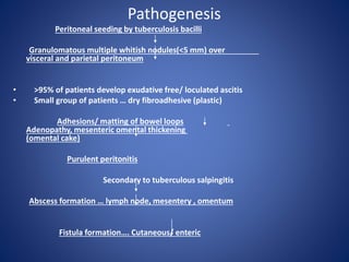
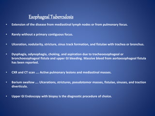
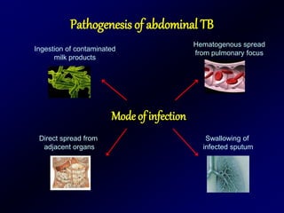
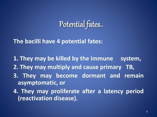
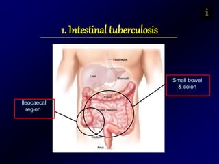
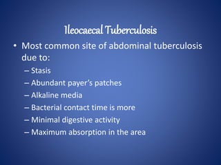
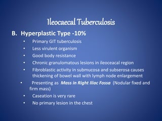
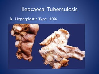
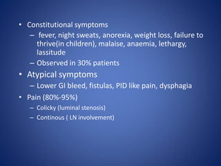
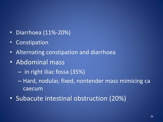
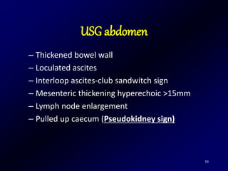
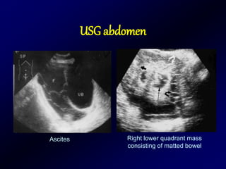
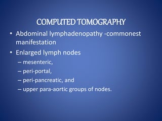
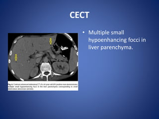
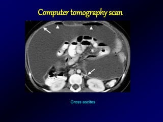
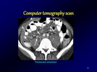
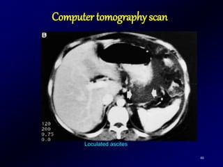
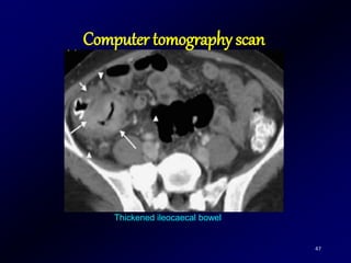
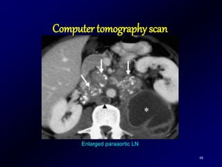
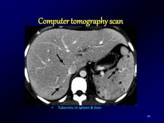
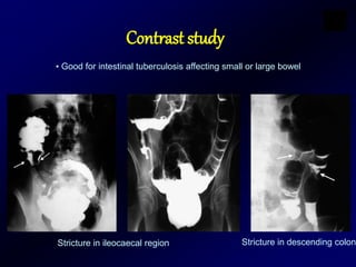
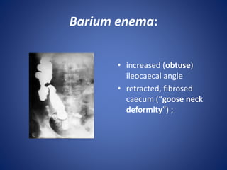
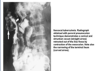
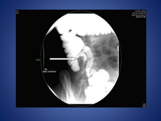
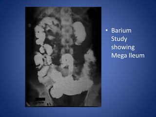
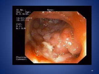
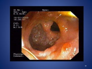
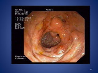
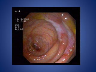
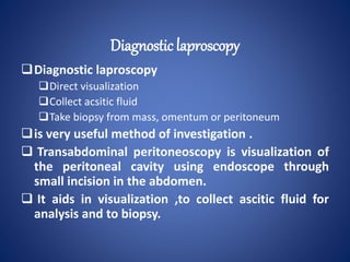
Abdominal tuberculosis is a common extrapulmonary manifestation of tuberculosis that most commonly involves the ileocaecal region of the small intestine. It can present with nonspecific constitutional symptoms like fever, weight loss, or abdominal pain. Diagnosis is challenging as findings can mimic other diseases like Crohn's disease, but imaging modalities like ultrasound, CT scan, and barium studies can reveal features suggestive of abdominal tuberculosis like enlarged lymph nodes, bowel wall thickening, strictures, and ascites. Blood tests are often nonspecific but may show elevated ESR or mild anemia. Sputum tests have low yield for diagnosis but help evaluate for concurrent pulmonary tuberculosis. Tissue sampling is often needed for confirmation.


![72
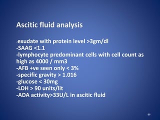
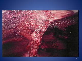
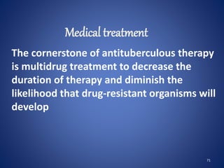
Antituberculosis Drugs
Drug/Formulation Adult Dosage (Daily) Main Adverse Effects
First-Line Drugs
Isoniazid (INH)[*] 5 mg/kg (max 300 mg) PO, IM, IV
Hepatic toxicity,
peripheral neuropathy
100, 300 mg tabs
50 mg/5 mL syrup
100 mg/mL injection
Rifampin (Rifadin, Rimactane) 10 mg/kg (max 600 mg) PO, IV Hepatic toxicity,
flulike syndrome,
pruritus
150, 300 mg caps
600 mg injection
powder](https://image.slidesharecdn.com/abdominaltuberculosis-171229125201/85/Abdominal-tuberculosis-dr-syed-obaid-72-320.jpg)
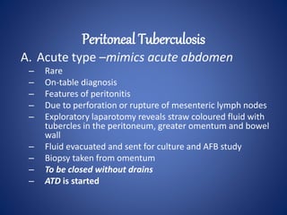
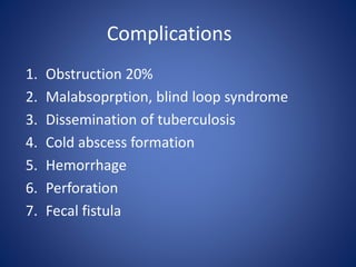
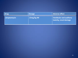
![73
Pyrazinamide 500 mg tabs 20-25 mg/kg PO Arthralgias, hepatic
toxicity, hyperuricemia,
gastrointestinal upset
Ethambutol[‡] (Myambutol) 100, 400
mg tabs
15-25 mg/kg PO
Decreased red-green color
discrimination, decreased
visual acuity
Drug/formulation Dosage Adverse effect](https://image.slidesharecdn.com/abdominaltuberculosis-171229125201/85/Abdominal-tuberculosis-dr-syed-obaid-73-320.jpg)
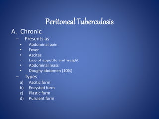
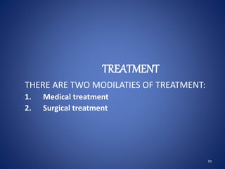
![75
Second-Line Drugs
Capreomycin (Capastat) 15 mg/kg IM (max 1 g) Auditory and vestibular
toxicity, renal damage
Kanamycin (Kantrex and others) 15 mg/kg IM, IV (max 1 g) Auditory toxicity, renal
damage
Amikacin (Amikin) 15 mg/kg IM, IV (max 1 g) Auditory toxicity, renal
damage
Cycloserine[¶] (Seromycin and others)
10-15 mg/kg in two doses
(max 500 mg bid) PO
Psychiatric symptoms,
seizures
Ethionamide (Trecator-SC) 15-20 mg/kg in two doses
(max 500 mg bid) PO
Gastrointestinal and
hepatic toxicity,
hypothyroidism
Ciprofloxacin (Cipro and others) 750-1500 mg PO, IV Nausea, abdominal pain,
restlessness, confusion
Ofloxacin (Floxin) 600-800 mg PO, IV Nausea, abdominal pain,
restlessness, confusion
Drug Dosage Adverse effect](https://image.slidesharecdn.com/abdominaltuberculosis-171229125201/85/Abdominal-tuberculosis-dr-syed-obaid-75-320.jpg)
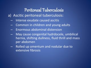
![76
Levofloxacin (Levaquin) 500-1000 mg PO, IV Nausea, abdominal pain,
restlessness, confusion
Gatifloxacin[¶] (Tequin) 400 mg PO, IV
Nausea, abdominal pain,
restlessness, confusion
Moxifloxacin[¶¶] (Avelox) 400 mg PO, IV
Nausea, abdominal pain,
restlessness, confusion
Aminosalicylic acid (PAS; Paser) 8-12 g in 2-3 doses PO Gastrointestinal
disturbance
Drug Dosage Adverse effect](https://image.slidesharecdn.com/abdominaltuberculosis-171229125201/85/Abdominal-tuberculosis-dr-syed-obaid-76-320.jpg)