PET
(Positron Emission Tomography)
BY:
Prikshit Kumar
2011CS1029
Contents
• Introduction
• How does it work?
• Applications
• Where it is used in Neurolinguistics?
• Summary
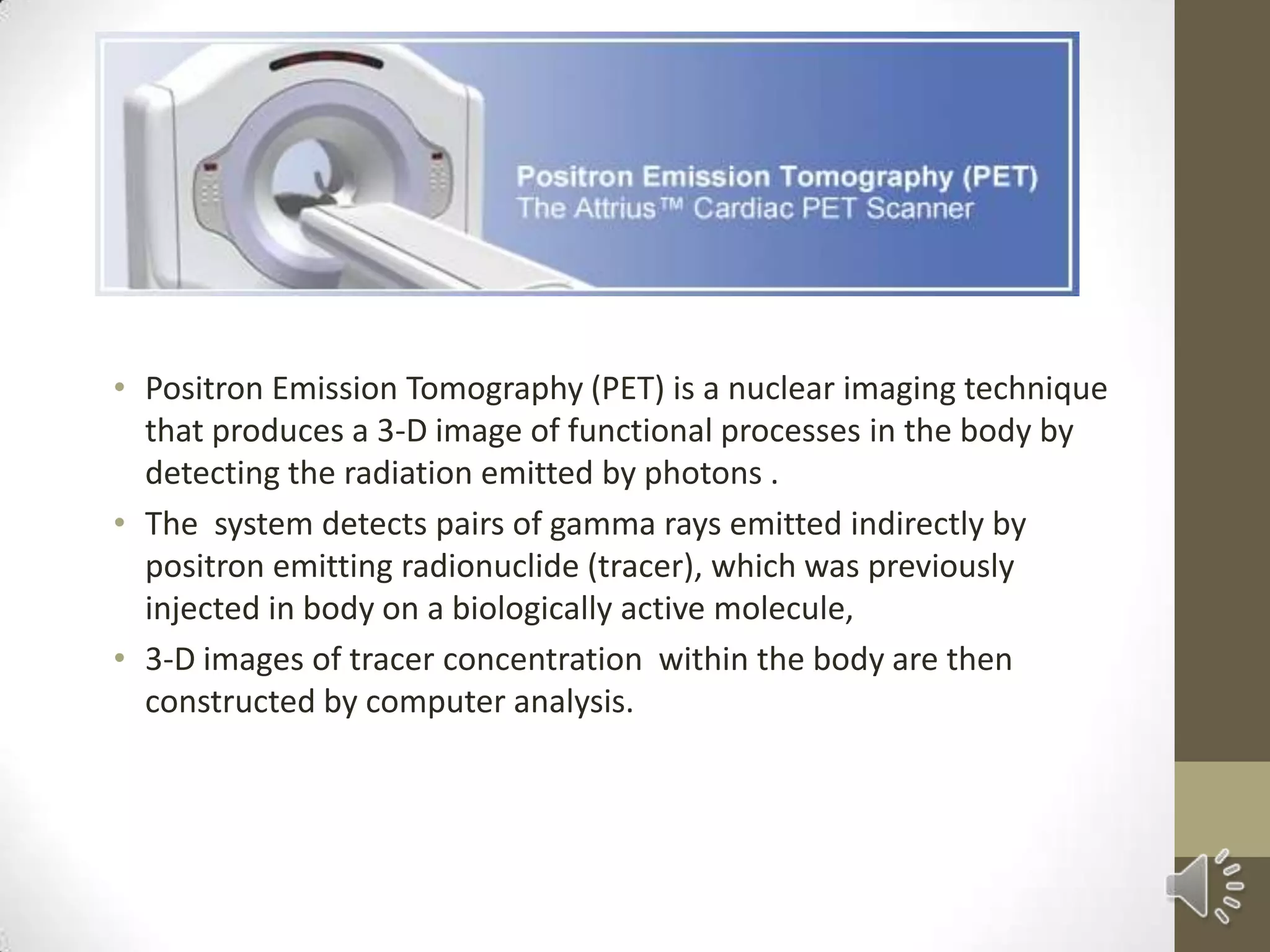
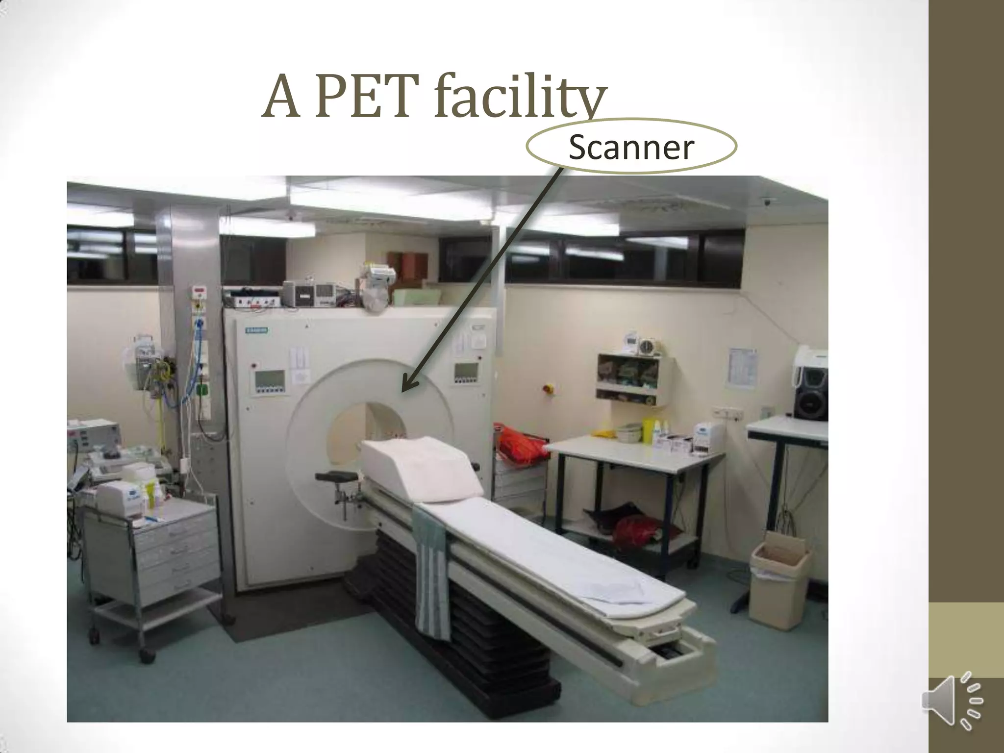
• Positron Emission Tomography (PET) is a nuclear imaging technique
that produces a 3-D image of functional processes in the body by
detecting the radiation emitted by photons .
• The system detects pairs of gamma rays emitted indirectly by
positron emitting radionuclide (tracer), which was previously
injected in body on a biologically active molecule,
• 3-D images of tracer concentration within the body are then
constructed by computer analysis.
A PET facility
Scanner
Working
• Injection of Short lived Radioactive Isotope in body.
most commonly used is FDG (fluoro-2-
deoxyglucose).
• Wait till tracer gets accumulated in tissues of interests.
• Subject is placed in the imaging scanner
• Tissue concentration is recorded with time.
• As isotope decays in body, it releases a positron in body.
On interaction with an electron, it produces a pair of
photons.
PETscannerdetectthesephotonsandwiththehelpofa
computercreatespicturesofferingdetailsonboththestructure
andfunction oforgansandtissuesinyourbody.
A whole body Pet
scan using FDG.
Applications of PET
• PET is used in following areas.
• Neuroimaging
• Clinical oncology (medical imaging of tumors).
• Musculo-skeletal imaging
• Cardiology
• Pharmacology
• Neuropsychology
Neuroimaging
• The greatest benefit of PET is that it can show blood and
oxygen flow and glucose metabolism to different tissues of
working brain. These measurements help in understanding
amount of brain activity and allow us to know more about
how brain works.
• PET is superior to all other imaging techniques because of its
resolution and speed of completion.
BRAINPETSCANSDURINGLINGUISTICSACTIVITIES
By analyzing the BRAIN PET scans , we can find out that
the person has difficulty in which areas.
For e.g.
Brain PET scans of different Addicts
Brain Diseases and PET
• PET scanning is also used for diagnosis of brain diseases most
notably because all the brain diseases like tumors, strokes and
neuron damaging diseases which cause Dementia (like
Alzheimer’s disease) all cause great change in brain
metabolism which causes easily detectable changes in PET
scans.
• PET scan is most commonly used in case of certain dementias
like( Alzheimer’s Disease and Pick’s Disease) where the early
damage is not detected by using CT and MRI scans.
Brain PET scans of Other Diseases
Pick’s Disease
PET scan of patient with Pick’s Disease showing Decreased Metabolism in
frontal lobe. (Activity is represented in black)
Neuropsychology
• PET is used in neuropsychology to examine links between
specific psychological processes or disorders and brain activity.
• Psychiatry:
• To cure mental disorders, patients are given some drugs that
selectively bind to Neuroreceptors of interest. To check the
states of these receptors , we need to use to PET SCANS.
Oncology
• Most widely used Application of PET
• Tracer used is FDG-18.
• Highly accurate as tumor cells consume lot of glucose.
• And Tracer is a glucose analog.
• Also, tracer is bound in tumor cell once it got there until it
decays.
• Thereby, giving more accurate results.
PETSCANofBrainTumor
White shows the areas of high Metabolic Activity
PETScanShowingLungCancer
Musculo –Skeletal Imagaing
• One of the main advantage using PET is that it can also
provide muscle activation data about deeper lying
muscles as compared to other muscle studying
techniques.
But…
• there is an disadvantage also as provides no timing
information about muscle activation because it has to be
measured after the exercise is complete.
Cardiology and Pharmacology
• In Cardiology, PET helps us to detect patients at the risk of
stroke.
• In pre-clinical trials, it is possible to radiolabel a new drug and
test it on animals. The drug accumulates in tissues of interest
and hence, providing information about the effectiveness of
drug.
Summary
• PET scan is very good imaging technique used in both medical
oncology and brain imaging.
• This scan is most effective due to its lesser time of completion
and better resolution and also less exposing to radioactivity
than CT scan, high accuracy in detecting brain diseases (at very
early stage too )and prediction of various strokes.
• But its high cost is a major drawback.
• In future, if we are able to reduce its cost, than this technique
can of great use to us.
References
• [[PET]] Wikipedia.com
• Science Direct
• Google
• House.Wikia
• Medicalnewstoday.com
Thank You

Positron Emission Tomography

  • 1.
  • 2.
    Contents • Introduction • Howdoes it work? • Applications • Where it is used in Neurolinguistics? • Summary
  • 3.
    • Positron EmissionTomography (PET) is a nuclear imaging technique that produces a 3-D image of functional processes in the body by detecting the radiation emitted by photons . • The system detects pairs of gamma rays emitted indirectly by positron emitting radionuclide (tracer), which was previously injected in body on a biologically active molecule, • 3-D images of tracer concentration within the body are then constructed by computer analysis.
  • 4.
  • 5.
    Working • Injection ofShort lived Radioactive Isotope in body. most commonly used is FDG (fluoro-2- deoxyglucose). • Wait till tracer gets accumulated in tissues of interests. • Subject is placed in the imaging scanner • Tissue concentration is recorded with time. • As isotope decays in body, it releases a positron in body. On interaction with an electron, it produces a pair of photons.
  • 6.
  • 7.
    A whole bodyPet scan using FDG.
  • 8.
    Applications of PET •PET is used in following areas. • Neuroimaging • Clinical oncology (medical imaging of tumors). • Musculo-skeletal imaging • Cardiology • Pharmacology • Neuropsychology
  • 9.
    Neuroimaging • The greatestbenefit of PET is that it can show blood and oxygen flow and glucose metabolism to different tissues of working brain. These measurements help in understanding amount of brain activity and allow us to know more about how brain works. • PET is superior to all other imaging techniques because of its resolution and speed of completion.
  • 10.
    BRAINPETSCANSDURINGLINGUISTICSACTIVITIES By analyzing theBRAIN PET scans , we can find out that the person has difficulty in which areas. For e.g.
  • 11.
    Brain PET scansof different Addicts
  • 12.
    Brain Diseases andPET • PET scanning is also used for diagnosis of brain diseases most notably because all the brain diseases like tumors, strokes and neuron damaging diseases which cause Dementia (like Alzheimer’s disease) all cause great change in brain metabolism which causes easily detectable changes in PET scans. • PET scan is most commonly used in case of certain dementias like( Alzheimer’s Disease and Pick’s Disease) where the early damage is not detected by using CT and MRI scans.
  • 13.
    Brain PET scansof Other Diseases
  • 14.
    Pick’s Disease PET scanof patient with Pick’s Disease showing Decreased Metabolism in frontal lobe. (Activity is represented in black)
  • 15.
    Neuropsychology • PET isused in neuropsychology to examine links between specific psychological processes or disorders and brain activity. • Psychiatry: • To cure mental disorders, patients are given some drugs that selectively bind to Neuroreceptors of interest. To check the states of these receptors , we need to use to PET SCANS.
  • 16.
    Oncology • Most widelyused Application of PET • Tracer used is FDG-18. • Highly accurate as tumor cells consume lot of glucose. • And Tracer is a glucose analog. • Also, tracer is bound in tumor cell once it got there until it decays. • Thereby, giving more accurate results.
  • 17.
    PETSCANofBrainTumor White shows theareas of high Metabolic Activity
  • 18.
  • 19.
    Musculo –Skeletal Imagaing •One of the main advantage using PET is that it can also provide muscle activation data about deeper lying muscles as compared to other muscle studying techniques. But… • there is an disadvantage also as provides no timing information about muscle activation because it has to be measured after the exercise is complete.
  • 20.
    Cardiology and Pharmacology •In Cardiology, PET helps us to detect patients at the risk of stroke. • In pre-clinical trials, it is possible to radiolabel a new drug and test it on animals. The drug accumulates in tissues of interest and hence, providing information about the effectiveness of drug.
  • 21.
    Summary • PET scanis very good imaging technique used in both medical oncology and brain imaging. • This scan is most effective due to its lesser time of completion and better resolution and also less exposing to radioactivity than CT scan, high accuracy in detecting brain diseases (at very early stage too )and prediction of various strokes. • But its high cost is a major drawback. • In future, if we are able to reduce its cost, than this technique can of great use to us.
  • 22.
    References • [[PET]] Wikipedia.com •Science Direct • Google • House.Wikia • Medicalnewstoday.com
  • 23.