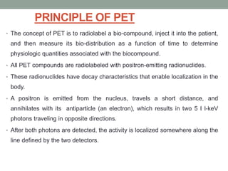
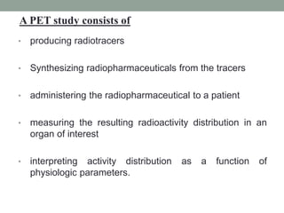
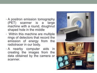
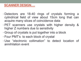
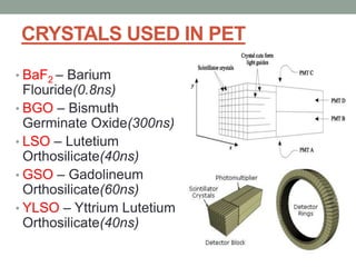
SlideShare a Scribd company logo
SPECT & PET
NAGARAJU.B
• Radionuclides are unstable nuclei
• Having a neutron excess or deficit, are radioactive → 'decay'
to become stable nuclei, with the emission of any combination
of alpha, beta, and gamma radiation.
Production of radionuclides:
• Natural radionuclides: few, sufficiently long lived, e.g.
uranium, radium, and radon.
• Radionuclides in medical imaging → produced artificially, in
the following way
• Some radionucleids produced by additional neutron
forced into a stable nucleus:
• Nucleus has a neutron excess → unstable.
• In a nuclear reactor,
• e.g. with Molybdenum (Mo):
98Mo + n → 99Mo
• Same atomic number.
• Mass number increased by 1.
• Radionuclides produced by this process have same
chemical properties and called "an isotope" .
• Radionuclides produced by additional proton forced into a
stable nucleus → knocking out a neutron:
• Nucleus has a neutron deficit → unstable.
• In a cyclotron which accelerates positively charged
ions: e.g. protons or alpha particles.
• e.g. with Boron (B):
11B + p → 11C + n
• Atomic number increased by 1.
• Same mass number.
Radioactive decay
• Nuclides with a neutron excess:
β - Decay
• Neutron change into a proton & an electron → the electron is
ejected from the nucleus with high energy “-ve beta particle”
n → p + β -
• For example, Iodine-131 (131I, Z= 53) → Xenon-131
(131Xe, Z= 54).
• Same mass number.
• Atomic number increased by 1 .
•The daughter nucleus mostly produced with excess energy
with immediate loss of energy with the emission of one or more
γ photons.
• Isomeric transition: in this some radionucleids the
gamma ray is not emitted until an appreciable time after
the emission of beta particle.
• For ex:99Mo decays by emission of –ve beta particle, the
daughter nucleus Tc remains in exited stable for variable
length of time, it is said to be metastable and is written as
99mTc.
• Its decay to ground state , 99Tc, is most often with
emission of gamma ray of energy 140kev.
• Nuclides with a neutron deficit:
β + Decay
• A proton change into a neutron and a positive electron
→ the latter is ejected from the
nucleus with high energy "+ve β particle".
p → n + β+
• For example,
Carbon-11 (11C, Z= 6) → Boron-11 (11B, Z= 5).
• Same Mass and charge.
• The atomic number decreased by 1 .
•The daughter nucleus, if excited → loses excess energy
by the emission of gamma photons till reaches the ground
state.
• K-electron capture (EC)
• The nucleus capturing an extra-nuclear electron from the
nearest (K) shell → increase its
number of neutrons relative to the number of protons.
p + e → n
• Same Mass and charge.
The atomic number decreased by 1 .
•The daughter nuclide will emit characteristic X-rays
when the hole so created in the Kshell is filled by an
electron from an outer shell ± gamma rays if still excited.
• Iodine-123 (123I) decays by electron capture and emits
160 keV gamma and 28 keV Xrays, but no β-particles.
Radiopharmaceuticals
• Radiopharmaceuticals are the radioactive substances or
radioactive drugs for diagnostic or therapeutic
interventions.it contains
• a radioactive isotope that can be injected safely into the
body, and
• a carrier molecule which delivers the isotope to the area
to be treated or examined.
• Isotope during its conversion to stable forms emits
radiation.
SPECT
• Single-photon emission computed tomography (SPECT)
provides three-dimensional (3D) image information about
the distribution of a radiopharmaceutical injected into the
patient for diagnostic purposes.
• By combining conventional scintigraphic and computed
tomographic methods, SPECT images present 3D
functional information about the patient in more detail and
higher contrast than found in planar scintigrams.
• A typical SPECT system consists of one or more
scintillation cameras that acquire multiple two-dimensional
planar projection images around the patient. The
projection data are reconstructed into 3D images. The
collimator of the scintillation camera has substantial
effects on the spatial resolution and detection efficiency of
the SPECT system.
• Physical factors such as photon attenuation and scatter
affect the quantitative accuracy and quality of SPECT
images, and various methods have been developed to
compensate for these image-degrading effects.
• SPECT studies use standard radionuclides (eg,
technetium-99m or iodine-123) and simpler imaging
instrumentation. These standard radionuclides commonly
emit gamma-ray photons with energies that are much
lower than 511 keV. A typical example is Tc-99m, which
emits 140 keV photons.
• An exception is the widely used myocardial agent,
thallium-201, whose decay to mercury-201 results in the
emission of characteristic mercury x rays with an average
energy of about 72 keV.
In a typical conventional scintigraphy system which is used
in SPECT. A given radioisotope-labeled pharmaceutical is
injected into the patient.
Depending on the biodistribution properties of the
pharmaceutical, the radioactivity is distributed in specific
organs and normal or abnormal tissues.
Gamma-ray photons emitted from the radiopharmaceutical
interact with matter inside the patient’s body. Those
photons that exit the patient’s body and pass through the
holes of a collimator are detected by a position-sensitive
detector.
• Typically a scintillation (or Anger) camera mounted on a
rotating gantry. A typical large field-of-view scintillation camera
consists of a 0.95-cm-thick, 40-cm-collimeter sodium iodide
crystal and an array of photomultiplier tubes at its back. The
detected photons are transformed into visible light (ie,
scintillations), which is converted into electrical signals by the
photomultiplier tubes.
• The magnitudes of the electrical signals are proportional to the
energies of the photons. A pulse height analyzer evaluates the
energies of the detected photons and accepts only those that
fall within a preset energy window, which is centered at the
energy peak of the primary photons; thus, scattered photons
and their adversive effects are rejected.
• The effectiveness of this scatter rejection depends on the
energy resolution of the detector and the width of the
energy window. Finally, a two-dimensional(2D) projection
image of the three-dimensional (3D) radioactivity
distribution is formed from the registered photon counts .
PHYSICALAND INSTRUMENTATION
FACTORS THATAFFECT SPECT IMAGES
• A number of factors substantially affect the quality and
quantitative accuracy of SPECT images.
• They include physical factors, which are related to the
interaction of photons emitted from the
radiopharmaceuticals with matter inside the patient, and
instrumentation factors, which are related to the SPECT
imaging system.
• For photon energies commonly used in scintigraphy and
SPECT are photoelectric and Compton scatter are the
major interactions of radiation with matter.
• In a photoelectric interaction, the photon is totally
absorbed by an atom. In a Compton scatter interaction,
the photon changes its direction of travel, with partial loss
of its original energy.
• Photon attenuation can be defined as the reduction of the
number of primary photons that pass through a given
thickness of material. Photon attenuation is caused by
photoelectric and Compton scatter interactions in the
material.
• The amount of attenuation depends on the incident
photon energy and on the thickness and attenuation
coefficient of the material. For example, the 140-keV
photons from Tc-99m lose half of their original number in
about 4.5 cm of water or soft tissue. As a result, photon
attenuation substantially affects the quantitative accuracy
of scintigraphic and SPECT images.
• Image reconstruction is done from the data acquired.
• There are the two reconstruction algorithma used in
SPECT.
• FILTERED BACK PROJECTION
• ITERATIVE RECONSTRUCTION
Common clinical applications
PRINCIPLE OF PET
• The concept of PET is to radiolabel a bio-compound, inject it into the patient,
and then measure its bio-distribution as a function of time to determine
physiologic quantities associated with the biocompound.
• All PET compounds are radiolabeled with positron-emitting radionuclides.
• These radionuclides have decay characteristics that enable localization in the
body.
• A positron is emitted from the nucleus, travels a short distance, and
annihilates with its antiparticle (an electron), which results in two 5 I l-keV
photons traveling in opposite directions.
• After both photons are detected, the activity is localized somewhere along the
line defined by the two detectors.
• producing radiotracers
• Synthesizing radiopharmaceuticals from the tracers
• administering the radiopharmaceutical to a patient
• measuring the resulting radioactivity distribution in an
organ of interest
• interpreting activity distribution as a function of
physiologic parameters.
A PET study consists of
PRODUCTION OF RADIONUCLIDE
• PET radionuclides are positron emitters.
• There are 5 convenient nuclides-
HALF LIFE (min)
Rubidium- 82 1.23
Fluorine – 18 109
Oxygen- 15 2
Nitrogen- 13 10
Carbon- 11 20
Commonly produced isotopes : “F O N C”
SYNTHESIS OF F-18 &FDG
• Over 500 PET compounds have been synthesized since
1970.
• Natural substrates such as amino acids, analogues
,fluorinated glucose &drugs.
• MC used is a glucose analogue, 2-[F18]fluoro-2-deoxy-D-
glucose (FDG).
Why is 18 F the most used positron
emitter?
• 18F is a small atom.
• its addition to a molecule does not deform it to the point
where it is not recognized by the body anymore
• has a half-life of 109 minutes.
• This is long enough to perform a complicated chemistry
(labelling) , and to allow transport over some distance.
• It is also not long enough to keep the radiation burden to
patient low
USES
Fluorodeoxyglucose
[18F]-labeled 2-deoxyglucose (FDG) is used in neurology,
cardiology and oncology to study glucose metabolism. FDG is
potentially useful in differentiating benign from malignant forms
of lesions because of the high metabolic activity of many types
of aggressive tumors.
Oxygen
[15O]-labeled water is used to evaluate myocardial oxygen
consumption and oxygen extraction fraction. It can also be used
to measure tumor necrosis.
• Ammonia
[13N]-labeled ammonia can be used to measure blood
flow.
• Leucine
[11C]-labeled methionine and leucine can be used to
evaluate amino acid uptake and protein synthesis,
providing an indicator of tumor viability.
• Fluorine Ion
Radiolabeled fluorine ion [18F-] was once a standard
agent for clinical bone scanning.
Principle of metabolic imaging with FDG
Glucose 18 FDG Blood vessel
Cell
Glucose 6 Phosphate 18FDG 6 Phosphate
Fructose 6 Phosphate
Pyruvate
Anaerobic resp Citric acid cycle
G LUT
SCANNER DESIGN
• A positron emission tomography
(PET) scanner is a large
machine with a round, doughnut
shaped hole in the middle
• Within this machine are multiple
rings of detectors that record the
emission of energy from the
radiotracer in our body.
• A nearby computer aids in
creating the images from the
data obtained by the camera or
scanner.
SCANNER DESIGN….
• Detectors are 18-40 rings of crystals forming a
cylindrical field of view about 15cm long that can
acquire many slices of coincidence data
• PET scanners use crystals with higher density &
higher Z numbers due to sensitivity
• Group of crystals is put together into a block
• Four PMT’s to each block of crystal
• Use “electronic collimation” to detect location of
annihilation event
SCANNER DESIGN….
• Localizing the site of impact is achieved by measuring
the light detected in each PMT
• Signal is then amplified
• System must be able to determine which signals come
from paired 511keV photons and record the time of
detection (timing discriminator)
• Coincidence circuit then examines signals to confirm it if
it occurred with in the time window
CRYSTALS USED IN PET
• BaF2 – Barium
Flouride(0.8ns)
• BGO – Bismuth
Germinate Oxide(300ns)
• LSO – Lutetium
Orthosilicate(40ns)
• GSO – Gadolineum
Orthosilicate(60ns)
• YLSO – Yttrium Lutetium
Orthosilicate(40ns)
COINCIDENCE DETECTION
• Photons should arrive with in a certain time of one another
• A coincidence timing window allows detection of the PMT
electrical signal from photon pair (4-12ns)
• If it falls within timing window it is registered as a true event
• When two different annihilation events are detected with in
the timing window is known as “random event”
DATA ACQUSITION
• The detection of photon pairs by opposing crystals create
one event .
• Millions of these event will be stored with in sinograms
and used to reconstruct the image
• Spatial resolution is determined by the size of crystal and
their separation and is typically 3-5mm
• PET is 50-100 times more sensitive and produces higher
quality than a SPECT
• Reconstruction is similar to SPECT
RECONSTRUCTION
• PET reconstruction can be performed with a variety
of algorithms
• Filtered back projection
• Iterative reconstruction(ordered subset
reconstruction )
ATTENUATION CORRECTION
• Mathematical attenuation correction techniques
may be used if tissue attenuation is the same at
all areas within a transaxial slice .
• Measured attenuation may be performed by two
methods:
• Transmission scan using a radioactive source rotating it
around the patient
• CT scan to measure tissue density
• The ability to correct for attenuation improves
quality and permits absolute quantification of
radioactivity in the body
• Resolution in PET is determined by three factors:
• distance the positron travels before it annihilates with an electron,
• variation in angle between the two annihilation photons, and
• physical size of the detectors.
• A positron will travel between 0.5 and 2 mm in tissue
before annihilation, depending on its energy.
• Typical detector sizes are 1 -3 mm.
• The best possible resolution of a PET scanner is 1-2mm.
Typical clinical scanners have a resolution of
approximately 4-7 mm.
PET vs. CT & MRI
PET CT and MRI
Shows extent of disease
Detects changes in body
structure
Can help in monitoring
treatment and shows it’s
effectiveness
Simply confirms the presence
of a mass
Reveals disease earlier, can
diagnose faster
Can detect whether a mass is
benign or malignant
Can detect abnomalities before
there is an anatomical change
SAFETYASPECTS OF PET
• PET has 511Kev gamma rays energy, that is 3 times of 140Kev
gamma ray energy of Technitium99m
• Due to their high energy 16 times more lead is required to obtain the
same stopping effect for 511Kev photons as compared to 140Kev
photons
So Tungsten shielding
is used for Positron
emitting radionuclide.
It provides 1.4 times
the shielding capability
for the same thickness
of Lead
CONTRA INDICATIONS
• Pregnancy
• Use of caffeine, tobacco, or alcohol in
past 24hours before scan
• Using sedatives
• Using medicines that change
metabolism ex: INSULIN.
PET-CT FUSION
• FDG PET is a strictly functional modality and lacks anatomic landmarks.
• Unless anatomic correlation is available to delineate normal structures,
pathologic sites of FDG accumulation can easily be confused with normal
physiologic uptake, leading to false-positive or false-negative findings.
• Coregistration of PET scans with CT using a combined PET-CT scanner
improves the overall sensitivity and specificity of information provided by
PET or CT alone .
• advantage is ability to correlate findings at two complementary imaging
modalities in a comprehensive examination. Hence, PET-CT provides
more precise anatomic definition for both the physiologic and
pathologic uptake seen at FDG PET
Max coverage during combined study
Distance betwn pet and ct scanner
SCANNING TECHNIQUE
• Nil orally for approximately 4–6 hours
• avoid caffeinated or alcoholic beverages but can have water
during this period.
• blood glucose level of less than 150 mg/dL is desirable.
• Avoid strenuous activity to avoid physiologic muscle uptake
of FDG
• water-soluble iodinated contrast media orally for bowel
opacification except for head and neck study.
• 10 mCi injected intravenously
• Patient activity and speech are limited for 20 minutes
immediately following injection
• Pet study is started 60 mins after injection.
CT TECHNIQUE
• Contrast material–enhanced helical CT is performed following
injection of 125 mL of a contrast medium at a rate of 4 mL/sec by
using a power injector .
• Whole-body PET-CT study scanning begins at the level of the skull
base and extends caudally to the level of the symphysis pubis.
PET TECHNIQUE
• The PET scanner is located behind the CT scanner and housed in the
same extended-length gantry. PET is performed following the CT
study without moving the patient in the caudocranial direction, starting
at the thighs to limit artifacts from the FDG metabolite excretion into
the urinary system
Typical scout image obtained during an FDG PET-CT study. The blue-purple rectangle
represents CT coverage during the study, and each overlapping green rectangle
represents PET coverage.
INTERPRETATION OF IMAGES
• PET provides images of quantitative uptake of the
radionuclide injected that can give the concentration of
radiotracer activity in kilobecquerels per milliliter .
• Methods for assessment of radiotracer uptake –
• visual inspection
• standardized uptake value (SUV)
• glucose metabolic rate
LIMITATIONS AND ARTIFACTS OF PET-CT
• 1.Patient motion may cause confusion as to the correct position of the
origin of the detected photon.
• Patient motion is minimized by –
• carefully instructing patients not to move during the study;
• placing them in a comfortable position before the start of the
study;
• ensuring that they are not taking diuretics, which may
otherwise require them to evacuate the bladder during the
study;
• having patients empty their bladder before the start of the
study or catheterizing the bladder.
ADVANTAGES OF PET-CT
1. helpful in accurate localization of small areas of increased
radiotracer activity that would have been difficult or not possible
to localize on PET images alone .
2. helps in distinguishing structures that normally show high
metabolic activity from those with abnormally increased activity.
3. PET-CT combines the advantages of the excellent functional
information provided by PET and the superb spatial and
contrast resolution of CT
4. Finally, attenuation correction for quantitative or semi
quantitative assessment of data is possible by using the CT
data,
PET/MRI: TECHNICAL EVOLUTION
• The idea to combine PET and MRI arose as early as the
mid 1990s, even before PET/CT was introduced.
• The PET/MRI combination requires 3 risky technologic
steps that modify state-of-the-art PET and MRI.
1. First, the photomultiplier technology must be replaced with
magnetic field–insensitive photodiodes .
2. Second, compact PET detectors must be constructed so that it
shouldn't interfere with the field gradients or MR radiofrequency.
3. Finally, the MRI scanner must be adapted to accommodate the
PET detectors and to allow simultaneous data acquisition without
mutual interference.
• Based on the technologic challenges to combine PET and MRI
into a single gantry, Philips and Siemens proposed 2
fundamentally different prototype PET/MRI designs.
• In the Siemens prototypes include 4 dedicated brain PET
scanners that fit into a standard 3-T clinical MRI scanner.
• The PET/MRI system, together with a dedicated radiofrequency
head coil, allows simultaneous PET/MRI data acquisition of the
human brain or body extremities.
• Philips developed a PET/MRI design in which the gantries are
approximately 2.5 m apart but share a common patient handling
system. This implementation does not allow for simultaneous
data acquisition and, therefore, results in longer examination
times.
Scanner Design
CLINICAL POTENTIAL OF PET/MRI
• It is reasonable to expect that brain PET/MRI will provide new
insights in the field of neuroscience and neurologic disorders,
such as neuro degeneration, brain ischemia, neuro oncologyor
seizures .
• It is feasible with current prototypes and future-generation
systems to simultaneously study brain function, metabolism,
oxygen consumption, and perfusion.
• In oncology, an accurate spatial match between PET and MRI
data is mandatory for both radiation therapy planning and biopsy
guidance.
• Combining PET with cardiac MRI may enable detection and
differentiation of vulnerable plaques and diseased myocardium.
Advantage of PET/MRI over PET/CT
1. is not associated with significant radiation exposure
2. has a much higher soft tissue contrast.
3. MRI allows for additional techniques - such as angiography, functional
MRI ,diffusion ,spectroscopy and perfusion techniques within one
single examination.
PHYSIOLOGIC VERSES PATHOLOGIC FDG UPTAKE
• There are several sites of normal physiologic accumulation of FDG.
FDG accumulation is most intense in the cerebral cortex, basal
ganglia, thalamus, and cerebellum. The myocardium expresses
insulin-sensitive glucose transporters, which facilitate the transport of
glucose into muscle. A recent meal often causes intense myocardial
FDG uptake because of the associated elevated serum insulin levels
• Because FDG appears in the glomerular filtrate and, unlike glucose, is
not reabsorbed in the tubules, intense FDG activity is seen in the
intrarenal collecting systems, ureters, and bladder
Physiologic FDG uptake
• Some clinical applications where PET used:
• The Role of PET/CT in Lymphoma
• Assess response to therapy/residual disease
• Identify recurrent disease
• Initial diagnosis and staging
• Identify suitable sites for biopsy
• Disease surveillance
• Radiotherapy planning
An example of stage 4 disease.
NHL with disease in the
mediastinum, neck, and
abdomen
• ASSESSMENT OF TREATMENT RESPONSE
Pretherapy and post
therapy studies
showing a complete
metabolic response
to therapy.
NEURO-ONCOLOGY
Tumor recurrence versus radiation necrosis
Diagnosis
Grading
Monitoring response to therapy
Radiotherapy planning
Biopsy planning
SEIZURE FOCUS IDENTIFICATION
STROKE
DEMENTIAS
OTHER APPLICATIONS
Brain injury- vascular, trauma
Psychiatry- depression, schizophrenia,anxiety
Movement disorders with 18F-dopa
Miscellaneous- infection, substance abuse,eating
disorders
PET
CT
PET/CT
Biopsy Planning
Localizing
the most
viable tissue
in the lesion
PET in Brain Disorders
Simultaneous PET/MRI study in
Alzheimer disease
Hypo metabolism in left temporal lobe secondary to epilepsy
CARDIAC PET and PET CT IMAGING
The stress images
show a severe
perfusion defect
throughout the
anterolateral wall
that is completely
reversible at rest
PET vs. SPECT
• PET have superior sensitivity and resolution
• Greater flexibility of incorporating positron labels into
biomolecules
• PET is more expensive and requires the presence of an
onsite cyclotron
PEM
• Positron Emission Mammography
• PEM is a specialized & improved
form of PET for imaging breasts and
other small body parts.
• Camera and detectors are closer to
the area affected with cancer which
produces a very sharp detailed
image of tumors and cancerous
tissue.
• Can see cancers as small as 1.5 –
2mm about the width of a grain of
rice.
• Also allows for the earlier detection
of elusive cancers such as DCIS
(ductal carcinoma in situ).
PEM
• Differences between whole-body PET and PEM
• better spatial resolution (1-2 mm vs. 5-10 mm). This
comes at the cost of field-of-view
• Photon-Detection Sensitivity
• Closer proximity of PEM detectors increases geometric
sensitivity
• Allows lower dose/faster imaging/longer uptake time
spect and pet

More Related Content

What's hot

Fluoroscopy presentation
Fluoroscopy presentationFluoroscopy presentation
Fluoroscopy presentation
Huzaifa Oxford
 
CT artifact
CT artifact CT artifact
CT artifact
Ganesan Yogananthem
 
SPECT with clinical application
SPECT with clinical application SPECT with clinical application
SPECT with clinical application
عبدالله فهد
 
Quality Assurance Programme in Computed Tomography
Quality Assurance Programme in Computed TomographyQuality Assurance Programme in Computed Tomography
Quality Assurance Programme in Computed Tomography
Ramzee Small
 
CT Scan Image reconstruction
CT Scan Image reconstructionCT Scan Image reconstruction
CT Scan Image reconstructionGunjan Patel
 
Ct scan final (2)
Ct scan final (2)Ct scan final (2)
Ct scan final (2)
shekhar star
 
Basic Pulse Sequences In MRI
Basic Pulse Sequences In MRIBasic Pulse Sequences In MRI
Basic Pulse Sequences In MRI
Upakar Paudel
 
Nuclear imaging
Nuclear imagingNuclear imaging
Nuclear imaging
Shahid Younas
 
MRI
MRIMRI
Basic principles of mri
Basic principles of mriBasic principles of mri
Basic principles of mri
ramya prathyusha
 
MRI 3-Marks QUESTIONS AND ANSWERS
MRI 3-Marks QUESTIONS AND ANSWERSMRI 3-Marks QUESTIONS AND ANSWERS
MRI 3-Marks QUESTIONS AND ANSWERS
Ganesan Yogananthem
 
Nuclear Medicine - PET/CT
Nuclear Medicine - PET/CTNuclear Medicine - PET/CT
Nuclear Medicine - PET/CT
@Saudi_nmc
 
SPECT SCAN
SPECT SCANSPECT SCAN
SPECT SCAN
sensuiii
 
Nuclear medicine
Nuclear medicine Nuclear medicine
Nuclear medicine
Vharshini Manoharan
 
Basics of SPECT, PET and PET/CT imaging
Basics of SPECT, PET and PET/CT imagingBasics of SPECT, PET and PET/CT imaging
Basics of SPECT, PET and PET/CT imaging
@Saudi_nmc
 
Magnetic Resonance Imaging
Magnetic Resonance ImagingMagnetic Resonance Imaging
Magnetic Resonance Imaging
sailakshmi pullookkara
 
Image reconstrsuction in ct pdf
Image reconstrsuction in ct pdfImage reconstrsuction in ct pdf
Image reconstrsuction in ct pdf
mitians
 
Principles and application of PET CT & PET MR
Principles and application of PET CT & PET MRPrinciples and application of PET CT & PET MR
Principles and application of PET CT & PET MRcharusmita chaudhary
 
Computed Tomography Dose Index
Computed Tomography Dose IndexComputed Tomography Dose Index
Computed Tomography Dose Index
Anjan Dangal
 

What's hot (20)

Fluoroscopy presentation
Fluoroscopy presentationFluoroscopy presentation
Fluoroscopy presentation
 
CT artifact
CT artifact CT artifact
CT artifact
 
SPECT with clinical application
SPECT with clinical application SPECT with clinical application
SPECT with clinical application
 
Quality Assurance Programme in Computed Tomography
Quality Assurance Programme in Computed TomographyQuality Assurance Programme in Computed Tomography
Quality Assurance Programme in Computed Tomography
 
CT Scan Image reconstruction
CT Scan Image reconstructionCT Scan Image reconstruction
CT Scan Image reconstruction
 
Ct scan final (2)
Ct scan final (2)Ct scan final (2)
Ct scan final (2)
 
Basic Pulse Sequences In MRI
Basic Pulse Sequences In MRIBasic Pulse Sequences In MRI
Basic Pulse Sequences In MRI
 
Nuclear imaging
Nuclear imagingNuclear imaging
Nuclear imaging
 
MRI
MRIMRI
MRI
 
Basic principles of mri
Basic principles of mriBasic principles of mri
Basic principles of mri
 
MRI 3-Marks QUESTIONS AND ANSWERS
MRI 3-Marks QUESTIONS AND ANSWERSMRI 3-Marks QUESTIONS AND ANSWERS
MRI 3-Marks QUESTIONS AND ANSWERS
 
Nuclear Medicine - PET/CT
Nuclear Medicine - PET/CTNuclear Medicine - PET/CT
Nuclear Medicine - PET/CT
 
SPECT SCAN
SPECT SCANSPECT SCAN
SPECT SCAN
 
Nuclear medicine
Nuclear medicine Nuclear medicine
Nuclear medicine
 
Basics of SPECT, PET and PET/CT imaging
Basics of SPECT, PET and PET/CT imagingBasics of SPECT, PET and PET/CT imaging
Basics of SPECT, PET and PET/CT imaging
 
Magnetic Resonance Imaging
Magnetic Resonance ImagingMagnetic Resonance Imaging
Magnetic Resonance Imaging
 
Image reconstrsuction in ct pdf
Image reconstrsuction in ct pdfImage reconstrsuction in ct pdf
Image reconstrsuction in ct pdf
 
Principles and application of PET CT & PET MR
Principles and application of PET CT & PET MRPrinciples and application of PET CT & PET MR
Principles and application of PET CT & PET MR
 
Spect technology
Spect technologySpect technology
Spect technology
 
Computed Tomography Dose Index
Computed Tomography Dose IndexComputed Tomography Dose Index
Computed Tomography Dose Index
 

Similar to spect and pet

Nuclear imaging in dentistry
Nuclear imaging in dentistryNuclear imaging in dentistry
Nuclear imaging in dentistry
Dr Jinki Singha
 
Radioisotopes and their role in modern era imaging
Radioisotopes and their role in modern era imagingRadioisotopes and their role in modern era imaging
Radioisotopes and their role in modern era imaging
KhanAsma6
 
GAMMA SPECT PET.pptx
GAMMA SPECT PET.pptxGAMMA SPECT PET.pptx
GAMMA SPECT PET.pptx
VishwasLS
 
Radioactivity spectrum of diagnostic imaging and therapy X ray..pptx
Radioactivity spectrum of diagnostic imaging and therapy X ray..pptxRadioactivity spectrum of diagnostic imaging and therapy X ray..pptx
Radioactivity spectrum of diagnostic imaging and therapy X ray..pptx
Dr. Dheeraj Kumar
 
FOURIER TRANSFORM - INFRARED SPECTROSCOPY
FOURIER TRANSFORM - INFRARED SPECTROSCOPYFOURIER TRANSFORM - INFRARED SPECTROSCOPY
FOURIER TRANSFORM - INFRARED SPECTROSCOPY
SalmanLatif14
 
Radiopharmaceutical
Radiopharmaceutical Radiopharmaceutical
Radiopharmaceutical
laraib jameel
 
Unit 8
Unit 8Unit 8
Applications of Isotopes in Medicine
Applications of Isotopes in MedicineApplications of Isotopes in Medicine
Applications of Isotopes in Medicine
Pradeep Singh Narwat
 
Radiochemistry & Radiopharmaceuticals
Radiochemistry & RadiopharmaceuticalsRadiochemistry & Radiopharmaceuticals
Radiochemistry & Radiopharmaceuticals
Prachi Pathak
 
Instrumentation of uv visible spectroscopy
Instrumentation of uv visible spectroscopyInstrumentation of uv visible spectroscopy
Instrumentation of uv visible spectroscopy
Zainab&Sons
 
Nuclear medicine
Nuclear medicineNuclear medicine
Nuclear medicine
Shrikant Nagare
 
RADIOACTIVITY.pptx
RADIOACTIVITY.pptxRADIOACTIVITY.pptx
RADIOACTIVITY.pptx
AddwayChakraborty
 
Prabhakar singh ii sem-paper v-detection & measurement of radioactivity
Prabhakar singh  ii sem-paper v-detection & measurement of radioactivityPrabhakar singh  ii sem-paper v-detection & measurement of radioactivity
Prabhakar singh ii sem-paper v-detection & measurement of radioactivity
Department of Biochemistry, Veer Bahadur Singh Purvanchal Univarsity, Jaunpur
 
X-Ray Production.pptx
X-Ray Production.pptxX-Ray Production.pptx
X-Ray Production.pptx
Sachin Sharma
 
Pet
PetPet
Radiopharmaceutical
Radiopharmaceutical Radiopharmaceutical
Radiopharmaceutical
HimanshuJoshi255
 
Exploring structural and dynamical properties of microtubules by means of A...
Exploring structural and dynamical properties  of microtubules by means of  A...Exploring structural and dynamical properties  of microtubules by means of  A...
Exploring structural and dynamical properties of microtubules by means of A...
Rita Pizzi
 
Analys is techniques
Analys is techniquesAnalys is techniques
Analys is techniques
ShreyaNair37
 
xray generation and it's properties.pptx
xray generation and it's properties.pptxxray generation and it's properties.pptx
xray generation and it's properties.pptx
SHEENUPRIYA1
 

Similar to spect and pet (20)

Nuclear imaging in dentistry
Nuclear imaging in dentistryNuclear imaging in dentistry
Nuclear imaging in dentistry
 
Radioisotopes and their role in modern era imaging
Radioisotopes and their role in modern era imagingRadioisotopes and their role in modern era imaging
Radioisotopes and their role in modern era imaging
 
GAMMA SPECT PET.pptx
GAMMA SPECT PET.pptxGAMMA SPECT PET.pptx
GAMMA SPECT PET.pptx
 
Radioactivity spectrum of diagnostic imaging and therapy X ray..pptx
Radioactivity spectrum of diagnostic imaging and therapy X ray..pptxRadioactivity spectrum of diagnostic imaging and therapy X ray..pptx
Radioactivity spectrum of diagnostic imaging and therapy X ray..pptx
 
Uv spec
Uv specUv spec
Uv spec
 
FOURIER TRANSFORM - INFRARED SPECTROSCOPY
FOURIER TRANSFORM - INFRARED SPECTROSCOPYFOURIER TRANSFORM - INFRARED SPECTROSCOPY
FOURIER TRANSFORM - INFRARED SPECTROSCOPY
 
Radiopharmaceutical
Radiopharmaceutical Radiopharmaceutical
Radiopharmaceutical
 
Unit 8
Unit 8Unit 8
Unit 8
 
Applications of Isotopes in Medicine
Applications of Isotopes in MedicineApplications of Isotopes in Medicine
Applications of Isotopes in Medicine
 
Radiochemistry & Radiopharmaceuticals
Radiochemistry & RadiopharmaceuticalsRadiochemistry & Radiopharmaceuticals
Radiochemistry & Radiopharmaceuticals
 
Instrumentation of uv visible spectroscopy
Instrumentation of uv visible spectroscopyInstrumentation of uv visible spectroscopy
Instrumentation of uv visible spectroscopy
 
Nuclear medicine
Nuclear medicineNuclear medicine
Nuclear medicine
 
RADIOACTIVITY.pptx
RADIOACTIVITY.pptxRADIOACTIVITY.pptx
RADIOACTIVITY.pptx
 
Prabhakar singh ii sem-paper v-detection & measurement of radioactivity
Prabhakar singh  ii sem-paper v-detection & measurement of radioactivityPrabhakar singh  ii sem-paper v-detection & measurement of radioactivity
Prabhakar singh ii sem-paper v-detection & measurement of radioactivity
 
X-Ray Production.pptx
X-Ray Production.pptxX-Ray Production.pptx
X-Ray Production.pptx
 
Pet
PetPet
Pet
 
Radiopharmaceutical
Radiopharmaceutical Radiopharmaceutical
Radiopharmaceutical
 
Exploring structural and dynamical properties of microtubules by means of A...
Exploring structural and dynamical properties  of microtubules by means of  A...Exploring structural and dynamical properties  of microtubules by means of  A...
Exploring structural and dynamical properties of microtubules by means of A...
 
Analys is techniques
Analys is techniquesAnalys is techniques
Analys is techniques
 
xray generation and it's properties.pptx
xray generation and it's properties.pptxxray generation and it's properties.pptx
xray generation and it's properties.pptx
 

Recently uploaded

Polish students' mobility in the Czech Republic
Polish students' mobility in the Czech RepublicPolish students' mobility in the Czech Republic
Polish students' mobility in the Czech Republic
Anna Sz.
 
Sectors of the Indian Economy - Class 10 Study Notes pdf
Sectors of the Indian Economy - Class 10 Study Notes pdfSectors of the Indian Economy - Class 10 Study Notes pdf
Sectors of the Indian Economy - Class 10 Study Notes pdf
Vivekanand Anglo Vedic Academy
 
Cambridge International AS A Level Biology Coursebook - EBook (MaryFosbery J...
Cambridge International AS  A Level Biology Coursebook - EBook (MaryFosbery J...Cambridge International AS  A Level Biology Coursebook - EBook (MaryFosbery J...
Cambridge International AS A Level Biology Coursebook - EBook (MaryFosbery J...
AzmatAli747758
 
Phrasal Verbs.XXXXXXXXXXXXXXXXXXXXXXXXXX
Phrasal Verbs.XXXXXXXXXXXXXXXXXXXXXXXXXXPhrasal Verbs.XXXXXXXXXXXXXXXXXXXXXXXXXX
Phrasal Verbs.XXXXXXXXXXXXXXXXXXXXXXXXXX
MIRIAMSALINAS13
 
How libraries can support authors with open access requirements for UKRI fund...
How libraries can support authors with open access requirements for UKRI fund...How libraries can support authors with open access requirements for UKRI fund...
How libraries can support authors with open access requirements for UKRI fund...
Jisc
 
Ethnobotany and Ethnopharmacology ......
Ethnobotany and Ethnopharmacology ......Ethnobotany and Ethnopharmacology ......
Ethnobotany and Ethnopharmacology ......
Ashokrao Mane college of Pharmacy Peth-Vadgaon
 
2024.06.01 Introducing a competency framework for languag learning materials ...
2024.06.01 Introducing a competency framework for languag learning materials ...2024.06.01 Introducing a competency framework for languag learning materials ...
2024.06.01 Introducing a competency framework for languag learning materials ...
Sandy Millin
 
Chapter 3 - Islamic Banking Products and Services.pptx
Chapter 3 - Islamic Banking Products and Services.pptxChapter 3 - Islamic Banking Products and Services.pptx
Chapter 3 - Islamic Banking Products and Services.pptx
Mohd Adib Abd Muin, Senior Lecturer at Universiti Utara Malaysia
 
aaaaaaaaaaaaaaaaaaaaaaaaaaaaaaaaaaaaaaaaaaaaaaaaaaaaaaa
aaaaaaaaaaaaaaaaaaaaaaaaaaaaaaaaaaaaaaaaaaaaaaaaaaaaaaaaaaaaaaaaaaaaaaaaaaaaaaaaaaaaaaaaaaaaaaaaaaaaaaaaaaaaaa
aaaaaaaaaaaaaaaaaaaaaaaaaaaaaaaaaaaaaaaaaaaaaaaaaaaaaaa
siemaillard
 
Students, digital devices and success - Andreas Schleicher - 27 May 2024..pptx
Students, digital devices and success - Andreas Schleicher - 27 May 2024..pptxStudents, digital devices and success - Andreas Schleicher - 27 May 2024..pptx
Students, digital devices and success - Andreas Schleicher - 27 May 2024..pptx
EduSkills OECD
 
MARUTI SUZUKI- A Successful Joint Venture in India.pptx
MARUTI SUZUKI- A Successful Joint Venture in India.pptxMARUTI SUZUKI- A Successful Joint Venture in India.pptx
MARUTI SUZUKI- A Successful Joint Venture in India.pptx
bennyroshan06
 
TESDA TM1 REVIEWER FOR NATIONAL ASSESSMENT WRITTEN AND ORAL QUESTIONS WITH A...
TESDA TM1 REVIEWER  FOR NATIONAL ASSESSMENT WRITTEN AND ORAL QUESTIONS WITH A...TESDA TM1 REVIEWER  FOR NATIONAL ASSESSMENT WRITTEN AND ORAL QUESTIONS WITH A...
TESDA TM1 REVIEWER FOR NATIONAL ASSESSMENT WRITTEN AND ORAL QUESTIONS WITH A...
EugeneSaldivar
 
1.4 modern child centered education - mahatma gandhi-2.pptx
1.4 modern child centered education - mahatma gandhi-2.pptx1.4 modern child centered education - mahatma gandhi-2.pptx
1.4 modern child centered education - mahatma gandhi-2.pptx
JosvitaDsouza2
 
The Art Pastor's Guide to Sabbath | Steve Thomason
The Art Pastor's Guide to Sabbath | Steve ThomasonThe Art Pastor's Guide to Sabbath | Steve Thomason
The Art Pastor's Guide to Sabbath | Steve Thomason
Steve Thomason
 
Instructions for Submissions thorugh G- Classroom.pptx
Instructions for Submissions thorugh G- Classroom.pptxInstructions for Submissions thorugh G- Classroom.pptx
Instructions for Submissions thorugh G- Classroom.pptx
Jheel Barad
 
The Roman Empire A Historical Colossus.pdf
The Roman Empire A Historical Colossus.pdfThe Roman Empire A Historical Colossus.pdf
The Roman Empire A Historical Colossus.pdf
kaushalkr1407
 
GIÁO ÁN DẠY THÊM (KẾ HOẠCH BÀI BUỔI 2) - TIẾNG ANH 8 GLOBAL SUCCESS (2 CỘT) N...
GIÁO ÁN DẠY THÊM (KẾ HOẠCH BÀI BUỔI 2) - TIẾNG ANH 8 GLOBAL SUCCESS (2 CỘT) N...GIÁO ÁN DẠY THÊM (KẾ HOẠCH BÀI BUỔI 2) - TIẾNG ANH 8 GLOBAL SUCCESS (2 CỘT) N...
GIÁO ÁN DẠY THÊM (KẾ HOẠCH BÀI BUỔI 2) - TIẾNG ANH 8 GLOBAL SUCCESS (2 CỘT) N...
Nguyen Thanh Tu Collection
 
Welcome to TechSoup New Member Orientation and Q&A (May 2024).pdf
Welcome to TechSoup   New Member Orientation and Q&A (May 2024).pdfWelcome to TechSoup   New Member Orientation and Q&A (May 2024).pdf
Welcome to TechSoup New Member Orientation and Q&A (May 2024).pdf
TechSoup
 
How to Break the cycle of negative Thoughts
How to Break the cycle of negative ThoughtsHow to Break the cycle of negative Thoughts
How to Break the cycle of negative Thoughts
Col Mukteshwar Prasad
 
Mule 4.6 & Java 17 Upgrade | MuleSoft Mysore Meetup #46
Mule 4.6 & Java 17 Upgrade | MuleSoft Mysore Meetup #46Mule 4.6 & Java 17 Upgrade | MuleSoft Mysore Meetup #46
Mule 4.6 & Java 17 Upgrade | MuleSoft Mysore Meetup #46
MysoreMuleSoftMeetup
 

Recently uploaded (20)

Polish students' mobility in the Czech Republic
Polish students' mobility in the Czech RepublicPolish students' mobility in the Czech Republic
Polish students' mobility in the Czech Republic
 
Sectors of the Indian Economy - Class 10 Study Notes pdf
Sectors of the Indian Economy - Class 10 Study Notes pdfSectors of the Indian Economy - Class 10 Study Notes pdf
Sectors of the Indian Economy - Class 10 Study Notes pdf
 
Cambridge International AS A Level Biology Coursebook - EBook (MaryFosbery J...
Cambridge International AS  A Level Biology Coursebook - EBook (MaryFosbery J...Cambridge International AS  A Level Biology Coursebook - EBook (MaryFosbery J...
Cambridge International AS A Level Biology Coursebook - EBook (MaryFosbery J...
 
Phrasal Verbs.XXXXXXXXXXXXXXXXXXXXXXXXXX
Phrasal Verbs.XXXXXXXXXXXXXXXXXXXXXXXXXXPhrasal Verbs.XXXXXXXXXXXXXXXXXXXXXXXXXX
Phrasal Verbs.XXXXXXXXXXXXXXXXXXXXXXXXXX
 
How libraries can support authors with open access requirements for UKRI fund...
How libraries can support authors with open access requirements for UKRI fund...How libraries can support authors with open access requirements for UKRI fund...
How libraries can support authors with open access requirements for UKRI fund...
 
Ethnobotany and Ethnopharmacology ......
Ethnobotany and Ethnopharmacology ......Ethnobotany and Ethnopharmacology ......
Ethnobotany and Ethnopharmacology ......
 
2024.06.01 Introducing a competency framework for languag learning materials ...
2024.06.01 Introducing a competency framework for languag learning materials ...2024.06.01 Introducing a competency framework for languag learning materials ...
2024.06.01 Introducing a competency framework for languag learning materials ...
 
Chapter 3 - Islamic Banking Products and Services.pptx
Chapter 3 - Islamic Banking Products and Services.pptxChapter 3 - Islamic Banking Products and Services.pptx
Chapter 3 - Islamic Banking Products and Services.pptx
 
aaaaaaaaaaaaaaaaaaaaaaaaaaaaaaaaaaaaaaaaaaaaaaaaaaaaaaa
aaaaaaaaaaaaaaaaaaaaaaaaaaaaaaaaaaaaaaaaaaaaaaaaaaaaaaaaaaaaaaaaaaaaaaaaaaaaaaaaaaaaaaaaaaaaaaaaaaaaaaaaaaaaaa
aaaaaaaaaaaaaaaaaaaaaaaaaaaaaaaaaaaaaaaaaaaaaaaaaaaaaaa
 
Students, digital devices and success - Andreas Schleicher - 27 May 2024..pptx
Students, digital devices and success - Andreas Schleicher - 27 May 2024..pptxStudents, digital devices and success - Andreas Schleicher - 27 May 2024..pptx
Students, digital devices and success - Andreas Schleicher - 27 May 2024..pptx
 
MARUTI SUZUKI- A Successful Joint Venture in India.pptx
MARUTI SUZUKI- A Successful Joint Venture in India.pptxMARUTI SUZUKI- A Successful Joint Venture in India.pptx
MARUTI SUZUKI- A Successful Joint Venture in India.pptx
 
TESDA TM1 REVIEWER FOR NATIONAL ASSESSMENT WRITTEN AND ORAL QUESTIONS WITH A...
TESDA TM1 REVIEWER  FOR NATIONAL ASSESSMENT WRITTEN AND ORAL QUESTIONS WITH A...TESDA TM1 REVIEWER  FOR NATIONAL ASSESSMENT WRITTEN AND ORAL QUESTIONS WITH A...
TESDA TM1 REVIEWER FOR NATIONAL ASSESSMENT WRITTEN AND ORAL QUESTIONS WITH A...
 
1.4 modern child centered education - mahatma gandhi-2.pptx
1.4 modern child centered education - mahatma gandhi-2.pptx1.4 modern child centered education - mahatma gandhi-2.pptx
1.4 modern child centered education - mahatma gandhi-2.pptx
 
The Art Pastor's Guide to Sabbath | Steve Thomason
The Art Pastor's Guide to Sabbath | Steve ThomasonThe Art Pastor's Guide to Sabbath | Steve Thomason
The Art Pastor's Guide to Sabbath | Steve Thomason
 
Instructions for Submissions thorugh G- Classroom.pptx
Instructions for Submissions thorugh G- Classroom.pptxInstructions for Submissions thorugh G- Classroom.pptx
Instructions for Submissions thorugh G- Classroom.pptx
 
The Roman Empire A Historical Colossus.pdf
The Roman Empire A Historical Colossus.pdfThe Roman Empire A Historical Colossus.pdf
The Roman Empire A Historical Colossus.pdf
 
GIÁO ÁN DẠY THÊM (KẾ HOẠCH BÀI BUỔI 2) - TIẾNG ANH 8 GLOBAL SUCCESS (2 CỘT) N...
GIÁO ÁN DẠY THÊM (KẾ HOẠCH BÀI BUỔI 2) - TIẾNG ANH 8 GLOBAL SUCCESS (2 CỘT) N...GIÁO ÁN DẠY THÊM (KẾ HOẠCH BÀI BUỔI 2) - TIẾNG ANH 8 GLOBAL SUCCESS (2 CỘT) N...
GIÁO ÁN DẠY THÊM (KẾ HOẠCH BÀI BUỔI 2) - TIẾNG ANH 8 GLOBAL SUCCESS (2 CỘT) N...
 
Welcome to TechSoup New Member Orientation and Q&A (May 2024).pdf
Welcome to TechSoup   New Member Orientation and Q&A (May 2024).pdfWelcome to TechSoup   New Member Orientation and Q&A (May 2024).pdf
Welcome to TechSoup New Member Orientation and Q&A (May 2024).pdf
 
How to Break the cycle of negative Thoughts
How to Break the cycle of negative ThoughtsHow to Break the cycle of negative Thoughts
How to Break the cycle of negative Thoughts
 
Mule 4.6 & Java 17 Upgrade | MuleSoft Mysore Meetup #46
Mule 4.6 & Java 17 Upgrade | MuleSoft Mysore Meetup #46Mule 4.6 & Java 17 Upgrade | MuleSoft Mysore Meetup #46
Mule 4.6 & Java 17 Upgrade | MuleSoft Mysore Meetup #46
 

spect and pet

  • 2. • Radionuclides are unstable nuclei • Having a neutron excess or deficit, are radioactive → 'decay' to become stable nuclei, with the emission of any combination of alpha, beta, and gamma radiation. Production of radionuclides: • Natural radionuclides: few, sufficiently long lived, e.g. uranium, radium, and radon. • Radionuclides in medical imaging → produced artificially, in the following way
  • 3. • Some radionucleids produced by additional neutron forced into a stable nucleus: • Nucleus has a neutron excess → unstable. • In a nuclear reactor, • e.g. with Molybdenum (Mo): 98Mo + n → 99Mo • Same atomic number. • Mass number increased by 1. • Radionuclides produced by this process have same chemical properties and called "an isotope" .
  • 4. • Radionuclides produced by additional proton forced into a stable nucleus → knocking out a neutron: • Nucleus has a neutron deficit → unstable. • In a cyclotron which accelerates positively charged ions: e.g. protons or alpha particles. • e.g. with Boron (B): 11B + p → 11C + n • Atomic number increased by 1. • Same mass number.
  • 5. Radioactive decay • Nuclides with a neutron excess: β - Decay • Neutron change into a proton & an electron → the electron is ejected from the nucleus with high energy “-ve beta particle” n → p + β - • For example, Iodine-131 (131I, Z= 53) → Xenon-131 (131Xe, Z= 54). • Same mass number. • Atomic number increased by 1 . •The daughter nucleus mostly produced with excess energy with immediate loss of energy with the emission of one or more γ photons.
  • 6. • Isomeric transition: in this some radionucleids the gamma ray is not emitted until an appreciable time after the emission of beta particle. • For ex:99Mo decays by emission of –ve beta particle, the daughter nucleus Tc remains in exited stable for variable length of time, it is said to be metastable and is written as 99mTc. • Its decay to ground state , 99Tc, is most often with emission of gamma ray of energy 140kev.
  • 7. • Nuclides with a neutron deficit: β + Decay • A proton change into a neutron and a positive electron → the latter is ejected from the nucleus with high energy "+ve β particle". p → n + β+ • For example, Carbon-11 (11C, Z= 6) → Boron-11 (11B, Z= 5). • Same Mass and charge. • The atomic number decreased by 1 . •The daughter nucleus, if excited → loses excess energy by the emission of gamma photons till reaches the ground state.
  • 8. • K-electron capture (EC) • The nucleus capturing an extra-nuclear electron from the nearest (K) shell → increase its number of neutrons relative to the number of protons. p + e → n • Same Mass and charge. The atomic number decreased by 1 . •The daughter nuclide will emit characteristic X-rays when the hole so created in the Kshell is filled by an electron from an outer shell ± gamma rays if still excited. • Iodine-123 (123I) decays by electron capture and emits 160 keV gamma and 28 keV Xrays, but no β-particles.
  • 9. Radiopharmaceuticals • Radiopharmaceuticals are the radioactive substances or radioactive drugs for diagnostic or therapeutic interventions.it contains • a radioactive isotope that can be injected safely into the body, and • a carrier molecule which delivers the isotope to the area to be treated or examined. • Isotope during its conversion to stable forms emits radiation.
  • 10. SPECT • Single-photon emission computed tomography (SPECT) provides three-dimensional (3D) image information about the distribution of a radiopharmaceutical injected into the patient for diagnostic purposes. • By combining conventional scintigraphic and computed tomographic methods, SPECT images present 3D functional information about the patient in more detail and higher contrast than found in planar scintigrams.
  • 11. • A typical SPECT system consists of one or more scintillation cameras that acquire multiple two-dimensional planar projection images around the patient. The projection data are reconstructed into 3D images. The collimator of the scintillation camera has substantial effects on the spatial resolution and detection efficiency of the SPECT system. • Physical factors such as photon attenuation and scatter affect the quantitative accuracy and quality of SPECT images, and various methods have been developed to compensate for these image-degrading effects.
  • 12. • SPECT studies use standard radionuclides (eg, technetium-99m or iodine-123) and simpler imaging instrumentation. These standard radionuclides commonly emit gamma-ray photons with energies that are much lower than 511 keV. A typical example is Tc-99m, which emits 140 keV photons. • An exception is the widely used myocardial agent, thallium-201, whose decay to mercury-201 results in the emission of characteristic mercury x rays with an average energy of about 72 keV.
  • 13. In a typical conventional scintigraphy system which is used in SPECT. A given radioisotope-labeled pharmaceutical is injected into the patient. Depending on the biodistribution properties of the pharmaceutical, the radioactivity is distributed in specific organs and normal or abnormal tissues. Gamma-ray photons emitted from the radiopharmaceutical interact with matter inside the patient’s body. Those photons that exit the patient’s body and pass through the holes of a collimator are detected by a position-sensitive detector.
  • 14. • Typically a scintillation (or Anger) camera mounted on a rotating gantry. A typical large field-of-view scintillation camera consists of a 0.95-cm-thick, 40-cm-collimeter sodium iodide crystal and an array of photomultiplier tubes at its back. The detected photons are transformed into visible light (ie, scintillations), which is converted into electrical signals by the photomultiplier tubes. • The magnitudes of the electrical signals are proportional to the energies of the photons. A pulse height analyzer evaluates the energies of the detected photons and accepts only those that fall within a preset energy window, which is centered at the energy peak of the primary photons; thus, scattered photons and their adversive effects are rejected.
  • 15. • The effectiveness of this scatter rejection depends on the energy resolution of the detector and the width of the energy window. Finally, a two-dimensional(2D) projection image of the three-dimensional (3D) radioactivity distribution is formed from the registered photon counts .
  • 16.
  • 17. PHYSICALAND INSTRUMENTATION FACTORS THATAFFECT SPECT IMAGES • A number of factors substantially affect the quality and quantitative accuracy of SPECT images. • They include physical factors, which are related to the interaction of photons emitted from the radiopharmaceuticals with matter inside the patient, and instrumentation factors, which are related to the SPECT imaging system. • For photon energies commonly used in scintigraphy and SPECT are photoelectric and Compton scatter are the major interactions of radiation with matter.
  • 18. • In a photoelectric interaction, the photon is totally absorbed by an atom. In a Compton scatter interaction, the photon changes its direction of travel, with partial loss of its original energy.
  • 19. • Photon attenuation can be defined as the reduction of the number of primary photons that pass through a given thickness of material. Photon attenuation is caused by photoelectric and Compton scatter interactions in the material. • The amount of attenuation depends on the incident photon energy and on the thickness and attenuation coefficient of the material. For example, the 140-keV photons from Tc-99m lose half of their original number in about 4.5 cm of water or soft tissue. As a result, photon attenuation substantially affects the quantitative accuracy of scintigraphic and SPECT images.
  • 20. • Image reconstruction is done from the data acquired. • There are the two reconstruction algorithma used in SPECT. • FILTERED BACK PROJECTION • ITERATIVE RECONSTRUCTION
  • 21.
  • 22.
  • 24. PRINCIPLE OF PET • The concept of PET is to radiolabel a bio-compound, inject it into the patient, and then measure its bio-distribution as a function of time to determine physiologic quantities associated with the biocompound. • All PET compounds are radiolabeled with positron-emitting radionuclides. • These radionuclides have decay characteristics that enable localization in the body. • A positron is emitted from the nucleus, travels a short distance, and annihilates with its antiparticle (an electron), which results in two 5 I l-keV photons traveling in opposite directions. • After both photons are detected, the activity is localized somewhere along the line defined by the two detectors.
  • 25.
  • 26. • producing radiotracers • Synthesizing radiopharmaceuticals from the tracers • administering the radiopharmaceutical to a patient • measuring the resulting radioactivity distribution in an organ of interest • interpreting activity distribution as a function of physiologic parameters. A PET study consists of
  • 27. PRODUCTION OF RADIONUCLIDE • PET radionuclides are positron emitters. • There are 5 convenient nuclides- HALF LIFE (min) Rubidium- 82 1.23 Fluorine – 18 109 Oxygen- 15 2 Nitrogen- 13 10 Carbon- 11 20 Commonly produced isotopes : “F O N C”
  • 28. SYNTHESIS OF F-18 &FDG • Over 500 PET compounds have been synthesized since 1970. • Natural substrates such as amino acids, analogues ,fluorinated glucose &drugs. • MC used is a glucose analogue, 2-[F18]fluoro-2-deoxy-D- glucose (FDG).
  • 29. Why is 18 F the most used positron emitter? • 18F is a small atom. • its addition to a molecule does not deform it to the point where it is not recognized by the body anymore • has a half-life of 109 minutes. • This is long enough to perform a complicated chemistry (labelling) , and to allow transport over some distance. • It is also not long enough to keep the radiation burden to patient low
  • 30. USES Fluorodeoxyglucose [18F]-labeled 2-deoxyglucose (FDG) is used in neurology, cardiology and oncology to study glucose metabolism. FDG is potentially useful in differentiating benign from malignant forms of lesions because of the high metabolic activity of many types of aggressive tumors. Oxygen [15O]-labeled water is used to evaluate myocardial oxygen consumption and oxygen extraction fraction. It can also be used to measure tumor necrosis.
  • 31. • Ammonia [13N]-labeled ammonia can be used to measure blood flow. • Leucine [11C]-labeled methionine and leucine can be used to evaluate amino acid uptake and protein synthesis, providing an indicator of tumor viability. • Fluorine Ion Radiolabeled fluorine ion [18F-] was once a standard agent for clinical bone scanning.
  • 32. Principle of metabolic imaging with FDG Glucose 18 FDG Blood vessel Cell Glucose 6 Phosphate 18FDG 6 Phosphate Fructose 6 Phosphate Pyruvate Anaerobic resp Citric acid cycle G LUT
  • 33. SCANNER DESIGN • A positron emission tomography (PET) scanner is a large machine with a round, doughnut shaped hole in the middle • Within this machine are multiple rings of detectors that record the emission of energy from the radiotracer in our body. • A nearby computer aids in creating the images from the data obtained by the camera or scanner.
  • 34. SCANNER DESIGN…. • Detectors are 18-40 rings of crystals forming a cylindrical field of view about 15cm long that can acquire many slices of coincidence data • PET scanners use crystals with higher density & higher Z numbers due to sensitivity • Group of crystals is put together into a block • Four PMT’s to each block of crystal • Use “electronic collimation” to detect location of annihilation event
  • 35. SCANNER DESIGN…. • Localizing the site of impact is achieved by measuring the light detected in each PMT • Signal is then amplified • System must be able to determine which signals come from paired 511keV photons and record the time of detection (timing discriminator) • Coincidence circuit then examines signals to confirm it if it occurred with in the time window
  • 36. CRYSTALS USED IN PET • BaF2 – Barium Flouride(0.8ns) • BGO – Bismuth Germinate Oxide(300ns) • LSO – Lutetium Orthosilicate(40ns) • GSO – Gadolineum Orthosilicate(60ns) • YLSO – Yttrium Lutetium Orthosilicate(40ns)
  • 37. COINCIDENCE DETECTION • Photons should arrive with in a certain time of one another • A coincidence timing window allows detection of the PMT electrical signal from photon pair (4-12ns) • If it falls within timing window it is registered as a true event • When two different annihilation events are detected with in the timing window is known as “random event”
  • 38. DATA ACQUSITION • The detection of photon pairs by opposing crystals create one event . • Millions of these event will be stored with in sinograms and used to reconstruct the image • Spatial resolution is determined by the size of crystal and their separation and is typically 3-5mm • PET is 50-100 times more sensitive and produces higher quality than a SPECT • Reconstruction is similar to SPECT
  • 39. RECONSTRUCTION • PET reconstruction can be performed with a variety of algorithms • Filtered back projection • Iterative reconstruction(ordered subset reconstruction )
  • 40. ATTENUATION CORRECTION • Mathematical attenuation correction techniques may be used if tissue attenuation is the same at all areas within a transaxial slice . • Measured attenuation may be performed by two methods: • Transmission scan using a radioactive source rotating it around the patient • CT scan to measure tissue density • The ability to correct for attenuation improves quality and permits absolute quantification of radioactivity in the body
  • 41. • Resolution in PET is determined by three factors: • distance the positron travels before it annihilates with an electron, • variation in angle between the two annihilation photons, and • physical size of the detectors. • A positron will travel between 0.5 and 2 mm in tissue before annihilation, depending on its energy. • Typical detector sizes are 1 -3 mm. • The best possible resolution of a PET scanner is 1-2mm. Typical clinical scanners have a resolution of approximately 4-7 mm.
  • 42. PET vs. CT & MRI PET CT and MRI Shows extent of disease Detects changes in body structure Can help in monitoring treatment and shows it’s effectiveness Simply confirms the presence of a mass Reveals disease earlier, can diagnose faster Can detect whether a mass is benign or malignant Can detect abnomalities before there is an anatomical change
  • 43. SAFETYASPECTS OF PET • PET has 511Kev gamma rays energy, that is 3 times of 140Kev gamma ray energy of Technitium99m • Due to their high energy 16 times more lead is required to obtain the same stopping effect for 511Kev photons as compared to 140Kev photons
  • 44. So Tungsten shielding is used for Positron emitting radionuclide. It provides 1.4 times the shielding capability for the same thickness of Lead
  • 45. CONTRA INDICATIONS • Pregnancy • Use of caffeine, tobacco, or alcohol in past 24hours before scan • Using sedatives • Using medicines that change metabolism ex: INSULIN.
  • 46. PET-CT FUSION • FDG PET is a strictly functional modality and lacks anatomic landmarks. • Unless anatomic correlation is available to delineate normal structures, pathologic sites of FDG accumulation can easily be confused with normal physiologic uptake, leading to false-positive or false-negative findings. • Coregistration of PET scans with CT using a combined PET-CT scanner improves the overall sensitivity and specificity of information provided by PET or CT alone . • advantage is ability to correlate findings at two complementary imaging modalities in a comprehensive examination. Hence, PET-CT provides more precise anatomic definition for both the physiologic and pathologic uptake seen at FDG PET
  • 47. Max coverage during combined study Distance betwn pet and ct scanner
  • 48. SCANNING TECHNIQUE • Nil orally for approximately 4–6 hours • avoid caffeinated or alcoholic beverages but can have water during this period. • blood glucose level of less than 150 mg/dL is desirable. • Avoid strenuous activity to avoid physiologic muscle uptake of FDG • water-soluble iodinated contrast media orally for bowel opacification except for head and neck study. • 10 mCi injected intravenously • Patient activity and speech are limited for 20 minutes immediately following injection • Pet study is started 60 mins after injection.
  • 49. CT TECHNIQUE • Contrast material–enhanced helical CT is performed following injection of 125 mL of a contrast medium at a rate of 4 mL/sec by using a power injector . • Whole-body PET-CT study scanning begins at the level of the skull base and extends caudally to the level of the symphysis pubis. PET TECHNIQUE • The PET scanner is located behind the CT scanner and housed in the same extended-length gantry. PET is performed following the CT study without moving the patient in the caudocranial direction, starting at the thighs to limit artifacts from the FDG metabolite excretion into the urinary system
  • 50. Typical scout image obtained during an FDG PET-CT study. The blue-purple rectangle represents CT coverage during the study, and each overlapping green rectangle represents PET coverage.
  • 51. INTERPRETATION OF IMAGES • PET provides images of quantitative uptake of the radionuclide injected that can give the concentration of radiotracer activity in kilobecquerels per milliliter . • Methods for assessment of radiotracer uptake – • visual inspection • standardized uptake value (SUV) • glucose metabolic rate
  • 52. LIMITATIONS AND ARTIFACTS OF PET-CT • 1.Patient motion may cause confusion as to the correct position of the origin of the detected photon. • Patient motion is minimized by – • carefully instructing patients not to move during the study; • placing them in a comfortable position before the start of the study; • ensuring that they are not taking diuretics, which may otherwise require them to evacuate the bladder during the study; • having patients empty their bladder before the start of the study or catheterizing the bladder.
  • 53. ADVANTAGES OF PET-CT 1. helpful in accurate localization of small areas of increased radiotracer activity that would have been difficult or not possible to localize on PET images alone . 2. helps in distinguishing structures that normally show high metabolic activity from those with abnormally increased activity. 3. PET-CT combines the advantages of the excellent functional information provided by PET and the superb spatial and contrast resolution of CT 4. Finally, attenuation correction for quantitative or semi quantitative assessment of data is possible by using the CT data,
  • 54. PET/MRI: TECHNICAL EVOLUTION • The idea to combine PET and MRI arose as early as the mid 1990s, even before PET/CT was introduced. • The PET/MRI combination requires 3 risky technologic steps that modify state-of-the-art PET and MRI. 1. First, the photomultiplier technology must be replaced with magnetic field–insensitive photodiodes . 2. Second, compact PET detectors must be constructed so that it shouldn't interfere with the field gradients or MR radiofrequency. 3. Finally, the MRI scanner must be adapted to accommodate the PET detectors and to allow simultaneous data acquisition without mutual interference.
  • 55. • Based on the technologic challenges to combine PET and MRI into a single gantry, Philips and Siemens proposed 2 fundamentally different prototype PET/MRI designs. • In the Siemens prototypes include 4 dedicated brain PET scanners that fit into a standard 3-T clinical MRI scanner. • The PET/MRI system, together with a dedicated radiofrequency head coil, allows simultaneous PET/MRI data acquisition of the human brain or body extremities. • Philips developed a PET/MRI design in which the gantries are approximately 2.5 m apart but share a common patient handling system. This implementation does not allow for simultaneous data acquisition and, therefore, results in longer examination times. Scanner Design
  • 56.
  • 57. CLINICAL POTENTIAL OF PET/MRI • It is reasonable to expect that brain PET/MRI will provide new insights in the field of neuroscience and neurologic disorders, such as neuro degeneration, brain ischemia, neuro oncologyor seizures . • It is feasible with current prototypes and future-generation systems to simultaneously study brain function, metabolism, oxygen consumption, and perfusion. • In oncology, an accurate spatial match between PET and MRI data is mandatory for both radiation therapy planning and biopsy guidance. • Combining PET with cardiac MRI may enable detection and differentiation of vulnerable plaques and diseased myocardium.
  • 58. Advantage of PET/MRI over PET/CT 1. is not associated with significant radiation exposure 2. has a much higher soft tissue contrast. 3. MRI allows for additional techniques - such as angiography, functional MRI ,diffusion ,spectroscopy and perfusion techniques within one single examination.
  • 59. PHYSIOLOGIC VERSES PATHOLOGIC FDG UPTAKE • There are several sites of normal physiologic accumulation of FDG. FDG accumulation is most intense in the cerebral cortex, basal ganglia, thalamus, and cerebellum. The myocardium expresses insulin-sensitive glucose transporters, which facilitate the transport of glucose into muscle. A recent meal often causes intense myocardial FDG uptake because of the associated elevated serum insulin levels • Because FDG appears in the glomerular filtrate and, unlike glucose, is not reabsorbed in the tubules, intense FDG activity is seen in the intrarenal collecting systems, ureters, and bladder
  • 61. • Some clinical applications where PET used: • The Role of PET/CT in Lymphoma • Assess response to therapy/residual disease • Identify recurrent disease • Initial diagnosis and staging • Identify suitable sites for biopsy • Disease surveillance • Radiotherapy planning
  • 62. An example of stage 4 disease. NHL with disease in the mediastinum, neck, and abdomen
  • 63. • ASSESSMENT OF TREATMENT RESPONSE Pretherapy and post therapy studies showing a complete metabolic response to therapy.
  • 64. NEURO-ONCOLOGY Tumor recurrence versus radiation necrosis Diagnosis Grading Monitoring response to therapy Radiotherapy planning Biopsy planning SEIZURE FOCUS IDENTIFICATION STROKE DEMENTIAS OTHER APPLICATIONS Brain injury- vascular, trauma Psychiatry- depression, schizophrenia,anxiety Movement disorders with 18F-dopa Miscellaneous- infection, substance abuse,eating disorders
  • 66. PET in Brain Disorders Simultaneous PET/MRI study in Alzheimer disease
  • 67. Hypo metabolism in left temporal lobe secondary to epilepsy
  • 68. CARDIAC PET and PET CT IMAGING The stress images show a severe perfusion defect throughout the anterolateral wall that is completely reversible at rest
  • 69. PET vs. SPECT • PET have superior sensitivity and resolution • Greater flexibility of incorporating positron labels into biomolecules • PET is more expensive and requires the presence of an onsite cyclotron
  • 70. PEM • Positron Emission Mammography • PEM is a specialized & improved form of PET for imaging breasts and other small body parts. • Camera and detectors are closer to the area affected with cancer which produces a very sharp detailed image of tumors and cancerous tissue. • Can see cancers as small as 1.5 – 2mm about the width of a grain of rice. • Also allows for the earlier detection of elusive cancers such as DCIS (ductal carcinoma in situ).
  • 71. PEM
  • 72. • Differences between whole-body PET and PEM • better spatial resolution (1-2 mm vs. 5-10 mm). This comes at the cost of field-of-view • Photon-Detection Sensitivity • Closer proximity of PEM detectors increases geometric sensitivity • Allows lower dose/faster imaging/longer uptake time