Download as PDF, PPTX

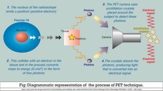
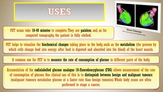

The document discusses Positron Emission Tomography (PET), a non-toxic imaging technique used in nuclear medicine that provides insights into tissue biochemistry and helps evaluate various medical conditions. It involves the injection of a radioactive tracer, which emits radiation detectable by a PET camera, and is commonly used for diagnosing neurological diseases, cancer, and assessing heart function. PET scans are typically painless, take 10-40 minutes, and may be combined with other imaging methods like CT or MRI for enhanced diagnostic accuracy.
![PET - Production of [18F] PET tracers: Beyond [18F]FDG](https://cdn.slidesharecdn.com/ss_thumbnails/jq5oqe2qnkyw3vzn1pyb-signature-e2021a44809bb314ac99609dc68de0b69b8ecda2aded008bfe49c080b80c183b-poli-180221142953-thumbnail.jpg?width=640&height=640&fit=bounds)


