Downloaded 309 times


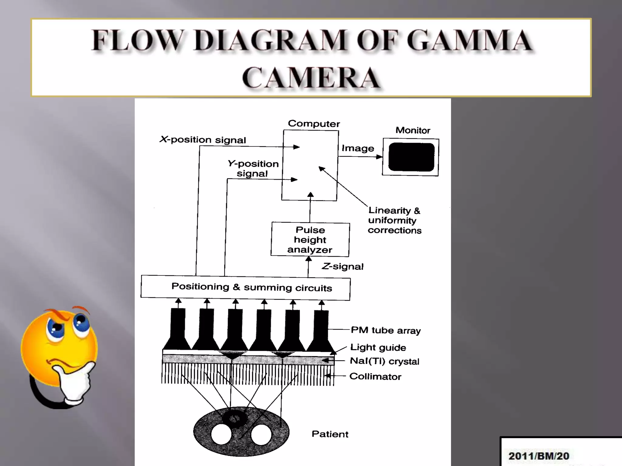


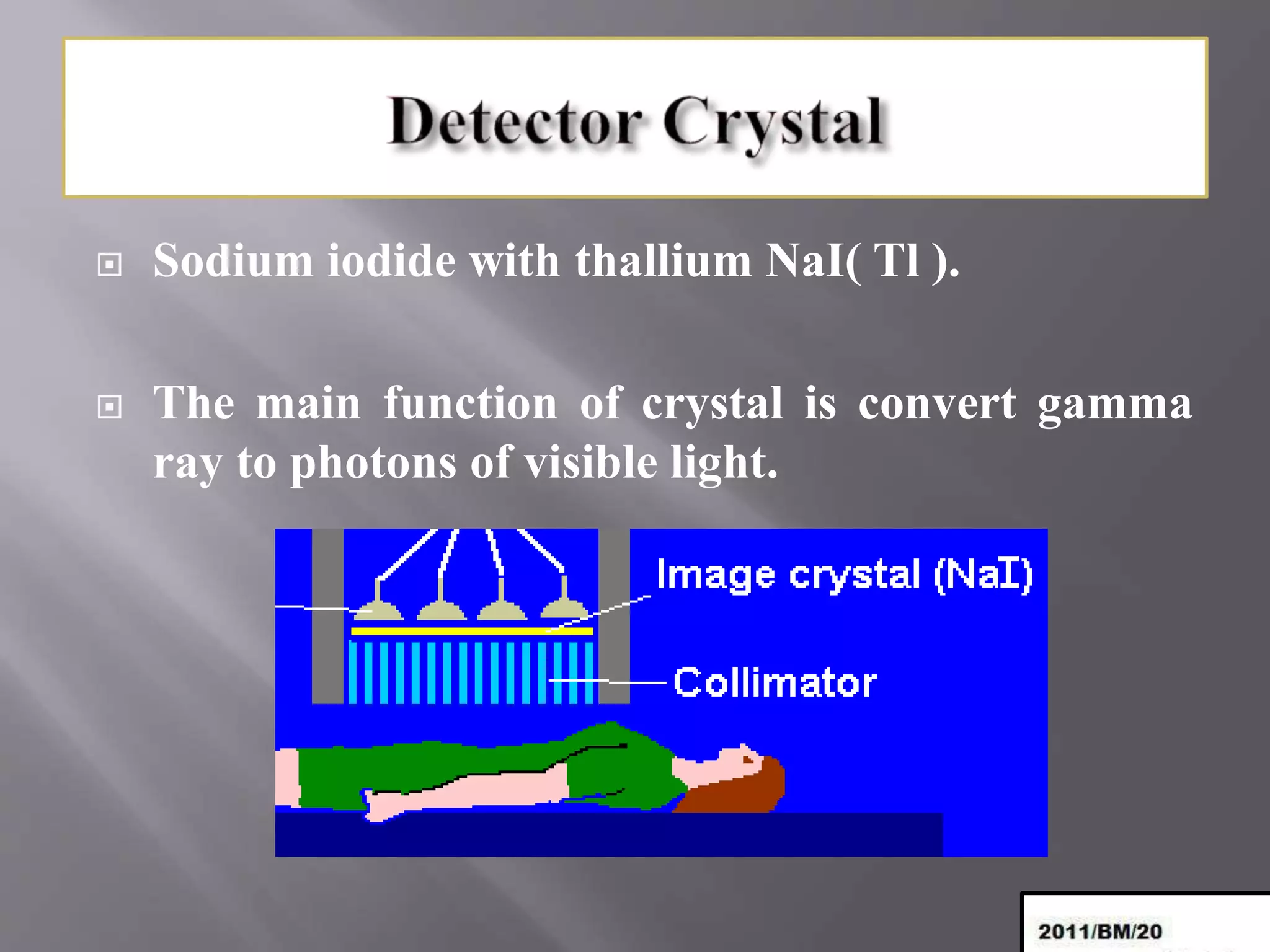


The gamma camera, also known as the Anger camera, was developed in 1957 to detect gamma rays emitted from radiotracers introduced into the body. It uses a collimator, sodium iodide crystal, photomultiplier tubes, preamplifiers, and other components to detect gamma rays and determine their position, which can then be plotted and displayed. The gamma camera is used to scan the whole body and produce anatomical images using computer reconstruction of the gamma ray emission data.


