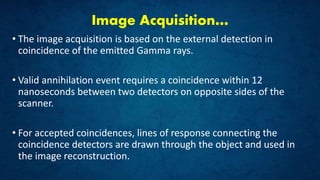
Downloaded 754 times

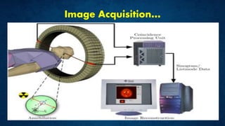
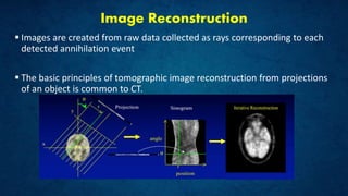
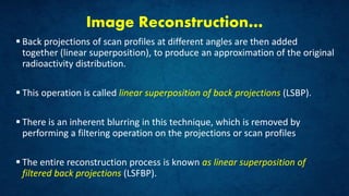
![Image Reconstruction…
Process…
The detectors collect a series of lines of responses LORs,
A profile of counts versus distance is produced for each angle of LORs
Each profile maps the location of the source in the direction parallel to the
scan profile (however, the source can lie at any depth along the line
perpendicular to that profile)
The source distribution can be obtained by projecting the data from each
scan profile back across the entire image grid. [backprojection]](https://image.slidesharecdn.com/positronemissiontomographypowerpoint-180501025107/85/Positron-Emission-Tomography-19-320.jpg)
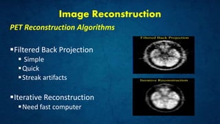
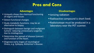


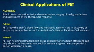
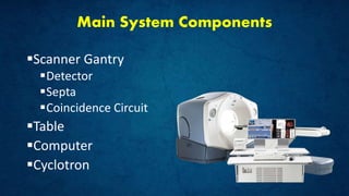
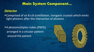
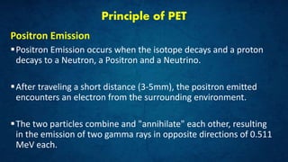
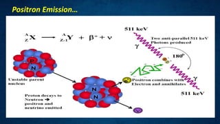
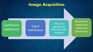
Positron emission tomography (PET) is an imaging technique that uses radiolabeled compounds for disease diagnosis through image acquisition from tracer distribution in the body. Its clinical applications include oncology, neurology, and cardiology, with systems comprising components like detectors, a cyclotron, and a computer for image reconstruction. While offering unique insights into organ function and metabolic activity, PET has disadvantages such as exposure to ionizing radiation and the need for short-lived radioisotopes.


![Pet appilcation[1]](https://cdn.slidesharecdn.com/ss_thumbnails/petappilcation1-191002015502-thumbnail.jpg?width=640&height=640&fit=bounds)


