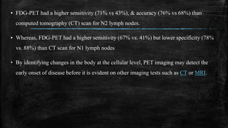
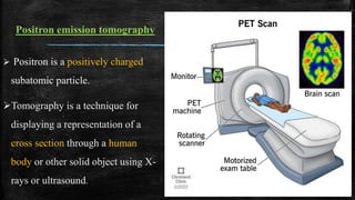
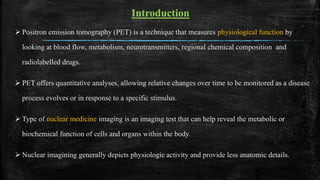
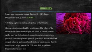
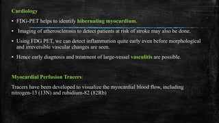
PET is a technique that measures physiological function by looking at blood flow, metabolism, neurotransmitters, regional chemical composition and radiolabelled drugs.
2. Positron emission tomography
Positron is a positively charged
subatomic particle.
Tomography is a technique for
displaying a representation of a
cross section through a human
body or other solid object using X-
rays or ultrasound.
3. Introduction
Positron emission tomography (PET) is a technique that measures physiological function by
looking at blood flow, metabolism, neurotransmitters, regional chemical composition and
radiolabelled drugs.
PET offers quantitative analyses, allowing relative changes over time to be monitored as a disease
process evolves or in response to a specific stimulus.
Type of nuclear medicine imaging is an imaging test that can help reveal the metabolic or
biochemical function of cells and organs within the body.
Nuclear imagining generally depicts physiologic activity and provide less anatomic details.
4. Procedure
The patient preparation protocol is as follows:
1. NPO for 4 hr prior to PET scan.
2. Patient well hydrated.
3. Move patient to quiet waiting/prep area 15 to 30 min before PET scan.
5. Start slow i. v. drip of normal saline.
6. Keep patient comfortable and calm until moved into the scanning room.
7. Avoid the use of sedation, if possible.
5. ▪ Radio-tracers can be injected, swallowed, or inhaled depending upon the site of the body
being examined, and the tracer gets trapped in various tissues of the body depending upon
the affinity.
▪ Areas of higher activity show higher uptake and brighter spots on images.
▪ The tracer is administered as an intravenous injection usually labelled with oxygen-15,
fluorine-18, carbon-11, or nitrogen-13.
▪ PET scans take 10-40 minutes to complete. They are painless, and, as for computed
tomography, the patient is fully clothed.
6. How it works?
Radiotracers are molecules linked to, or "labeled"
with, a small amount of radioactive material. They
accumulate in tumors or regions of inflammation.
They can also bind to specific proteins in the body..
For e.g. When radioactive substance is tagged to
glucose derivative known as FDG by this areas off
the body containing such cells can be imagined.
When positrons which are subatomic particles
attached to FDG decay, collide with electrons which
produces gamma rays in opposite reaction.
7. Special cameras detect this energy and, with the
help of a computer, create pictures that detail how
your organs and tissues look and function.
Areas of greater intensity are called “hot spots.”
These may show large concentrations of the
radiotracer and where there is a high level of
chemical or metabolic activity.
Less intense areas, or “cold spots,” indicate a
smaller concentration of radiotracer and less
activity.
So this technique is based on the detection of
radioactivity emitted after a small amount of a
radioactive tracer is injected into a peripheral vein.
8.
9. The use of standardized uptake values (SUVs) is now common place in clinical
FDG-PET/CT oncology imaging, and has a specific role in assessing patient
response to cancer therapy.
SUVs for the main organs in a whole-body 11 C-PK11195 PET scan
10. Oncology
▪ Tracers used commonly include fluorine-18 (18F) fluoro-
deoxyglucose (FDG), called FDG PET.
▪ FDG being a glucose analog, gets picked up by the cells.
▪ Glucose gets phosphorylated by hexokinases. The values of the
mitochondrial form of this enzyme are raised in cancers that are
rapidly growing. In locations of cancer, the metabolic activity is
quite high; hence the glucose uptake is quite high. So this 18F-FDG
also gets taken up quite significantly in these locations, and then this
shows up as a bright spot on the PET scan. This helps in the
detection of metastasis also.
11. ▪ FDG-PET is used for diagnosis, staging, and monitoring cancers particularly in Hodgkin's
lymphoma, non-Hodgkin lymphoma, esophagus, lung, cervical and gastroesophageal
cancer, solitary pulmonary nodule.
▪ The 68-gallium DOTA peptide is used to detect primary and metastatic neuroendocrine
tumors (NETs).
▪ Other tracers being used include C-labelled metomidate for detecting adrenocortical
tumors.
▪ F-DOPA PET-CT is shown to be a sensitive alternative to find and localize
pheochromocytoma
12. detect cancer and/or make a diagnosis.
determine whether a cancer has spread in the body.
assess the effectiveness of treatment.
determine if a cancer has returned after treatment.
evaluate prognosis.
assess tissue metabolism and viability
13.
14. Neurology
▪ Areas of high radiotracer uptake are associated with higher brain activity.
▪ It measures indirectly the flow of blood through the brain, which has been correlated with areas
of higher brain activity.
▪ Oxygen-15 is used for this.
▪ FDG PET of the brain is used to differentiate Alzheimer disease from other dementias as a
Perfusion, glucose metabolism, and Aβ imaging have an established role and are included in the
revised diagnostic criteria as important biomarkers.
▪ Pet scan shows temporoparietal glucose hypometabolism. With progression of disease,
there may also be frontal involvement in Alzheimer diseases.
15.
16. ▪ FDG PET in dementia with Lewy bodies (DLB) reveals changes similar to
those seen in AD, plus additional hypometabolism in the primary and
associative visual cortices.
▪ FDG PET in multi-infarct dementia shows multiple focal areas of
hypometabolism,
▪ Protein kinase C promotes the induction of alpha secretase or a disintegrin and
metalloprotease (non-amyloidogenic cleavage of amyloid precursor protein) and
hence have an important role in the maintenance of memory in Alzheimer disease.
▪ Deficits in PKC are seen early in the course of the disease.
17. ▪ P-Glycoprotein (P-gp) in the blood-brain barrier (BBB) has been thought to play a role in Aβ clearance
from the brain.
▪ The cholinergic deficit has been studied with PET, the radio-labelled analog of acetylcholine, such as N-
[(11) C]-methyl-4-piperidyl acetate (MP4A).
▪ PET scanning has resulted in improvement in the knowledge of the pathophysiology of atypical
Parkinsonism disorders and may be used as supportive criteria for differential diagnosis of these
conditions.
▪ Tracers that bind to presynaptic dopamine transporters, such as C-methylphenidate, and
dopamine terminal vesicle monoamine transporters, such as C-dihydrotetrabenazine, have been
developed as markers of presynaptic dopaminergic function. Dopaminergic neurons in these
regions project to the putamen and head of the caudate nucleus, respectively.
18.
19. ▪ These changes are detected by F-dopa PET, as evidenced by progressive decline in F-dopa Ki in
the putamen and in a caudal-rostral pattern.
▪ Brain PET imaging with FDG may be useful in seizure focus localization.
▪ γ-Aminobutyric acid (GABA) is the principal inhibitory neurotransmitter in the brain, acting at up
to 40% of synapses. A decreased number of GABAergic inhibitory interneurons was found in
epileptogenic cortex.
▪ Flumazenil (FMZ) is a specific reversible antagonist that binds to the benzodiazepine binding site
of the GABAA–central benzodiazepine receptor complex.
▪ C-FMZ PET therefore provides an in vivo marker of GABAA receptor binding. C-FMZ binding is
reduced by 30% in epileptogenic foci.
20. ▪ Knowledge acquired from PET cerebral blood flow and metabolism studies has
contributed significantly to the development of thrombolysis as a therapeutic
approach in ischemic stroke. Following stroke, PET can identify a “core” region of
irreversibly damaged tissue with profoundly depressed cerebral blood flow and
metabolism.
21. Neuropsychology:
▪ PET scanning helps delineate a link between specific processes and brain activity.
Psychiatry:
Radiotracers binding to dopamine, serotonin, opioid, and cholinergic receptors are used to
study their roles in various psychological disorders.
Stereotactic Surgery and Radiosurgery:
▪ PET-image guided surgeries are now being done.
22. Cardiology
▪ FDG-PET helps to identify hibernating myocardium.
▪ Imaging of atherosclerosis to detect patients at risk of stroke may also be done.
▪ Using FDG PET, we can detect inflammation quite early even before morphological
and irreversible vascular changes are seen.
▪ Hence early diagnosis and treatment of large-vessel vasculitis are possible.
Myocardial Perfusion Tracers:
Tracers have been developed to visualize the myocardial blood flow, including
nitrogen-13 (13N) and rubidium-82 (82Rb)
23.
24. Infectious Diseases
▪ PET can be used to image bacterial infections via 18F-FDG, by identifying infection-
associated inflammatory response.
▪ Agents include [18F]maltose,[18F]maltohexaose & [18F]2-fluorodeoxysorbitol (FDS).
▪ FDS importantly targets only Enterobacteriaceae.
▪ Applications include FUO, vascular graft infections, musculoskeletal infections
including osteomyelitis, joint prosthesis infections & diabetic foot infection.
▪ graft imaging, colonic inflammation & peritoneal tuberculosis.
26. Autoimmune Diseases
The upcoming role of PET is included in the new group of diseases called IgG4
diseases.
18F-FDG-PET can differentiate normal thyroid parenchyma from diffuse inflammatory
changes of the thyroid gland in patients with autoimmune thyroid diseases (AITD)
FDG uptake in Rheumatoid arthritis in affected joints reflects disease activity.
FDG-PET/CT shows a high diagnostic value for polymyalgia rheumatica in
differential diagnosis from rheumatoid arthritis.
27. Musculoskeletal System Diseases
PET provides muscle activation data about deep-lying muscles compared with
techniques like electromyography, which is useful in superficial muscles only.
[18F]-NaF is used to measure regional bone metabolism and blood flow.
[18F]NaF is recently being used in studying bone metastasis also
28. Interfering Factors
Strenuous exercises can lead to a considerable increase in radiotracer uptake by
various tissues & hence should be avoided prior to imaging.
Twenty-four hours before the scan, a low-carbohydrate, no-sugar diet is advised.
Allowable foods include meat, cheese, egg, and vegetables without starch.
Foods not allowed include cereals, pasta, milk, bread, and sugar.
29. Six hours before the scan, not eating or drinking anything is recommended.
Metal may interfere; hence it is preferably removed.
Other interfering factors include high blood glucose levels in diabetics; caffeine,
alcohol, or tobacco within 24 hours of the procedure
excessive anxiety which affects brain function; medicines like insulin,
tranquilizers, and sedatives; neurological or psychiatric conditions, which prevent
the ability to lie still.
30. Complications
▪ PET-CT has the complications involved with contrast administration, including possible
anaphylaxis, contrast-induced nephropathy, etc.
▪ Generally, the radiotracers used don’t cause any significant side effects.
31. Patient Safety and Education
▪ PET-CT involves radioactive material, and exposure to radiation is there. 18F-FDG has an
effective radiation dose of 14 c. Potential side effects of radiation as they hold for other
imaging modalities are seen in PET scan also. In a standard PET scan amount of radiation is
quite less, of the range of 8 mSv, about equivalent to that received from natural sources like
the sun. PET-CT uses higher levels of radiation of the range of 24mSv. Pregnant ladies
shouldn’t undergo a PET scan unless absolutely necessary as radioactivity can affect the
fetus. A breastfeeding woman should limit close contact with infants or pregnant women for
up to 12 hours. Breast milk may be discarded until 12 hours & after about 24 hours, it is
considered safe to breastfeed again.
32. Clinical Significance
▪ As mentioned above, PET scanning has a lot of potential in newer and newer
fields of medicine, including the already established oncology & the upcoming
fields like neurology, cardiology, psychiatry, and immunology. Many new uses
are being discovered every day, and PET scanning is becoming a radiological
test, which is quite sought after.
33. Contraindications
▪ Pregnancy is a relative contraindication.
• Expose your child to radiation if you are breastfeeding
• Cause an allergic reaction, although this is rare
▪ A very obese person may not fit into the opening
▪ The image resolution of nuclear medicine images may not be as high as that of CT or
MRI. However, nuclear medicine scans are more sensitive for a variety of indications.
The functional information they yield is often unobtainable using other imaging
techniques
34. ▪ Altered blood sugar or blood insulin levels may adversely affect the test results of diabetic
patients or patients who have eaten a few hours prior to the exam.
▪ The radiotracer decays quickly and is effective for only a short time. Therefore, it is
important for you to be on time for your appointment and to receive the radioactive
material at the scheduled time. Late arrival for an appointment may require you to
reschedule the procedure.
35. ▪ CT is useful for size and accurately localizing the cancer and is especially important for evaluating vital
structures adjacent to the cancer. But CT cannot detect metabolic activity.
▪ A PET scan can often detect the abnormal metabolism of the tracer in diseases before the disease shows up
on other imaging tests, such as computerized tomography (CT) and magnetic resonance imaging (MRI).
▪ PET/CT allows clinicians to see a fused image of physiologic and anatomic information.
▪ The tracer will then collect into areas of your body that have higher levels of metabolic or biochemical
activity, which often pinpoints the location of the disease. The PET images are typically combined
with CT or MRI and are called PET-CT or PET-MRI scans.
CT, MRI and PET SCAN
36. ▪ FDG-PET had a higher sensitivity (71% vs 43%), & accuracy (76% vs 68%) than
computed tomography (CT) scan for N2 lymph nodes.
▪ Whereas, FDG-PET had a higher sensitivity (67% vs. 41%) but lower specificity (78%
vs. 88%) than CT scan for N1 lymph nodes
• By identifying changes in the body at the cellular level, PET imaging may detect the
early onset of disease before it is evident on other imaging tests such as CT or MRI.