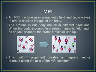
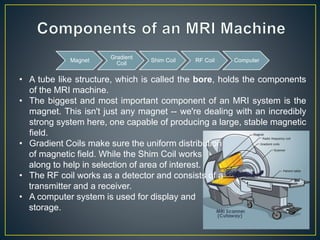
Syed Mustafa Jamal
• MRI stands for Magnetic Resonance Imaging.
• Magnetic resonance imaging (MRI) is a technique that
uses a magnetic field and radio waves to create detailed
images of the organs and tissues within your body.
• An MRI machine uses a magnetic field and radio waves
to create detailed images of the body.
• The protons in our body are all in different directions.
When the body is placed in a strong magnetic field, such
as an MRI scanner, the protons' axes all line up.
• This uniform alignment creates a magnetic vector
oriented along the axis of the MRI scanner
When additional energy (in the form of a radio wave) is
added to the magnetic field, the magnetic vector is
deflected.
• The magnetic field soon realigns the magnetic field,
causing the emission of energy signals.
• Video
• A tube like structure, which is called the bore, holds the components
of the MRI machine.
• The biggest and most important component of an MRI system is the
magnet. This isn't just any magnet -- we're dealing with an incredibly
strong system here, one capable of producing a large, stable magnetic
field.
• Gradient Coils make sure the uniform distribution
of magnetic field. While the Shim Coil works
along to help in selection of area of interest.
• The RF coil works as a detector and consists of a
transmitter and a receiver.
• A computer system is used for display and
storage.
Magnet
Gradient
Coil
Shim Coil RF Coil Computer
Most MRI systems use a superconducting magnet, which consists of many
coils or windings of wire through which a current of electricity is passed,
creating a magnetic field of up to 2.0 tesla.
Two other magnets are used in MRI systems to a much lesser extent.
Resistive magnets and Permanent magnets.
Gradient magnets are also present inside the MRI machine. These magnets
are much lower strength compared to the main magnetic field. While the main
magnet creates an intense, stable magnetic field around the patient, the
gradient magnets create a variable field, which
allows different parts of the body to be
scanned.
• NMR uses oxygen atoms
for the visualization.
• Oxygen has a higher
atomic number (16),
therefore requires higher
energy.
• The required magnetic field
strength for oxygen is 10 –
15 Tesla.
• MRI uses hydrogen atoms
for visualization.
• Hydrogen has a lower
atomic number (1),
therefore requires less
energy.
• The required magnetic field
strength for hydrogen is 0.5
– 3.5 Tesla.
• MRI can be used for both; Physiology and Anatomy.
• They are particularly useful for showing soft tissue
structures, such as ligaments and cartilage, and organs
such as the brain, heart, and eyes.
• They can provide information about how the blood moves
through certain organs and blood vessels, allowing
problems with blood circulation, such as blockages, to be
identified.
• It is a non ionizing technique, which means no radiations.
• The image is a 3D image.

MRI

  • 1.
  • 2.
    • MRI standsfor Magnetic Resonance Imaging. • Magnetic resonance imaging (MRI) is a technique that uses a magnetic field and radio waves to create detailed images of the organs and tissues within your body.
  • 3.
    • An MRImachine uses a magnetic field and radio waves to create detailed images of the body. • The protons in our body are all in different directions. When the body is placed in a strong magnetic field, such as an MRI scanner, the protons' axes all line up. • This uniform alignment creates a magnetic vector oriented along the axis of the MRI scanner
  • 4.
    When additional energy(in the form of a radio wave) is added to the magnetic field, the magnetic vector is deflected. • The magnetic field soon realigns the magnetic field, causing the emission of energy signals.
  • 5.
  • 6.
    • A tubelike structure, which is called the bore, holds the components of the MRI machine. • The biggest and most important component of an MRI system is the magnet. This isn't just any magnet -- we're dealing with an incredibly strong system here, one capable of producing a large, stable magnetic field. • Gradient Coils make sure the uniform distribution of magnetic field. While the Shim Coil works along to help in selection of area of interest. • The RF coil works as a detector and consists of a transmitter and a receiver. • A computer system is used for display and storage. Magnet Gradient Coil Shim Coil RF Coil Computer
  • 7.
    Most MRI systemsuse a superconducting magnet, which consists of many coils or windings of wire through which a current of electricity is passed, creating a magnetic field of up to 2.0 tesla. Two other magnets are used in MRI systems to a much lesser extent. Resistive magnets and Permanent magnets. Gradient magnets are also present inside the MRI machine. These magnets are much lower strength compared to the main magnetic field. While the main magnet creates an intense, stable magnetic field around the patient, the gradient magnets create a variable field, which allows different parts of the body to be scanned.
  • 8.
    • NMR usesoxygen atoms for the visualization. • Oxygen has a higher atomic number (16), therefore requires higher energy. • The required magnetic field strength for oxygen is 10 – 15 Tesla. • MRI uses hydrogen atoms for visualization. • Hydrogen has a lower atomic number (1), therefore requires less energy. • The required magnetic field strength for hydrogen is 0.5 – 3.5 Tesla.
  • 9.
    • MRI canbe used for both; Physiology and Anatomy. • They are particularly useful for showing soft tissue structures, such as ligaments and cartilage, and organs such as the brain, heart, and eyes. • They can provide information about how the blood moves through certain organs and blood vessels, allowing problems with blood circulation, such as blockages, to be identified. • It is a non ionizing technique, which means no radiations. • The image is a 3D image.