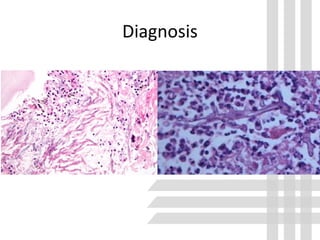
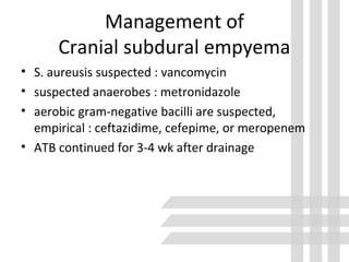
This document discusses brain abscess, cranial subdural empyema, and epidural abscess. It covers the epidemiology, etiology, pathogenesis, clinical findings, diagnosis, and management of these conditions. Brain abscesses are typically caused by bacteria spreading from contiguous sites of infection or through the bloodstream. Clinical findings depend on the location and size of the abscess. Diagnosis involves neuroimaging and culture of aspirated contents. Treatment involves antibiotics and sometimes surgery. Outcomes depend on early diagnosis and treatment.
This presentation briefly summarizes pathophysiology, clinical features, diagnosis and treatment of different types of tuberculosis of brain and spinal cord.
Intracerebral hemorhage Diagnosis and managementRamesh Babu
About ICH - Diagnosis and management, Discussed the clinical presentation, evaluation, radiological features and management including recent guidelines
Meningitis is a severe CNS pathology and early and appropriate intervention is needed to prevent adverse outcome including mortality and long term complications. This presentation focuses on the different types of meningitis and the appropriate management options
Anti ulcer drugs and their Advance pharmacology ||
Anti-ulcer drugs are medications used to prevent and treat ulcers in the stomach and upper part of the small intestine (duodenal ulcers). These ulcers are often caused by an imbalance between stomach acid and the mucosal lining, which protects the stomach lining.
||Scope: Overview of various classes of anti-ulcer drugs, their mechanisms of action, indications, side effects, and clinical considerations.
Ozempic: Preoperative Management of Patients on GLP-1 Receptor Agonists Saeid Safari
Preoperative Management of Patients on GLP-1 Receptor Agonists like Ozempic and Semiglutide
ASA GUIDELINE
NYSORA Guideline
2 Case Reports of Gastric Ultrasound
Title: Sense of Smell
Presenter: Dr. Faiza, Assistant Professor of Physiology
Qualifications:
MBBS (Best Graduate, AIMC Lahore)
FCPS Physiology
ICMT, CHPE, DHPE (STMU)
MPH (GC University, Faisalabad)
MBA (Virtual University of Pakistan)
Learning Objectives:
Describe the primary categories of smells and the concept of odor blindness.
Explain the structure and location of the olfactory membrane and mucosa, including the types and roles of cells involved in olfaction.
Describe the pathway and mechanisms of olfactory signal transmission from the olfactory receptors to the brain.
Illustrate the biochemical cascade triggered by odorant binding to olfactory receptors, including the role of G-proteins and second messengers in generating an action potential.
Identify different types of olfactory disorders such as anosmia, hyposmia, hyperosmia, and dysosmia, including their potential causes.
Key Topics:
Olfactory Genes:
3% of the human genome accounts for olfactory genes.
400 genes for odorant receptors.
Olfactory Membrane:
Located in the superior part of the nasal cavity.
Medially: Folds downward along the superior septum.
Laterally: Folds over the superior turbinate and upper surface of the middle turbinate.
Total surface area: 5-10 square centimeters.
Olfactory Mucosa:
Olfactory Cells: Bipolar nerve cells derived from the CNS (100 million), with 4-25 olfactory cilia per cell.
Sustentacular Cells: Produce mucus and maintain ionic and molecular environment.
Basal Cells: Replace worn-out olfactory cells with an average lifespan of 1-2 months.
Bowman’s Gland: Secretes mucus.
Stimulation of Olfactory Cells:
Odorant dissolves in mucus and attaches to receptors on olfactory cilia.
Involves a cascade effect through G-proteins and second messengers, leading to depolarization and action potential generation in the olfactory nerve.
Quality of a Good Odorant:
Small (3-20 Carbon atoms), volatile, water-soluble, and lipid-soluble.
Facilitated by odorant-binding proteins in mucus.
Membrane Potential and Action Potential:
Resting membrane potential: -55mV.
Action potential frequency in the olfactory nerve increases with odorant strength.
Adaptation Towards the Sense of Smell:
Rapid adaptation within the first second, with further slow adaptation.
Psychological adaptation greater than receptor adaptation, involving feedback inhibition from the central nervous system.
Primary Sensations of Smell:
Camphoraceous, Musky, Floral, Pepperminty, Ethereal, Pungent, Putrid.
Odor Detection Threshold:
Examples: Hydrogen sulfide (0.0005 ppm), Methyl-mercaptan (0.002 ppm).
Some toxic substances are odorless at lethal concentrations.
Characteristics of Smell:
Odor blindness for single substances due to lack of appropriate receptor protein.
Behavioral and emotional influences of smell.
Transmission of Olfactory Signals:
From olfactory cells to glomeruli in the olfactory bulb, involving lateral inhibition.
Primitive, less old, and new olfactory systems with different path
Prix Galien International 2024 Forum ProgramLevi Shapiro
June 20, 2024, Prix Galien International and Jerusalem Ethics Forum in ROME. Detailed agenda including panels:
- ADVANCES IN CARDIOLOGY: A NEW PARADIGM IS COMING
- WOMEN’S HEALTH: FERTILITY PRESERVATION
- WHAT’S NEW IN THE TREATMENT OF INFECTIOUS,
ONCOLOGICAL AND INFLAMMATORY SKIN DISEASES?
- ARTIFICIAL INTELLIGENCE AND ETHICS
- GENE THERAPY
- BEYOND BORDERS: GLOBAL INITIATIVES FOR DEMOCRATIZING LIFE SCIENCE TECHNOLOGIES AND PROMOTING ACCESS TO HEALTHCARE
- ETHICAL CHALLENGES IN LIFE SCIENCES
- Prix Galien International Awards Ceremony
TEST BANK for Operations Management, 14th Edition by William J. Stevenson, Ve...kevinkariuki227
TEST BANK for Operations Management, 14th Edition by William J. Stevenson, Verified Chapters 1 - 19, Complete Newest Version.pdf
Pulmonary Thromboembolism - etilogy, types, medical- Surgical and nursing man...VarunMahajani
Disruption of blood supply to lung alveoli due to blockage of one or more pulmonary blood vessels is called as Pulmonary thromboembolism. In this presentation we will discuss its causes, types and its management in depth.
These lecture slides, by Dr Sidra Arshad, offer a quick overview of physiological basis of a normal electrocardiogram.
Learning objectives:
1. Define an electrocardiogram (ECG) and electrocardiography
2. Describe how dipoles generated by the heart produce the waveforms of the ECG
3. Describe the components of a normal electrocardiogram of a typical bipolar leads (limb II)
4. Differentiate between intervals and segments
5. Enlist some common indications for obtaining an ECG
Study Resources:
1. Chapter 11, Guyton and Hall Textbook of Medical Physiology, 14th edition
2. Chapter 9, Human Physiology - From Cells to Systems, Lauralee Sherwood, 9th edition
3. Chapter 29, Ganong’s Review of Medical Physiology, 26th edition
4. Electrocardiogram, StatPearls - https://www.ncbi.nlm.nih.gov/books/NBK549803/
5. ECG in Medical Practice by ABM Abdullah, 4th edition
6. ECG Basics, http://www.nataliescasebook.com/tag/e-c-g-basics
Title: Sense of Taste
Presenter: Dr. Faiza, Assistant Professor of Physiology
Qualifications:
MBBS (Best Graduate, AIMC Lahore)
FCPS Physiology
ICMT, CHPE, DHPE (STMU)
MPH (GC University, Faisalabad)
MBA (Virtual University of Pakistan)
Learning Objectives:
Describe the structure and function of taste buds.
Describe the relationship between the taste threshold and taste index of common substances.
Explain the chemical basis and signal transduction of taste perception for each type of primary taste sensation.
Recognize different abnormalities of taste perception and their causes.
Key Topics:
Significance of Taste Sensation:
Differentiation between pleasant and harmful food
Influence on behavior
Selection of food based on metabolic needs
Receptors of Taste:
Taste buds on the tongue
Influence of sense of smell, texture of food, and pain stimulation (e.g., by pepper)
Primary and Secondary Taste Sensations:
Primary taste sensations: Sweet, Sour, Salty, Bitter, Umami
Chemical basis and signal transduction mechanisms for each taste
Taste Threshold and Index:
Taste threshold values for Sweet (sucrose), Salty (NaCl), Sour (HCl), and Bitter (Quinine)
Taste index relationship: Inversely proportional to taste threshold
Taste Blindness:
Inability to taste certain substances, particularly thiourea compounds
Example: Phenylthiocarbamide
Structure and Function of Taste Buds:
Composition: Epithelial cells, Sustentacular/Supporting cells, Taste cells, Basal cells
Features: Taste pores, Taste hairs/microvilli, and Taste nerve fibers
Location of Taste Buds:
Found in papillae of the tongue (Fungiform, Circumvallate, Foliate)
Also present on the palate, tonsillar pillars, epiglottis, and proximal esophagus
Mechanism of Taste Stimulation:
Interaction of taste substances with receptors on microvilli
Signal transduction pathways for Umami, Sweet, Bitter, Sour, and Salty tastes
Taste Sensitivity and Adaptation:
Decrease in sensitivity with age
Rapid adaptation of taste sensation
Role of Saliva in Taste:
Dissolution of tastants to reach receptors
Washing away the stimulus
Taste Preferences and Aversions:
Mechanisms behind taste preference and aversion
Influence of receptors and neural pathways
Impact of Sensory Nerve Damage:
Degeneration of taste buds if the sensory nerve fiber is cut
Abnormalities of Taste Detection:
Conditions: Ageusia, Hypogeusia, Dysgeusia (parageusia)
Causes: Nerve damage, neurological disorders, infections, poor oral hygiene, adverse drug effects, deficiencies, aging, tobacco use, altered neurotransmitter levels
Neurotransmitters and Taste Threshold:
Effects of serotonin (5-HT) and norepinephrine (NE) on taste sensitivity
Supertasters:
25% of the population with heightened sensitivity to taste, especially bitterness
Increased number of fungiform papillae
3. Epidemiology
• 0.3 -1.3 cases per 100,000 person
• Male : Female 2:1
• Median age 30-40 years
– Otitic focus : < 20 Yrs , >40 Yrs
– Paranasal sinus : 30 – 40 Yrs
• After cranial operation : 0.2 % of 1587 , 10 of 16540
• Commonly in Immunocompromised : infected with
HIV, receiving chemotherapy for cancer, receiving
immunosuppressive therapy after organ
transplantation, or after prolonged use of
corticosteroids.
4. Risk factor
• Pulmonary abnormality
– Infection
– AV fistula
• Congenital cyanotic heart disease
• Bacterial endocarditis
• Penetrating trauma
• Chronic sinusitis or otitis media
• AIDS
6. Pathogenesis
• Contiguous source : middle ear, mastoid cells,
paranasal sinuses
• From purulent sinusitis : spread by osteomyelitis or
by phlebitis of emissary vein
• Rare in infant because they lack aerated paranasal
and mastoid air cells
• Localize of Contiguous source
– Otitis media : temporal lobe or cerebellum
– Paranasal sinusitis : frontal lobe
– Sphenoid sinusitis(less common for sinusitis ) : temporal
lobe, sella turcica
7. Pathogenesis
• Hematogenous dissemination : multiple,
multiloculated abscesses, higher mortality rate
• Most common source in adult
– Most common : pyogenic lung diseases (especially lung
abscess)
– bronchiectasis
– Empyema
– cystic fibrosis
– Skin infection, osteomyelitis, pelvic infections, and intra-
abdominal infections
8. Pathogenesis
• In children
• Cyanotic congenital heart disease (tetralogy of Fallot)
because increase Hct and low PO2 provide and hypoxic
environment suitable for abscess proliferation
• Right to left shunt
• Streptococcal oral flora from oral dental procedure
9. Pathogenesis
• Trauma : open cranial fracture with dural breach or
foreign body injury or as a sequela of neurosurgery
• Nosocomial brain abscess : halo pin insertion,after
electrode insertion to localize seizure foci,and in
malignant glioma patients treated by placement
of Gliadel wafers in the tumor bed to release
carmustine
10. Etiology
• Bacterial : most common, streptococci (aerobic,
anaerobic, and microaerophilic), 70%
• Streptococcus anginosus (milleri) : oral cavity,
appendix, and female genital tract
• Staphylococcus aureus : cranial trauma or infective
endocarditis
• Enteric gram-negative bacilli ,25-30% (e.g., Proteus
spp., Escherichia coli, Klebsiellaspp., Pseudomonas
aeruginosa, and Enterobacterspp.) : otitis media,
bacteremia, neurosurgical procedures, and the
immunocompromised state
11. • Negative culture : 0-43% (previous use ATB)
• Listeria monocytogenesis uncommon (<1% of cases),
considered in patient who are immunocompromised
(e.g., leukemia, lymphoma, HIV infection, and
conditions requiring corticosteroids or other agents
that cause immunosuppression)
• Salmonella, Nocardia, Streptococcus pneumoniae,
Haemophilus influenzae, Burkholderia pseudomallei,
and Actinomyces species
Etiology
12. Etiology
• the incidence of fungal brain abscess has been
rising as a result of the increased use of
corticosteroid therapy, broad-spectrum
antimicrobial therapy, and immunosuppressive
agents
• Candida : microabscesses, macroabscesses,
noncaseating granulomas, and diffuse glial nodule
13. Etiology
• Risk factors for candidal brain abscess include the
use of broad-spectrum antimicrobial agents,
corticosteroids, and hyperalimentation; premature
birth; malignancy; neutropenia; chronic
granulomatous disease; diabetes mellitus; thermal
injury; and the presence of a central venous catheter
• Aspergillosis : neutropenia, hepatic disease, diabetes
mellitus, chronic granulomatous disease, Cushing’s
syndrome, HIV infection, injection drug use, organ
transplantation, and bone marrow transplantation
14. Etiology
• Mucorales group : Diabetes mellitus, patients with
acidemia from profound systemic illness (e.g., sepsis,
severe dehydration, severe diarrhea, chronic
kidney disease)
• Scedosporium : immunocompetent and
immunocompromised hosts
• Cryptococcus neoformans, the endemic mycoses
(Coccidioides spp., Histoplasma spp., and
Blastomyces dermatitidis)
16. Initiation of infection
• The brain appears to be significantly more sensitive
to infection than many other tissues.
• The brain may also be more susceptible to
infection by different organisms
19. Host defence mechanism
• Although the brain is generally protected from
infection by an intact blood-brain barrier, once
infection is established, immune defenses are
usually inadequate to control the infection
• Encapsulate bacteria : E.coli, B.fragilis
23. Clinical finding
• Intraventricular rupture of brain abscess : severe
headaches and signs of meningeal irritation were
prominent findings before rupture
• Intraventricular rupture appears to be more likely
if the abscess is deep-seated, multiloculated, and in
close proximity to the ventricular wall
24. Clinical finding
• Related to pathogen
– Nocardia : concomitant pulmonary, skin, or muscle
lesions
– Aspergillus brain abscess : commonly manifest signs of a
stroke syndrome as a result of ischemia or intracerebral
hemorrhage, or both
– rhinocerebral mucormycosis : symptoms referable to
the eyes or sinuses and complaints of headache, facial
pain, diplopia, lacrimation, and nasal stuffiness or epistaxis
cranial nerve involvement blindness
25. Clinical finding
• Related to pathogen
– Scedosporium apiospermum brain abscess :
• occur in immunocompromised patients or in
individuals 15 to 30 days after an episode of near-
drowning
• cerebrum, cerebellum, or brainstem
• Clinical finding : seizures, altered consciousness,
headache, meningeal irritation, focal neurological
deficits, abnormal behavior, and aphasi
26. Diagnosis
• WBC : mildly elevate (>10,000)
• Blood C/S : should be obtain (usually negative)
• ESR : may be normal
• CRP : may arise
• LP : dubious, OP increase, WBC count, Protein
elevate, risk for tantentorial herniation
27. Diagnosis
• MRI is more sensitivity than CT, early detection of
cerebritis, more conspicuous spread of inflammation
into the ventricles and subarachnoid space, and
earlier detection of satellite lesions
• T1 : the abscess capsule often appears as a discrete
rim that is isointense to mildly hyperintense
• T1 c Gad : rim enhancement, cerebral edema
• T2 : zone of edema : hyperintensity, capsule : ill-
defined hypointensity
• DWI : restricted diffusion (bright signal) may be seen
• ADC : Dark
29. Diagnosis
• Aspergillosis : finding of a cerebral infarct, which
typically develops into either single or multiple
abscesses, in immunocompromised patients, there
may be little or no contrast enhancement on MRI
• Rhinocerebral mucormycosis : sinus opacification,
erosion of bone, and obliteration of deep fascial
planes; cavernous sinus thrombosis
30. Diagnosis
• stereotactic MRI- or CT-guided aspiration
• Specimens sent for : Gram stain, routine aerobic
and anaerobic culture, modified acid-fast smears,
acidfast smears and culture, and fungal smears and
culture
• Aspergillus brain abscess : septate hyphae with
acute-angle, dichotomous branching
• Mucormycosis : irregular hyphae with right-angle
branching.
32. Diagnosis
• Scedosporiumspecies are indistinguishable from
those caused by Aspergillus species. The hyphae of
dematiaceous fungi may be brownish on
hematoxylin-eosin staining but are not
distinguishable from those of other molds.
33.
34. Management
• Multidisciplinary, neuroradiologist, neurosurgeon,
and infectious disease specialist
• Larger than 2.5 cm : excision
• Smaller than 2.5 cm : aspiration for definite diagnosis
• Empirical ATB : metronidazole + third-generation
cephalosporin
• S.aureus consider : add vancomycin
• gram-negative bacilli such as P. aeruginosa :
ceftazidime, cefepime, or meropenem
35. Management
• no clear predisposing factors : combination of
vancomycin, metronidazole, and a third- or fourth-
generation cephalosporin
• Once the infecting pathogen is isolated, antimicrobial
therapy can be modified for optimal treatment
36. Medical management alone
• Treatment began in cerebritis
• Small lesion 0.8-2.5 cm
• Duration of symptom < 2 Wk
• Patient show clinical improvement in 1 wk
37. Surgical treatment
• Significant mass effect exerted by lesion
• Difficult in diagnosis
• Proximity to ventricle
• Evedence of significane increase intracranial
pressure
• Poor neurological condition
• Traumatic abscees associated with foreign
material
• Fungal abscess
39. Bacterial brain abscess
• The principles of antimicrobial therapy for bacterial
brain abscess
– penetrate the abscess cavity
– in vitro activity against the isolated pathogen
• Metronidazole
– excellent in vitro activity against strict anaerobes
– an excellent pharmacokinetic profile
– good oral absorption
– penetration into brain abscess cavities
– must always be used in combination with an agent
effective against streptococci
40. Bacterial brain abscess
• Vancomycin, The third generation cephalosporins
are common used
• Imipenem : pyogenic and nocardial brain abscess,
beware seizure
• Meropenem : Enterobacter cloacae
41. Bacterial brain abscess
• Surgical therapy : aspiration after bur-hole
placement or complete excision after craniotomy
• Stereotactic aspiration : eloquent or inaccessible
regions
• Complete excision by craniotomy
– patients with multiloculated abscesses in whom
aspiration techniques have failed
– for abscesses containing gas
– for abscesses that fail to resolve
– posttraumatic abscesses that contain foreign bodies or
retained bone fragments to prevent recurrence
42. Bacterial brain abscess
• In patients with intraventricular rupture of a
purulent brain abscess :
– rapid evacuation and débridement of the abscess cavity
via urgent craniotomy and ventricular drainage
– intravenous or intrathecal ATB
• groups of patients may be treated with medical
therapy alone
– medical conditions that increase the risk associated with
surgery
– multiple abscesses
43. Bacterial brain abscess
• groups of patients may be treated with medical
therapy alone
– abscesses in a deep or dominant location
– the presence of coexisting meningitis or ependymitis
– early reduction of the abscess with clinical improvement
after antimicrobial therapy
– abscess size less than 3 cm
44. Bacterial brain abscess
• ATB optimal duration : 6-8 Wk IV then follow by oral
ATB for 2-3 Mo
• Repeat neuroimaging studies performed biweekly for
up to 3 months after completion of therapy has been
suggested to monitor for reexpansion of the abscess
or failure of resolution
45. Nocardial brain abscess
• Sulfonamide with or without trimethoprim is
recommend
• Alternative agent : minocycline, imipenem, amikacin,
third-generation cephalosporins, and linezolid
• In immunocompromised patients or those in whom
therapy fails : combination treatment with
regimens + a third-generation cephalosporin or
imipenem + a sulfonamide or amikacin
46. Nocardial brain abscess
• Craniotomy with total excision is difficult in
patients with Nocardia brain abscess because
these abscesses are often multiloculated
• ATB duration : 3-12 Mo but in immunocompromised
should be up to 1 Yr
47. Fungal brain abscess
• Candidal brain abscess : amphotericin B preparation
plus 5-flucytosine
• Aspergillusbrain abscess : voricazole
• CNS mucormycosis : amphotericin B deoxycholate
or a lipid formulation of amphotericin B with
aggressive surgical debridment
• Scedosporiumspecies : surgery
50. Adjunctive Therapy
• Steroid effect
– Reduced edema
– Decrese likehood of fibrous encapsulation
– May be reduced penetration of antibiotic into abscess
• Corticosteroid use
– edema and mass effect
– progressive neurological deterioration
– impending cerebral herniation
• High dose : dexamethasone 10 mg q 6 hr and
then tapered
52. Epidemiology and Etiology
• Cranial subdural empyema : collection of pus
between the space of the dura and arachnoid, 15-20
% intracranial lesion
• Less common than brain abscess
• SDE may be complicated by cerebral brain abcess,
cortical vein thrombosis, localized cerebritis
• Location : 70-80 % convexity, 10-20% parafalcine
53. Epidemiology and Etiology
• most common predisposing conditions :
otorhinologic infections, especially of the
paranasal sinuses
• Other predisposing conditions : skull trauma,
neurosurgical procedures, and infection of a
preexisting subdural hematoma
54. Epidemiology and Etiology
• Organisms
– aerobic streptococci (25% to 45%)
– staphylococci (10% to 15%)
– aerobic gram-negative bacilli (3% to 10%)
– anaerobic streptococci
– other anaerobes
• If the predisposing condition is postoperative or
posttraumatics : staphylococci and aerobic gram-
negative bacilli
55. Clinical finding
• Clinical manisfestation of subdural empyema
– rapidly progressive, with symptoms and signs related to
increased intracranial pressure (headache, vomiting)
– meningeal irritation
– focal cortical inflammation (hemiparesis and hemiplegia,
ocular palsies, dysphasia, homonymous hemianopia,
dilated pupils, and cerebellar signs
– Altered mental status
– Seizure
– Fever
56. Clinical finding
• Clinical manisfestation of cranial epidural abscess
– Insidious onset
– Abscess enlarge too slowly to produce a sudden onset of
major neurological deficits, as is seen in patients with
cranial subdural empyema
– Most common : fever, headache
– If the location of the epidural abscess is near the petrous
bone, Gradenigo’s syndrome may develop (involvement
of cranial nerves V and VI and manifested clinically as
unilateral facial pain and weakness of the lateral
rectus muscle)
57. Diagnosis
• Cranial subdural empyema should be suspected in
any patient with meningeal signs and a focal
neurological deficit
• CT brain : iv contrast : hypodense crescentric
• LP : potential hazardous for herniation
58. Diagnosis
• MRI in Cranial subdural empyema : crescentic or
elliptical area of hypointensity (on T1 images) below
the cranial vault or adjacent to the falx cerebri, high
signal on T2
• MRI in cranial epidural abscess : superficial,
circumscribed area of diminished intensity with
pachymeningeal enhancement.
59. Management of
Cranial subdural empyema
• Cranial subdural empyema is a surgical emergency
because antimicrobial therapy alone does not
reliably sterilize the empyema
• The goals of surgical therapy are to achieve adequate
decompression of the brain and to evacuate the
empyema completely.
• The optimal surgical approach is controversial.
60. Management of
Cranial subdural empyema
• Patients who underwent drainage via bur holes or
craniectomy required more frequent operations to
drain recurrent or remaining pus and exhibited
higher mortality rates and worse outcomes
• Drainage via bur holes or craniectomy is therefore
recommended only for
– patients in septic shock, those with localized parafalcine
collections
– children with subdural empyema secondary to
meningitis because there is usually no brain swelling and
the pus is thin
61. Management of
Cranial subdural empyema
• S. aureusis suspected : vancomycin
• suspected anaerobes : metronidazole
• aerobic gram-negative bacilli are suspected,
empirical : ceftazidime, cefepime, or meropenem
• ATB continued for 3-4 wk after drainage
62. Management of
Cranial subdural empyema
• ATB alone in
– patients with cranial subdural empyema who have
minimal or no impairment of consciousness
– no major neurological deficit
– limited extension of the empyema with no
midline shift
– early improvement with antimicrobial therapy
63. Management of
Cranial epidural abscess
• combined medical and surgical approach
• Empirical antimicrobial therapy is similar to that for
cranial subdural empyema
• For surgical drainage, craniotomy or craniectomy
is generally preferred over bur-hole placement or
aspiration of purulent material through the scalp
• ATN length 3-6 wk or longer in osteomyelitis