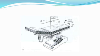
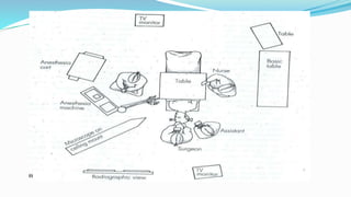
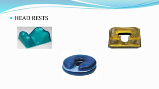
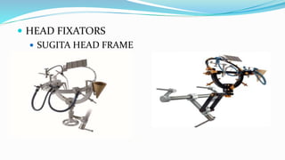
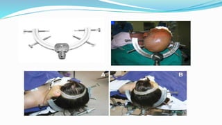
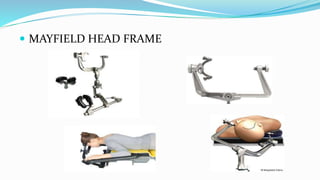
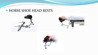
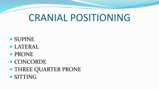
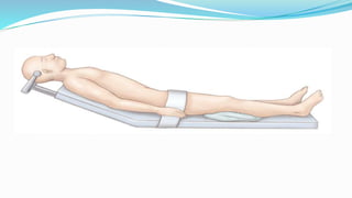
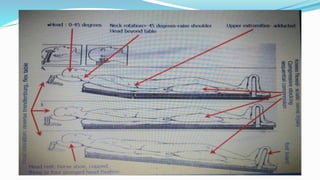
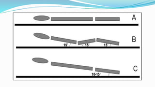
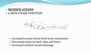
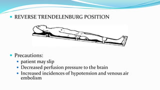
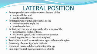
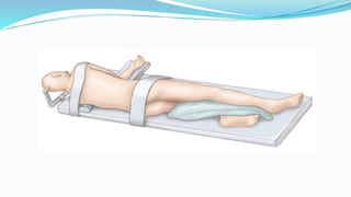
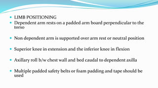
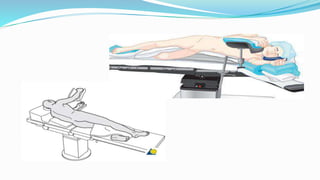
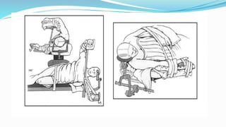
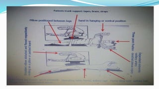
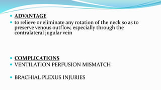
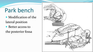
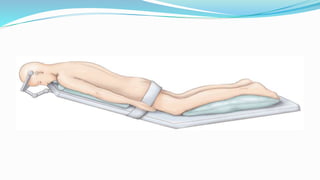
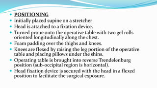
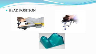
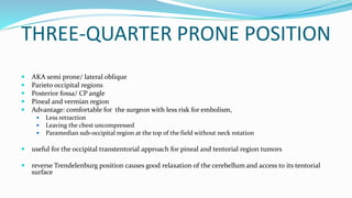
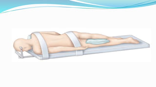
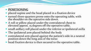
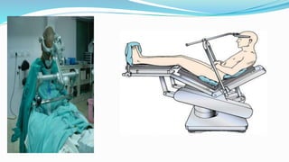
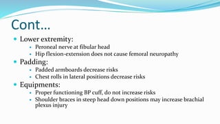
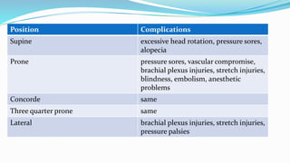
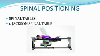
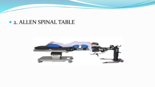
Abhinav Agrahari
31.7.18
 The term positioning refers to the position of the
surgeon as well as that of the patient.
 one of the most crucial steps in any cranial operation
The correct position
 In early years‐trial and error
 Today though standardized, not absolute
 Factors associated:
1. Age
2. Site and nature of lesion
3. Head position in relation to heart
4. Position of anesthesiologist/ nurse
5. Microscope and other imaging equipment
 Pediatric patients present a different set of considerations
 Some operations have more than one acceptable position
 Improve precision
 Better localization
 Patient and surgeon comfort
 Access to anesthetist
 Access for monitoring
 Ergonomical arrangement
AIMS/ ADVANTAGES
 most direct access to the surgical target and reduces the
working distance for the surgeon.
 avoids brain retraction
 minimizes bleeding into the operative field
 Intracranial pressure is reduced
 prevents pressure or traction injuries, including skin
breakdown, ocular injuries, and peripheral nerve injuries
 increases the likelihood of a successful operation
HEAD POSITION/ FIXATION
 HEAD PADS/ HEAD RINGS
 HEAD RESTS
 HEAD FIXATORS
 SUGITA HEAD FRAME
 MAYFIELD HEAD FRAME
 HORSE SHOE HEAD RESTS
CRANIAL POSITIONING
 SUPINE
 LATERAL
 PRONE
 CONCORDE
 THREE QUARTER PRONE
 SITTING
SUPINE POSITION
 common and versatile position
 Doesn’t require special instrumentation, is easily achievable
and doesn’t require disconnection of invasive monitors
 majority of brain lesions to be accessed with the patient
supine
 flexibility of the neck and
 ability to rotate the torso with a shoulder roll
 HEAD POSITIONS
 Head straight with flexion to access lesions of the frontal
lobes, anterior interhemispheric fissure, lateral and third
ventricles, bifrontal decompressive craniectomies.
 Head in slight extension for subfrontal approaches and
anterior skull base lesions allowing the frontal lobes to fall
away from the anterior cranial fossa
 The middle and posterior fossae accessed by turning the head
away from the side of the lesion.
 ARM POSITION
 MODIFICATIONS
 LAWN CHAIR POSITION
 Increased venous return from lower extremities
 Decreased stress on back, hips and knees
 Increased cerebral venous drainage
 REVERSE TRENDELENBURG POSITION
 Precautions:
 patient may slip
 Decreased perfusion pressure to the brain
 Increased incidences of hypotension and venous air
embolism
COMPLICATIONS
 PRESSURE ALOPECIA
 BACKACHE
 INCREASED RISK OF GASTRIC ASPIRATION
 PERIPHERAL NERVE INJURIES
 BRACHIAL PLEXUS INJURIES
LATERAL POSITION for temporal craniotomies to access lesions of the
 temporal lobes and
 middle cranial fossa,
 for lateral suboccipital approaches to the
 cerebellopontine angle and
 lateral cerebellum,
 for far/ extreme lateral approaches for lesions of the
 pineal region, posterior fossa,
 foramen magnum, and craniocervical junction
 Lateral approaches to the cervical spine
 Trans thoracic and retroperitoneal approaches to the spine
 Extremely obese or kyphotic patients
 Unilateral herniated discs‐offending side up
 Lumboperitoneal, syringoperitoneal shunts
 LIMB POSITIONING
 Dependent arm rests on a padded arm board perpendicular to the
torso
 Non dependent arm is supported over arm rest or neutral position
 Superior knee in extension and the inferior knee in flexion
 Axillary roll b/w chest wall and bed caudal to dependent axilla
 Multiple padded safety belts or foam padding and tape should be
used
 ADVANTAGE
 to relieve or eliminate any rotation of the neck so as to
preserve venous outflow, especially through the
contralateral jugular vein
 COMPLICATIONS
 VENTILATION PERFUSION MISMATCH
 BRACHIAL PLEXUS INJURIES
Park bench
 Modification of the
lateral position
 Better access to
the posterior fossa
PRONE POSITION
 The prone position is used for access to the
 occipital lobes,
 midline or paramedian cerebellum,
 pineal region,
 fourth ventricle, and
 upper cervical spine
 POSITIONING
 Initially placed supine on a stretcher
 Head is attached to a fixation device.
 Turned prone onto the operative table with two gel rolls
oriented longitudinally along the chest.
 Foam padding over the thighs and knees.
 Knees are flexed by raising the leg portion of the operative
table and placing pillows under the shins.
 Operating table is brought into reverse Trendelenburg
position (sub-occipital region is horizontal).
 Head fixation device is secured with the head in a flexed
position to facilitate the surgical exposure.
 HEAD POSITION
 ARM POSITION
 Arms not to be abducted and elbows not to be extended more than
90 degrees
 Elbows should be anterior to the shoulders to avoid wrapping of
brachial plexus
 ANESTHETIC CONCERNS
 Increased intra-thoracic and intra abdominal
pressures
 Decreased venous return and cardiac output
 Cephalad diaphragm- decreased pulmonary
compliance and FRC of lungs
 COMPLICATIONS
 POST OPERATIVE VISION LOSS
 RETINAL ISCHEMIA
 ISCHEMIC OPTIC NEUROPATHY
 MACROGLOSSIA
 POST EXTUBATION AIRWAY OBSTRUCTION
 COMPRESSION AND ISCHAEMIC INJURY TO
GENITALIA AND BREAST
Concorde position
 Modification of the prone position
 Best for occipital transtentorial and supracerebellar
infratentorial approaches
THREE-QUARTER PRONE POSITION
 AKA semi prone/ lateral oblique
 Parieto occipital regions
 Posterior fossa/ CP angle
 Pineal and vermian region
 Advantage: comfortable for the surgeon with less risk for embolism,
 Less retraction
 Leaving the chest uncompressed
 Paramedian sub-occipital region at the top of the field without neck rotation
 useful for the occipital transtentorial approach for pineal and tentorial region tumors
 reverse Trendelenburg position causes good relaxation of the cerebellum and access to its tentorial
surface
 POSITIONING
 placed supine and the head placed in a fixation device
 turned three-quarters prone onto the operating table, with
the shoulder on the operative side down.
 A roll or pillow placed under the contralateral chest to
elevated approx. 15 degrees off the operative table,
 small axillary roll placed under the inferior or ipsilateral axilla
 The ipsilateral arm placed behind the body
 contralateral arm placed against the patient’s side in a neutral
position down the long axis of the torso
 head fixation device is then secured to the operative table.
SITTING POSITION
 can be used for the retrosigmoid approach to the cerebellopontine
angle
 useful for parietooccipital and midline suboccipital craniotomies,
 supracerebellar infratentorial approach to the pineal region
 modified by many surgeons to the semi-sitting or “beach chair”
position which keeps most of its advantages but allows a rapid
Trendelenburg angling in the case of suspected air embolism
 ADVANTAGES
 Excellent surgical exposure
 Drier field and lesser blood loss
 Decreased facial swelling
 CONTRAINDICATIONS
 Patent ventriculo atrial shunts
 Patent foramen ovale
 Right to left cardiac shunts
 Cardiac instability
 POSITIONING
 initially placed supine on the operative table for anesthesia
induction and intubation
 Intraoperative transesophageal echocardiography (TEE) or
precordial Doppler ultrasonography and a central line
 The patient’s head is placed in a fixation device
 The back of the operative table is elevated and flexed so that
the patient is in a seated position with the hips in flexion
 The knees should also be placed in slight flexion
 The head fixation device is secured to the operative table
using a crossbar adaptor specifically designed for the sitting
position,
 head in a flexed position for optimal visualization.
 The goal is to try to reduce the angle of the tentorium
relative to the plane of the operating room floor
 Ideally, the neck would be flexed enough to make the
tentorium parallel to the floor
 The patient’s arms are typically secured in a neutral
position, padded, and flexed across the abdomen
 body is then secured to the operative table with a
padded safety belt or foam padding and tape
 critical points
 crossbar must be attached to the back of the table, the
same table segment to which the head frame is
attached
 care must be taken to ensure that the body is well
supported, so that the whole weight of the patient is
not hanging from the head in the clamp. Traction may
be placed on the neck; few reported cases of
quadriplegia after use of the sitting position.
 AIR EMBOLISM
 Incidence: upto 75% with TEE,20-30% with chest ECHO
 Sites: suboccipital venous plexus, occipital emissary veins, dural
sinus, diploic veins, veins inside tumor
 Signs: cyanosis and cardiovascular collapse
 Monitoring:
 Decreased end tidal CO2/ pO2
 Sudden hypotension
 QRS widening, ST changes
 Management
 Waxing the cut ends of bones
 Coagulate open veins
 Flood with irrigating solutions
 Pack the wound with wet guage
 Left lateral recumbent position right up
 Aspirate air
 Avoid nitrous oxide
 Cardiovascular support with inotropes
 Post op Pneumocephalus
Can be 100%
Nitric oxide
Tension pneumocephalus
 Quadriplegia
From cervical spine ischemia
avoid neck hyper flexion
Summary of task force consensus on the prevention of
perioperative peripheral neuropathies relevant to
positioning for neurosurgery
 Preop assessment:
 Check if patient can tolerate the anticipated position
 Upper extremity:
 Arm abduction should be limited to 90deg
 Prone may tolerate more than 90deg
 Decrease pressure on ulnar groove (humerus), neutral
forearms
 Radial nerve in spiral groove
 Elbow extension may injure the median nerve
Cont…
 Lower extremity:
 Peroneal nerve at fibular head
 Hip flexion-extension does not cause femoral neuropathy
 Padding:
 Padded armboards decrease risks
 Chest rolls in lateral positions decrease risks
 Equipments:
 Proper functioning BP cuff, do not increase risks
 Shoulder braces in steep head down positions may increase brachial
plexus injury
Cont…
 Post op assessment
 Simple assessment of extremity nerve function leads to early
recognition
 Documentation
 Improves care by helping practitioners focus attention on
relevant aspect of positioning
ASA task force on the prevention of perioperative peripheral neuropathies. Practice advisory for the prevention of perioperative
peripheral neuropathies. Anesthesiology 2000; 92: 1168-1182
Position Complications
Supine excessive head rotation, pressure sores,
alopecia
Prone pressure sores, vascular compromise,
brachial plexus injuries, stretch injuries,
blindness, embolism, anesthetic
problems
Concorde same
Three quarter prone same
Lateral brachial plexus injuries, stretch injuries,
pressure palsies
SPINAL POSITIONING
 SPINAL TABLES
 1. JACKSON SPINAL TABLE
 2. ALLEN SPINAL TABLE
 STANDARD TABLE
 bolsters, adjunctive strap-on frames, pillows, and/or
customized rolls are generally necessary to achieve the
desired patient position.
 WILSON FRAME
 adjunctive positioning frame for prone spinal surgery
 adapted to both spinal and standard tables
 The two laterally placed, adjustable longitudinal pads allow the
patient’s abdomen, breasts, and genitals to hang freely below the
padded supports
 induces varying degrees of kyphosis in the thoracolumbar spine,
thus better suiting it for procedures requiring only decompression
rather than those including instrumentation and/or arthrodesis.
 HEAD FIXATION DEVICES
 GARDNER-WELLS TONGS
 CRUTCHFIELD TONGS
 CASPAR HEAD HOLDER
 utilized for supine anterior cervical surgery
 A flexible rubber chin strap provides a modest degree of traction
and neck extension while stabilizing the head by pulling the
mandible away from the surgical field
THANK YOU

Positioning in neurosurgery

  • 1.
  • 2.
     The termpositioning refers to the position of the surgeon as well as that of the patient.  one of the most crucial steps in any cranial operation
  • 3.
  • 4.
     In earlyyears‐trial and error  Today though standardized, not absolute  Factors associated: 1. Age 2. Site and nature of lesion 3. Head position in relation to heart 4. Position of anesthesiologist/ nurse 5. Microscope and other imaging equipment  Pediatric patients present a different set of considerations  Some operations have more than one acceptable position
  • 5.
     Improve precision Better localization  Patient and surgeon comfort  Access to anesthetist  Access for monitoring  Ergonomical arrangement
  • 6.
    AIMS/ ADVANTAGES  mostdirect access to the surgical target and reduces the working distance for the surgeon.  avoids brain retraction  minimizes bleeding into the operative field  Intracranial pressure is reduced  prevents pressure or traction injuries, including skin breakdown, ocular injuries, and peripheral nerve injuries  increases the likelihood of a successful operation
  • 9.
    HEAD POSITION/ FIXATION HEAD PADS/ HEAD RINGS
  • 10.
  • 11.
     HEAD FIXATORS SUGITA HEAD FRAME
  • 13.
  • 14.
     HORSE SHOEHEAD RESTS
  • 15.
    CRANIAL POSITIONING  SUPINE LATERAL  PRONE  CONCORDE  THREE QUARTER PRONE  SITTING
  • 16.
    SUPINE POSITION  commonand versatile position  Doesn’t require special instrumentation, is easily achievable and doesn’t require disconnection of invasive monitors  majority of brain lesions to be accessed with the patient supine  flexibility of the neck and  ability to rotate the torso with a shoulder roll
  • 20.
     HEAD POSITIONS Head straight with flexion to access lesions of the frontal lobes, anterior interhemispheric fissure, lateral and third ventricles, bifrontal decompressive craniectomies.  Head in slight extension for subfrontal approaches and anterior skull base lesions allowing the frontal lobes to fall away from the anterior cranial fossa  The middle and posterior fossae accessed by turning the head away from the side of the lesion.
  • 21.
  • 22.
     MODIFICATIONS  LAWNCHAIR POSITION  Increased venous return from lower extremities  Decreased stress on back, hips and knees  Increased cerebral venous drainage
  • 23.
     REVERSE TRENDELENBURGPOSITION  Precautions:  patient may slip  Decreased perfusion pressure to the brain  Increased incidences of hypotension and venous air embolism
  • 24.
    COMPLICATIONS  PRESSURE ALOPECIA BACKACHE  INCREASED RISK OF GASTRIC ASPIRATION  PERIPHERAL NERVE INJURIES  BRACHIAL PLEXUS INJURIES
  • 25.
    LATERAL POSITION fortemporal craniotomies to access lesions of the  temporal lobes and  middle cranial fossa,  for lateral suboccipital approaches to the  cerebellopontine angle and  lateral cerebellum,  for far/ extreme lateral approaches for lesions of the  pineal region, posterior fossa,  foramen magnum, and craniocervical junction  Lateral approaches to the cervical spine  Trans thoracic and retroperitoneal approaches to the spine  Extremely obese or kyphotic patients  Unilateral herniated discs‐offending side up  Lumboperitoneal, syringoperitoneal shunts
  • 27.
     LIMB POSITIONING Dependent arm rests on a padded arm board perpendicular to the torso  Non dependent arm is supported over arm rest or neutral position  Superior knee in extension and the inferior knee in flexion  Axillary roll b/w chest wall and bed caudal to dependent axilla  Multiple padded safety belts or foam padding and tape should be used
  • 31.
     ADVANTAGE  torelieve or eliminate any rotation of the neck so as to preserve venous outflow, especially through the contralateral jugular vein  COMPLICATIONS  VENTILATION PERFUSION MISMATCH  BRACHIAL PLEXUS INJURIES
  • 32.
    Park bench  Modificationof the lateral position  Better access to the posterior fossa
  • 33.
    PRONE POSITION  Theprone position is used for access to the  occipital lobes,  midline or paramedian cerebellum,  pineal region,  fourth ventricle, and  upper cervical spine
  • 35.
     POSITIONING  Initiallyplaced supine on a stretcher  Head is attached to a fixation device.  Turned prone onto the operative table with two gel rolls oriented longitudinally along the chest.  Foam padding over the thighs and knees.  Knees are flexed by raising the leg portion of the operative table and placing pillows under the shins.  Operating table is brought into reverse Trendelenburg position (sub-occipital region is horizontal).  Head fixation device is secured with the head in a flexed position to facilitate the surgical exposure.
  • 36.
  • 38.
     ARM POSITION Arms not to be abducted and elbows not to be extended more than 90 degrees  Elbows should be anterior to the shoulders to avoid wrapping of brachial plexus
  • 39.
     ANESTHETIC CONCERNS Increased intra-thoracic and intra abdominal pressures  Decreased venous return and cardiac output  Cephalad diaphragm- decreased pulmonary compliance and FRC of lungs
  • 40.
     COMPLICATIONS  POSTOPERATIVE VISION LOSS  RETINAL ISCHEMIA  ISCHEMIC OPTIC NEUROPATHY  MACROGLOSSIA  POST EXTUBATION AIRWAY OBSTRUCTION  COMPRESSION AND ISCHAEMIC INJURY TO GENITALIA AND BREAST
  • 41.
    Concorde position  Modificationof the prone position  Best for occipital transtentorial and supracerebellar infratentorial approaches
  • 42.
    THREE-QUARTER PRONE POSITION AKA semi prone/ lateral oblique  Parieto occipital regions  Posterior fossa/ CP angle  Pineal and vermian region  Advantage: comfortable for the surgeon with less risk for embolism,  Less retraction  Leaving the chest uncompressed  Paramedian sub-occipital region at the top of the field without neck rotation  useful for the occipital transtentorial approach for pineal and tentorial region tumors  reverse Trendelenburg position causes good relaxation of the cerebellum and access to its tentorial surface
  • 44.
     POSITIONING  placedsupine and the head placed in a fixation device  turned three-quarters prone onto the operating table, with the shoulder on the operative side down.  A roll or pillow placed under the contralateral chest to elevated approx. 15 degrees off the operative table,  small axillary roll placed under the inferior or ipsilateral axilla  The ipsilateral arm placed behind the body  contralateral arm placed against the patient’s side in a neutral position down the long axis of the torso  head fixation device is then secured to the operative table.
  • 45.
    SITTING POSITION  canbe used for the retrosigmoid approach to the cerebellopontine angle  useful for parietooccipital and midline suboccipital craniotomies,  supracerebellar infratentorial approach to the pineal region  modified by many surgeons to the semi-sitting or “beach chair” position which keeps most of its advantages but allows a rapid Trendelenburg angling in the case of suspected air embolism
  • 48.
     ADVANTAGES  Excellentsurgical exposure  Drier field and lesser blood loss  Decreased facial swelling  CONTRAINDICATIONS  Patent ventriculo atrial shunts  Patent foramen ovale  Right to left cardiac shunts  Cardiac instability
  • 49.
     POSITIONING  initiallyplaced supine on the operative table for anesthesia induction and intubation  Intraoperative transesophageal echocardiography (TEE) or precordial Doppler ultrasonography and a central line  The patient’s head is placed in a fixation device  The back of the operative table is elevated and flexed so that the patient is in a seated position with the hips in flexion  The knees should also be placed in slight flexion  The head fixation device is secured to the operative table using a crossbar adaptor specifically designed for the sitting position,
  • 50.
     head ina flexed position for optimal visualization.  The goal is to try to reduce the angle of the tentorium relative to the plane of the operating room floor  Ideally, the neck would be flexed enough to make the tentorium parallel to the floor  The patient’s arms are typically secured in a neutral position, padded, and flexed across the abdomen  body is then secured to the operative table with a padded safety belt or foam padding and tape
  • 51.
     critical points crossbar must be attached to the back of the table, the same table segment to which the head frame is attached  care must be taken to ensure that the body is well supported, so that the whole weight of the patient is not hanging from the head in the clamp. Traction may be placed on the neck; few reported cases of quadriplegia after use of the sitting position.
  • 52.
     AIR EMBOLISM Incidence: upto 75% with TEE,20-30% with chest ECHO  Sites: suboccipital venous plexus, occipital emissary veins, dural sinus, diploic veins, veins inside tumor  Signs: cyanosis and cardiovascular collapse  Monitoring:  Decreased end tidal CO2/ pO2  Sudden hypotension  QRS widening, ST changes
  • 53.
     Management  Waxingthe cut ends of bones  Coagulate open veins  Flood with irrigating solutions  Pack the wound with wet guage  Left lateral recumbent position right up  Aspirate air  Avoid nitrous oxide  Cardiovascular support with inotropes
  • 54.
     Post opPneumocephalus Can be 100% Nitric oxide Tension pneumocephalus  Quadriplegia From cervical spine ischemia avoid neck hyper flexion
  • 55.
    Summary of taskforce consensus on the prevention of perioperative peripheral neuropathies relevant to positioning for neurosurgery  Preop assessment:  Check if patient can tolerate the anticipated position  Upper extremity:  Arm abduction should be limited to 90deg  Prone may tolerate more than 90deg  Decrease pressure on ulnar groove (humerus), neutral forearms  Radial nerve in spiral groove  Elbow extension may injure the median nerve
  • 56.
    Cont…  Lower extremity: Peroneal nerve at fibular head  Hip flexion-extension does not cause femoral neuropathy  Padding:  Padded armboards decrease risks  Chest rolls in lateral positions decrease risks  Equipments:  Proper functioning BP cuff, do not increase risks  Shoulder braces in steep head down positions may increase brachial plexus injury
  • 57.
    Cont…  Post opassessment  Simple assessment of extremity nerve function leads to early recognition  Documentation  Improves care by helping practitioners focus attention on relevant aspect of positioning ASA task force on the prevention of perioperative peripheral neuropathies. Practice advisory for the prevention of perioperative peripheral neuropathies. Anesthesiology 2000; 92: 1168-1182
  • 58.
    Position Complications Supine excessivehead rotation, pressure sores, alopecia Prone pressure sores, vascular compromise, brachial plexus injuries, stretch injuries, blindness, embolism, anesthetic problems Concorde same Three quarter prone same Lateral brachial plexus injuries, stretch injuries, pressure palsies
  • 59.
    SPINAL POSITIONING  SPINALTABLES  1. JACKSON SPINAL TABLE
  • 60.
     2. ALLENSPINAL TABLE
  • 61.
     STANDARD TABLE bolsters, adjunctive strap-on frames, pillows, and/or customized rolls are generally necessary to achieve the desired patient position.
  • 62.
     WILSON FRAME adjunctive positioning frame for prone spinal surgery  adapted to both spinal and standard tables  The two laterally placed, adjustable longitudinal pads allow the patient’s abdomen, breasts, and genitals to hang freely below the padded supports  induces varying degrees of kyphosis in the thoracolumbar spine, thus better suiting it for procedures requiring only decompression rather than those including instrumentation and/or arthrodesis.
  • 64.
     HEAD FIXATIONDEVICES  GARDNER-WELLS TONGS  CRUTCHFIELD TONGS
  • 65.
     CASPAR HEADHOLDER  utilized for supine anterior cervical surgery  A flexible rubber chin strap provides a modest degree of traction and neck extension while stabilizing the head by pulling the mandible away from the surgical field
  • 66.