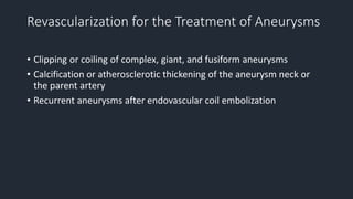
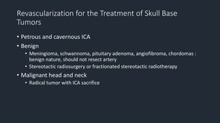
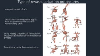
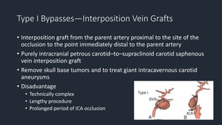
Revascularization Techniques
for Complex Aneurysms and
Skull Base Tumors
Youmans Chapter 380
Joshua R. Dusick
Nestor R. Gonzalez
Neil A. Martin
Outline
• Revascularization for the Treatment of Aneurysms
• Revascularization for the Treatment of Skull Base Tumors
• When Is the Collateral Circulation Inadequate and Bypass Necessary?
• Preoperative Planning and Preparation for Bypass Procedures
• Intraoperative Monitoring and Management
• Type of revascularization procedures
• Postoperative Management
• Complication
• Long-Term Graft Patency
Revascularization for the Treatment of Aneurysms
• Clipping or coiling of complex, giant, and fusiform aneurysms
• Calcification or atherosclerotic thickening of the aneurysm neck or
the parent artery
• Recurrent aneurysms after endovascular coil embolization
Revascularization for the Treatment of Skull Base
Tumors
• Petrous and cavernous ICA
• Benign
• Meningioma, schwannoma, pituitary adenoma, angiofibroma, chordomas :
benign nature, should not resect artery
• Stereotactic radiosurgery or fractionated stereotactic radiotherapy
• Malignant head and neck
• Radical tumor with ICA sacrifice
When Is the Collateral Circulation Inadequate and
Bypass Necessary?
• Anterior circulation
• Posterior circulation
• Distal arterial braches
Anterior circulation
• Selective approach
• angiographic evaluation of the competence of the circle of Willis
• balloon test occlusion coupled with measurement of cerebral blood flow
• patient pass balloon occlusion test : 20% chance of stroke with complete
occlusion without a bypass
• Universal approach
• advocate bypass for all patients who undergo ICA occlusion
Posterior circulation
• Unclipable and uncoilable posterior circulation
• Unilateral vertebral artery occlusion is well tolerated when
• the contralateral vertebral artery is not hypoplastic
• does not terminate in the posterior inferior cerebellar artery (PICA)
• Bilateral vertebral artery or basilar artery occlusion is associated with
a much higher risk for ischemia and should be considered only if
blood flow through both posterior communicating arteries is
sufficient (>1 mm)
Distal arterial braches
• Proximal occlusion : ICA, VA
• Occlusion of branch : MCA, PICA, AICA
Preoperative Planning and Preparation for Bypass
Procedures
• Optimal site of arterial occlusion, the collateral circulation, and the
size and location of the intended recipient and donor bypass vessels
• Anticonvulsants
• Aspirin (325 mg daily)
Intraoperative Monitoring and Management
• EEG : monitor burst suppression
• Evoked potential : activity of the sensory cortex and subcortical and
brainstem pathways during bypass procedures
• Mild hypothermic : 34-36 C
• Thiopental : cerebral protection during transient focal ischemia
• Local intraluminal anticoagulant irrigation is used
• Do not use systemic heparin
Type of revascularization procedures
Interposition Vein Grafts
Extracranial-to-Intracranial Bypass
with a Saphenous Vein Graft or
Radial Artery Graft
Scalp Artery (Superficial Temporal or
Occipital) Extracranial-to-Intracranial
Bypass
Direct Intracranial Revascularization
Type I Bypasses—Interposition Vein Grafts
• Interposition graft from the parent artery proximal to the site of the
occlusion to the point immediately distal to the parent artery
• Purely intracranial petrous carotid–to–supraclinoid carotid saphenous
vein interposition graft
• Remove skull base tumors and to treat giant intracavernous carotid
aneurysms
• Disadvantage
• Technically complex
• Lengthy procedure
• Prolonged period of ICA occlusion
Type II Bypasses—Extracranial-to-Intracranial Bypass
with a Saphenous Vein Graft or Radial Artery Graft
• Extracranial Carotid Artery–to–Middle Cerebral Artery Saphenous
Vein Interposition Graft
• External Carotid Artery–to–Posterior Cerebral Artery Saphenous Vein
Interposition Graft
Type II Bypasses—Extracranial-to-Intracranial Bypass
with a Saphenous Vein Graft or Radial Artery Graft
• Normal blood flow MCA : 250 mL/min, PCA moderate less
• STA graft position
• 15-30 mL/min, may increase with time
• not adequate to the circulation of major artery
• Saphenous vein graft
• 70-140 mL/min,can exceed 250 mL/min, 4-5 mm
• Lower longterm patency rate, higher risk of kinking, caliber mismatch
• Radial artery graft
• Smaller diameter, 3.5 mm
• 40-70 mL/min
Type II Bypasses—Extracranial-to-Intracranial Bypass
with a Saphenous Vein Graft or Radial Artery Graft
• Substiture for STA-MCA bypass
• scalp artery is hypoplastic, diseased, or occluded
• aneurysm that can be occluded only proximally, as in the case of some
dolichoectatic and fusiform aneurysms
• type II bypass may supply too much flow and can be dangerous
Extracranial Carotid Artery–to–Middle Cerebral
Artery Saphenous Vein Interposition Graft
External Carotid Artery–to–Posterior Cerebral
Artery Saphenous Vein Interposition Graft
• Basilar artery or bilateral vertebral arteries are occluded to treat an
unclippable basilar artery aneurysm
• Subtemporal approach
• The proximal 20 to 25 mm of the P2 segment is isolated, and a
segment free of brainstem perforating vessels is chosen for the
anastomosis
• Subdural hygroma : suggest routine subtemporal subdural-peritoneal
(or atrial) shunt
Type III Bypasses—Scalp Artery (Superficial Temporal or
Occipital) Extracranial-to-Intracranial Bypass
• Used
• a giant aneurysm requires occlusion of a single, crucial arterial branch
• carotid occlusion is required (for an aneurysm or tumor) and the circle of
Willis is only marginally inadequate
• Donor vessel : STA, occipital artery
• STA 15 – 30 mL/min
• STA-PCA, STA-SCA
• OA-PICA, OA-AICA
Superficial Temporal Artery–to–Middle Cerebral
Artery Bypass
• Parietal branch : Charter’s point (center, 6 cm, above EAC)
• Frontal branch : a second, vertically oriented incision above the ear is required
over Chater’s point
Superficial Temporal Artery–to–Middle
Cerebral Artery Bypass
• In patients who have two separate MCA branches that arise from the
dome of an aneurysm : double-barrel STA bypass
• Recipient a. ,> 1 mm diameter, 10 mm length
• Adventitia over the distal end of STA is removed
• Off temporary clip : distal MCA, proximal MCA, STA
Occipital Artery–to–Posterior Inferior Cerebellar
Artery Bypass
Superficial Temporal Artery–to–Superior Cerebellar
Artery Bypass and Superficial Temporal Artery–to–
Posterior Cerebral Artery Bypass
• Substituted for the saphenous vein graft–to-PCA bypass when some
collateral blood flow is available through small posterior
communicating arteries
Type IV Bypasses—Direct Intracranial Revascularization
• Anastomosis between two adjacent cerebral arteries
• Primary Reanastomosis
• intracranial arterial reconstruction involves excision of the aneurysm with
primary reconstruction of the parent artery
• Pericallosal-to-Pericallosal Bypass
• used to treat fusiform aneurysms of the proximal pericallosal artery or for
giant anterior communicating artery aneurysms that require trapping
• side-to-side anastomosis
Type IV Bypasses—Direct Intracranial Revascularization
• Posterior Inferior Cerebellar Artery–to–Posterior Inferior Cerebellar
Artery Anastomosis
• used when the occipital artery is small or has been damaged during a
previous surgical procedure
• side-to-side anastomosis
Techniques for Occluding or Trapping an Aneurysm
after Bypass
• Trapping
• combined proximal and distal parent artery occlusion
• it isolates the aneurysm from the circulation
• avoids the risk of rupture from retrograde filling
• allows immediate decompression of the aneurysm to relieve any mass effect.
• Proximal occlusion
• induce aneurysmal thrombosis
• Used in giant intracavernous aneurysms and large fusiform or dolichoectatic
vertebrobasilar aneurysms
Postoperative Management
• Palpating bypass pulse or by using a Doppler probe
• Continue ASA
• Euvolemia
• Normal SBP
• CTA
Complications
• Early graft occlusion
• careful avoidance of twisting, kinking, stretching, or tension of the graft
• avoidance of graft spasm by adventitial papaverine irrigation
• administration of perioperative antiplatelet therapy
• Post operative aneurysmal rupture
• High flow graft
• Emphasize the need to isolate the aneurysm completely from the circulation
by trapping whenever possible
Complications
• Ischemic neurological deficits
• Temporary arterial occlusion while the bypass anastomosis
• Cerebral protection with moderate hypothermia, induced arterial
hypertension, and barbiturate administration
• Subdural or epidural hematomas (or both) developed postoperatively
• given heparin in addition to aspirin
Long-Term Graft Patency
• Superior temporal artery graft
• Radial artery graft
• Saphenous vein graft

380 Revascularization techniques for complex aneurysms and skull base tumor

  • 1.
    Revascularization Techniques for ComplexAneurysms and Skull Base Tumors Youmans Chapter 380 Joshua R. Dusick Nestor R. Gonzalez Neil A. Martin
  • 2.
    Outline • Revascularization forthe Treatment of Aneurysms • Revascularization for the Treatment of Skull Base Tumors • When Is the Collateral Circulation Inadequate and Bypass Necessary? • Preoperative Planning and Preparation for Bypass Procedures • Intraoperative Monitoring and Management • Type of revascularization procedures • Postoperative Management • Complication • Long-Term Graft Patency
  • 3.
    Revascularization for theTreatment of Aneurysms • Clipping or coiling of complex, giant, and fusiform aneurysms • Calcification or atherosclerotic thickening of the aneurysm neck or the parent artery • Recurrent aneurysms after endovascular coil embolization
  • 4.
    Revascularization for theTreatment of Skull Base Tumors • Petrous and cavernous ICA • Benign • Meningioma, schwannoma, pituitary adenoma, angiofibroma, chordomas : benign nature, should not resect artery • Stereotactic radiosurgery or fractionated stereotactic radiotherapy • Malignant head and neck • Radical tumor with ICA sacrifice
  • 5.
    When Is theCollateral Circulation Inadequate and Bypass Necessary? • Anterior circulation • Posterior circulation • Distal arterial braches
  • 6.
    Anterior circulation • Selectiveapproach • angiographic evaluation of the competence of the circle of Willis • balloon test occlusion coupled with measurement of cerebral blood flow • patient pass balloon occlusion test : 20% chance of stroke with complete occlusion without a bypass • Universal approach • advocate bypass for all patients who undergo ICA occlusion
  • 7.
    Posterior circulation • Unclipableand uncoilable posterior circulation • Unilateral vertebral artery occlusion is well tolerated when • the contralateral vertebral artery is not hypoplastic • does not terminate in the posterior inferior cerebellar artery (PICA) • Bilateral vertebral artery or basilar artery occlusion is associated with a much higher risk for ischemia and should be considered only if blood flow through both posterior communicating arteries is sufficient (>1 mm)
  • 8.
    Distal arterial braches •Proximal occlusion : ICA, VA • Occlusion of branch : MCA, PICA, AICA
  • 9.
    Preoperative Planning andPreparation for Bypass Procedures • Optimal site of arterial occlusion, the collateral circulation, and the size and location of the intended recipient and donor bypass vessels • Anticonvulsants • Aspirin (325 mg daily)
  • 10.
    Intraoperative Monitoring andManagement • EEG : monitor burst suppression • Evoked potential : activity of the sensory cortex and subcortical and brainstem pathways during bypass procedures • Mild hypothermic : 34-36 C • Thiopental : cerebral protection during transient focal ischemia • Local intraluminal anticoagulant irrigation is used • Do not use systemic heparin
  • 11.
    Type of revascularizationprocedures Interposition Vein Grafts Extracranial-to-Intracranial Bypass with a Saphenous Vein Graft or Radial Artery Graft Scalp Artery (Superficial Temporal or Occipital) Extracranial-to-Intracranial Bypass Direct Intracranial Revascularization
  • 12.
    Type I Bypasses—InterpositionVein Grafts • Interposition graft from the parent artery proximal to the site of the occlusion to the point immediately distal to the parent artery • Purely intracranial petrous carotid–to–supraclinoid carotid saphenous vein interposition graft • Remove skull base tumors and to treat giant intracavernous carotid aneurysms • Disadvantage • Technically complex • Lengthy procedure • Prolonged period of ICA occlusion
  • 13.
    Type II Bypasses—Extracranial-to-IntracranialBypass with a Saphenous Vein Graft or Radial Artery Graft • Extracranial Carotid Artery–to–Middle Cerebral Artery Saphenous Vein Interposition Graft • External Carotid Artery–to–Posterior Cerebral Artery Saphenous Vein Interposition Graft
  • 14.
    Type II Bypasses—Extracranial-to-IntracranialBypass with a Saphenous Vein Graft or Radial Artery Graft • Normal blood flow MCA : 250 mL/min, PCA moderate less • STA graft position • 15-30 mL/min, may increase with time • not adequate to the circulation of major artery • Saphenous vein graft • 70-140 mL/min,can exceed 250 mL/min, 4-5 mm • Lower longterm patency rate, higher risk of kinking, caliber mismatch • Radial artery graft • Smaller diameter, 3.5 mm • 40-70 mL/min
  • 15.
    Type II Bypasses—Extracranial-to-IntracranialBypass with a Saphenous Vein Graft or Radial Artery Graft • Substiture for STA-MCA bypass • scalp artery is hypoplastic, diseased, or occluded • aneurysm that can be occluded only proximally, as in the case of some dolichoectatic and fusiform aneurysms • type II bypass may supply too much flow and can be dangerous
  • 16.
    Extracranial Carotid Artery–to–MiddleCerebral Artery Saphenous Vein Interposition Graft
  • 17.
    External Carotid Artery–to–PosteriorCerebral Artery Saphenous Vein Interposition Graft • Basilar artery or bilateral vertebral arteries are occluded to treat an unclippable basilar artery aneurysm • Subtemporal approach • The proximal 20 to 25 mm of the P2 segment is isolated, and a segment free of brainstem perforating vessels is chosen for the anastomosis • Subdural hygroma : suggest routine subtemporal subdural-peritoneal (or atrial) shunt
  • 18.
    Type III Bypasses—ScalpArtery (Superficial Temporal or Occipital) Extracranial-to-Intracranial Bypass • Used • a giant aneurysm requires occlusion of a single, crucial arterial branch • carotid occlusion is required (for an aneurysm or tumor) and the circle of Willis is only marginally inadequate • Donor vessel : STA, occipital artery • STA 15 – 30 mL/min • STA-PCA, STA-SCA • OA-PICA, OA-AICA
  • 19.
    Superficial Temporal Artery–to–MiddleCerebral Artery Bypass • Parietal branch : Charter’s point (center, 6 cm, above EAC) • Frontal branch : a second, vertically oriented incision above the ear is required over Chater’s point
  • 20.
    Superficial Temporal Artery–to–Middle CerebralArtery Bypass • In patients who have two separate MCA branches that arise from the dome of an aneurysm : double-barrel STA bypass • Recipient a. ,> 1 mm diameter, 10 mm length • Adventitia over the distal end of STA is removed • Off temporary clip : distal MCA, proximal MCA, STA
  • 21.
  • 22.
    Superficial Temporal Artery–to–SuperiorCerebellar Artery Bypass and Superficial Temporal Artery–to– Posterior Cerebral Artery Bypass • Substituted for the saphenous vein graft–to-PCA bypass when some collateral blood flow is available through small posterior communicating arteries
  • 23.
    Type IV Bypasses—DirectIntracranial Revascularization • Anastomosis between two adjacent cerebral arteries • Primary Reanastomosis • intracranial arterial reconstruction involves excision of the aneurysm with primary reconstruction of the parent artery • Pericallosal-to-Pericallosal Bypass • used to treat fusiform aneurysms of the proximal pericallosal artery or for giant anterior communicating artery aneurysms that require trapping • side-to-side anastomosis
  • 24.
    Type IV Bypasses—DirectIntracranial Revascularization • Posterior Inferior Cerebellar Artery–to–Posterior Inferior Cerebellar Artery Anastomosis • used when the occipital artery is small or has been damaged during a previous surgical procedure • side-to-side anastomosis
  • 25.
    Techniques for Occludingor Trapping an Aneurysm after Bypass • Trapping • combined proximal and distal parent artery occlusion • it isolates the aneurysm from the circulation • avoids the risk of rupture from retrograde filling • allows immediate decompression of the aneurysm to relieve any mass effect. • Proximal occlusion • induce aneurysmal thrombosis • Used in giant intracavernous aneurysms and large fusiform or dolichoectatic vertebrobasilar aneurysms
  • 26.
    Postoperative Management • Palpatingbypass pulse or by using a Doppler probe • Continue ASA • Euvolemia • Normal SBP • CTA
  • 27.
    Complications • Early graftocclusion • careful avoidance of twisting, kinking, stretching, or tension of the graft • avoidance of graft spasm by adventitial papaverine irrigation • administration of perioperative antiplatelet therapy • Post operative aneurysmal rupture • High flow graft • Emphasize the need to isolate the aneurysm completely from the circulation by trapping whenever possible
  • 28.
    Complications • Ischemic neurologicaldeficits • Temporary arterial occlusion while the bypass anastomosis • Cerebral protection with moderate hypothermia, induced arterial hypertension, and barbiturate administration • Subdural or epidural hematomas (or both) developed postoperatively • given heparin in addition to aspirin
  • 29.
    Long-Term Graft Patency •Superior temporal artery graft • Radial artery graft • Saphenous vein graft

Editor's Notes

  • #11 Burst suppression is an electroencephalography (EEG) pattern that is characterized by periods of high-voltage electrical activity alternating with periods of no activity in the brain. The pattern is found in patients with inactivated brain states, such as from general anesthesia, coma, orhypothermia.[1] This pattern can be physiological, as during early development, or pathological, as in diseases such as Ohtahara syndrome