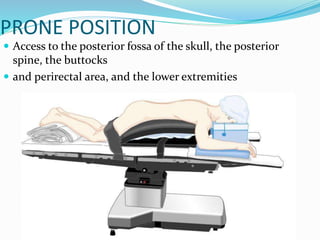
Patient positioning during anesthesia and surgery requires careful consideration of anatomical and physiological factors to prevent injury. The surgical position, procedure details, and patient characteristics and risks must be assessed to choose a position that allows procedure access while avoiding interference with breathing, circulation, nerve compression, or pressure points. Common positions like supine, lithotomy, lateral, and prone each have specific techniques, considerations, and potential risks that the anesthesia team must understand and address.
CONCLUSIONS:
- Cardiologist, obstetrician and anestesiologist should cooperate to each other
- The advantage of regional anesthesia is patients can communicate if symptoms occur
- If palpitations, chest pain and shortness of breath happened, immediate action should be performed
- RA should be given using lower dose of local anesthetics opioids and slow induction
- GA : standard technique “rapid sequence induction”
A patient with pacemaker presents a complex challenge to the attending anaesthesiologist. The mode of management will be according to the type of pacemaker implanted. This presentation discusses in brief the peri-operative consideration in a patient with pacemaker.
Anaesthetic management of ruptured ectopic pregnancy by Arowojolu BoluwajiArowojolu Samuel
anaesthetic management of a patient with ruptured ectopic pregnancy. helping anaesthetist to know what to do in emergency anaesthesia. this is an emergency case. salpingectomy. arowojolu boluwaji
One must be aware of the anatomic and physiologic changes associated with anesthesia, patient positioning, and the procedure.
• The following criteria should be met to prevent injury from pressure, obstruction or stretching:
– No interference with respiration
– No interference with circulation
– No pressure on peripheral nerves
– Minimal skin pressure
– Accessibility to operative site
– Accessibility for anesthetic administration
– No undue musculoskeletal discomfort
– Maintenance of individual requirements
Patient positioning and anaesthetic consideration. Explains how positioning affects your plan of anaesthesia
Also shows what kind of positioning is required for waht kind of surgery, and what are the implications that are associated with them, including the types of complications and their management.
MANAGEMENT OF ATRIOVENTRICULAR CONDUCTION BLOCK.pdfJim Jacob Roy
Cardiac conduction defects can occur due to various causes.
Atrioventricular conduction blocks ( AV blocks ) are classified into 3 types.
This document describes the acute management of AV block.
Couples presenting to the infertility clinic- Do they really have infertility...Sujoy Dasgupta
Dr Sujoy Dasgupta presented the study on "Couples presenting to the infertility clinic- Do they really have infertility? – The unexplored stories of non-consummation" in the 13th Congress of the Asia Pacific Initiative on Reproduction (ASPIRE 2024) at Manila on 24 May, 2024.
Title: Sense of Taste
Presenter: Dr. Faiza, Assistant Professor of Physiology
Qualifications:
MBBS (Best Graduate, AIMC Lahore)
FCPS Physiology
ICMT, CHPE, DHPE (STMU)
MPH (GC University, Faisalabad)
MBA (Virtual University of Pakistan)
Learning Objectives:
Describe the structure and function of taste buds.
Describe the relationship between the taste threshold and taste index of common substances.
Explain the chemical basis and signal transduction of taste perception for each type of primary taste sensation.
Recognize different abnormalities of taste perception and their causes.
Key Topics:
Significance of Taste Sensation:
Differentiation between pleasant and harmful food
Influence on behavior
Selection of food based on metabolic needs
Receptors of Taste:
Taste buds on the tongue
Influence of sense of smell, texture of food, and pain stimulation (e.g., by pepper)
Primary and Secondary Taste Sensations:
Primary taste sensations: Sweet, Sour, Salty, Bitter, Umami
Chemical basis and signal transduction mechanisms for each taste
Taste Threshold and Index:
Taste threshold values for Sweet (sucrose), Salty (NaCl), Sour (HCl), and Bitter (Quinine)
Taste index relationship: Inversely proportional to taste threshold
Taste Blindness:
Inability to taste certain substances, particularly thiourea compounds
Example: Phenylthiocarbamide
Structure and Function of Taste Buds:
Composition: Epithelial cells, Sustentacular/Supporting cells, Taste cells, Basal cells
Features: Taste pores, Taste hairs/microvilli, and Taste nerve fibers
Location of Taste Buds:
Found in papillae of the tongue (Fungiform, Circumvallate, Foliate)
Also present on the palate, tonsillar pillars, epiglottis, and proximal esophagus
Mechanism of Taste Stimulation:
Interaction of taste substances with receptors on microvilli
Signal transduction pathways for Umami, Sweet, Bitter, Sour, and Salty tastes
Taste Sensitivity and Adaptation:
Decrease in sensitivity with age
Rapid adaptation of taste sensation
Role of Saliva in Taste:
Dissolution of tastants to reach receptors
Washing away the stimulus
Taste Preferences and Aversions:
Mechanisms behind taste preference and aversion
Influence of receptors and neural pathways
Impact of Sensory Nerve Damage:
Degeneration of taste buds if the sensory nerve fiber is cut
Abnormalities of Taste Detection:
Conditions: Ageusia, Hypogeusia, Dysgeusia (parageusia)
Causes: Nerve damage, neurological disorders, infections, poor oral hygiene, adverse drug effects, deficiencies, aging, tobacco use, altered neurotransmitter levels
Neurotransmitters and Taste Threshold:
Effects of serotonin (5-HT) and norepinephrine (NE) on taste sensitivity
Supertasters:
25% of the population with heightened sensitivity to taste, especially bitterness
Increased number of fungiform papillae
micro teaching on communication m.sc nursing.pdfAnurag Sharma
Microteaching is a unique model of practice teaching. It is a viable instrument for the. desired change in the teaching behavior or the behavior potential which, in specified types of real. classroom situations, tends to facilitate the achievement of specified types of objectives.
Report Back from SGO 2024: What’s the Latest in Cervical Cancer?bkling
Are you curious about what’s new in cervical cancer research or unsure what the findings mean? Join Dr. Emily Ko, a gynecologic oncologist at Penn Medicine, to learn about the latest updates from the Society of Gynecologic Oncology (SGO) 2024 Annual Meeting on Women’s Cancer. Dr. Ko will discuss what the research presented at the conference means for you and answer your questions about the new developments.
Tom Selleck Health: A Comprehensive Look at the Iconic Actor’s Wellness Journeygreendigital
Tom Selleck, an enduring figure in Hollywood. has captivated audiences for decades with his rugged charm, iconic moustache. and memorable roles in television and film. From his breakout role as Thomas Magnum in Magnum P.I. to his current portrayal of Frank Reagan in Blue Bloods. Selleck's career has spanned over 50 years. But beyond his professional achievements. fans have often been curious about Tom Selleck Health. especially as he has aged in the public eye.
Follow us on: Pinterest
Introduction
Many have been interested in Tom Selleck health. not only because of his enduring presence on screen but also because of the challenges. and lifestyle choices he has faced and made over the years. This article delves into the various aspects of Tom Selleck health. exploring his fitness regimen, diet, mental health. and the challenges he has encountered as he ages. We'll look at how he maintains his well-being. the health issues he has faced, and his approach to ageing .
Early Life and Career
Childhood and Athletic Beginnings
Tom Selleck was born on January 29, 1945, in Detroit, Michigan, and grew up in Sherman Oaks, California. From an early age, he was involved in sports, particularly basketball. which played a significant role in his physical development. His athletic pursuits continued into college. where he attended the University of Southern California (USC) on a basketball scholarship. This early involvement in sports laid a strong foundation for his physical health and disciplined lifestyle.
Transition to Acting
Selleck's transition from an athlete to an actor came with its physical demands. His first significant role in "Magnum P.I." required him to perform various stunts and maintain a fit appearance. This role, which he played from 1980 to 1988. necessitated a rigorous fitness routine to meet the show's demands. setting the stage for his long-term commitment to health and wellness.
Fitness Regimen
Workout Routine
Tom Selleck health and fitness regimen has evolved. adapting to his changing roles and age. During his "Magnum, P.I." days. Selleck's workouts were intense and focused on building and maintaining muscle mass. His routine included weightlifting, cardiovascular exercises. and specific training for the stunts he performed on the show.
Selleck adjusted his fitness routine as he aged to suit his body's needs. Today, his workouts focus on maintaining flexibility, strength, and cardiovascular health. He incorporates low-impact exercises such as swimming, walking, and light weightlifting. This balanced approach helps him stay fit without putting undue strain on his joints and muscles.
Importance of Flexibility and Mobility
In recent years, Selleck has emphasized the importance of flexibility and mobility in his fitness regimen. Understanding the natural decline in muscle mass and joint flexibility with age. he includes stretching and yoga in his routine. These practices help prevent injuries, improve posture, and maintain mobilit
Lung Cancer: Artificial Intelligence, Synergetics, Complex System Analysis, S...Oleg Kshivets
RESULTS: Overall life span (LS) was 2252.1±1742.5 days and cumulative 5-year survival (5YS) reached 73.2%, 10 years – 64.8%, 20 years – 42.5%. 513 LCP lived more than 5 years (LS=3124.6±1525.6 days), 148 LCP – more than 10 years (LS=5054.4±1504.1 days).199 LCP died because of LC (LS=562.7±374.5 days). 5YS of LCP after bi/lobectomies was significantly superior in comparison with LCP after pneumonectomies (78.1% vs.63.7%, P=0.00001 by log-rank test). AT significantly improved 5YS (66.3% vs. 34.8%) (P=0.00000 by log-rank test) only for LCP with N1-2. Cox modeling displayed that 5YS of LCP significantly depended on: phase transition (PT) early-invasive LC in terms of synergetics, PT N0—N12, cell ratio factors (ratio between cancer cells- CC and blood cells subpopulations), G1-3, histology, glucose, AT, blood cell circuit, prothrombin index, heparin tolerance, recalcification time (P=0.000-0.038). Neural networks, genetic algorithm selection and bootstrap simulation revealed relationships between 5YS and PT early-invasive LC (rank=1), PT N0—N12 (rank=2), thrombocytes/CC (3), erythrocytes/CC (4), eosinophils/CC (5), healthy cells/CC (6), lymphocytes/CC (7), segmented neutrophils/CC (8), stick neutrophils/CC (9), monocytes/CC (10); leucocytes/CC (11). Correct prediction of 5YS was 100% by neural networks computing (area under ROC curve=1.0; error=0.0).
CONCLUSIONS: 5YS of LCP after radical procedures significantly depended on: 1) PT early-invasive cancer; 2) PT N0--N12; 3) cell ratio factors; 4) blood cell circuit; 5) biochemical factors; 6) hemostasis system; 7) AT; 8) LC characteristics; 9) LC cell dynamics; 10) surgery type: lobectomy/pneumonectomy; 11) anthropometric data. Optimal diagnosis and treatment strategies for LC are: 1) screening and early detection of LC; 2) availability of experienced thoracic surgeons because of complexity of radical procedures; 3) aggressive en block surgery and adequate lymph node dissection for completeness; 4) precise prediction; 5) adjuvant chemoimmunoradiotherapy for LCP with unfavorable prognosis.
3. One must be aware of the anatomic and physiologic
changes
associated with anesthesia, patient positioning, and the
procedure.
The following criteria should be met to prevent injury
from pressure, obstruction, or stretching:
– No interference with respiration
– No interference with circulation
– No pressure on peripheral nerves
– Minimal skin pressure
– Accessibility to operative site
– Accessibility for anesthetic administration
– No undue musculoskeletal discomfort
– Maintenance of individual requirements
4. The team should assess the following prior to
positioning of
the patient:
– Procedure length
– Surgeon’s preference of position
– Required position for procedure
– Anesthesia to be administered
– Patient’s risk factors
• age, weight, skin condition, mobility/limitations,
preexisting
conditions, airway etc.
– Patient’s privacy and medical needs
5.
6. PULMONARY CONCERNS
• Any position which limits movements of abdomen ,
chest wall
or diaphragm increase atelectasis and intrapulmonary
shunt
• Change from standing to supine ‐ decrease FRC due
to
cephalad displacement of the diaphragm
7. Surgical Positions
Four basic surgical positions
include:
– Supine
– Lateral
– Prone
– Lithotomy
• Variations include:
– Trendelenburg
– Reverse trendelenburg
– Fowler’s/semifowler
– Beach chair position
– Wattson jone position
– Position for robotic
surgeries
Variations include:
– Trendelenburg
– Reverse trendelenburg
– Fowler’s/semifowler
– Beach chair position
– Wattson jone position
– Position for robotic
surgeries
8. SUPINE
• Most common with the least amount of harm
• Placed on back with legs extended and uncrossed at the
ankles
• Arms either on arm boards abducted <90* with palms up or
tucked (not touching metal or constricted)
• Spinal column should be in alignment with legs parallel to the
OR bed
– Head in line with the spine and the face is upward
– Hips are parallel to the spine
• Padding is placed under the head, arms, and heels with a
pillow placed under the knees
• Safety belt placed 2” above the knees while not impeding
circulation
9. SUPINE CONCERNS
Greatest concerns are circulation and pressure points
• Most Common Nerve Damage:
– Brachial Plexus: positioning the arm >90*
– Radial and Ulnar: compression against the OR bed, metal
attachments, or when team members lean against the
arms during the procedure
– Peroneal and Tibial: Crossing of feet and plantar flexion of
ankles and feet
• Vulnerable Bony Prominences:
(due to rubbing and sustained pressure)
– Occiput, spine, scapula, Olecranon, Sacrum, Calcaneous
10. TRENDELENBURG
The patient is placed in the supine position while the OR bed
is modified to a head‐down tilt of 35 to 45 degrees resulting in
the head being lower than the pelvis
• Arms are in a comfortable position – either at the side or on
bilateral arm boards
• The foot of the OR bed is lowered to a desired angle
• ADVANTAGES
To increase V.R after spinal anesthesia
To increase central venous volume to facilitate central
cannulation
To minimise aspiration during regurgitation
12. REVERSE TRENDELENBURG
• The entire OR bed is tilted so the head is higher than the
feet
• Used for head and neck, laproscopic procedures
• Facilitates exposure, aids in breathing and decreases
blood
supply to the area
• A padded footboard is used to prevent the patient from
sliding toward the foot
• Reduces venous return therefore hypotension
• Laproscopic cholecystectomy : reverse trendelenburg
position with right up
13. Hemodynamic and Ventilation.(supine)
• Every 2.5 cm change of vertical ht. from the reference
point
at level of the heart leads to a change of MAP by 2
mmHg in
the opposite direction.
• V & Q are best in dependent lungs.
• Positive‐pressure ventilation provides the best
ventilation to
non‐dependent lung zones ‐V/Q mismatch.
14. LITHOTOMY
• With the patient in the supine position, the hips are flexed to
80‐100 o from the torso so that legs are parallel to it and legs
are abducted by 30‐45 o to expose the perineal region
• The patient’s buttocks are even with the lower break in the
OR bed (to prevent lumbosacral strain)
• The legs and feet are placed in stirrups that support the lower
extremities
• The legs are raised, positioned, and lowered slowly and
simultaneously, with the permission of the anesthesia care
provider
• Adequate padding and support for the legs/feet should
eliminate pressure on joints and nerve plexus
• The position must be symmetrical
• The perineum should be in line with the longitudinal axis of the
OR bed
16. PHYSIOLOGICAL CHANGES
Preload increases, causing a transient increase in CO ,
cerebral venous and intracranial pressure
• Reduce lung compliance
• If obesity or a large abdominal mass is present (tumor,
gravid
uterus)‐ VR to heart might decrease
• Normal lordotic curvature of the lumbar spine is lost
potentially aggravating any previous lower back pain
18. COMPARTMENT SYNDROME
• Rare
• Dorsiflexion of the ankle
• Excessive pressure of leg straps
• Surgeons leaning on suspended legs for long
durations
19. LATERAL
Anesthetized supine prior to turning
• Shoulder & hips turned simultaneously to prevent torsion of the spine
&
great vessels
• Lower leg is flexed at the hip; upper leg is straight
• Head must be in cervical alignment with the spine
• Breasts and genitalia to be free from torsion and pressure
• Axillary roll placed caudal to axilla of the downside arm (to
protect
brachial plexus)
• Padding placed under lower leg, to ankle and foot of upper leg, and to
lower arm (palm up) and upper arm
• Pillow placed lengthwise between
legs and between arms (if lateral
arm holder is not used)
• Stabilize patient with safety
strap and silk tape, if needed
20. Pulse should be monitored in the dependent arm for
early
detection of compression to axillary neurovascular
structures.
• Low saturation reading in pulse oximetry may be an
early
warning of compromised circulation.
• When a kidney rest is used, it must be properly placed
under
the dependent iliac crest to prevent inadvertent
compression
of the inferior vena cava
21. Park‐bench position: (SEMI‐PRONE POSITION)
– Modification of lat. position.
– Better access to posterior fossa.
– Upper arm positioned along lateral trunk & upper
shoulder is taped towards table.
Hemodynamic and Ventilation.
• In awake patient, Zone 3 West is occupying the dependent 18
cm of lung tissue. Lung tissue above 18 cm from bed level is
not perfused.
• During GA & positive pressure ventilation, the non‐dependent
lung zones are ventilated better ‐ worsening V/Q mismatch
22. PRONE POSITION
Access to the posterior fossa of the skull, the posterior
spine, the buttocks
and perirectal area, and the lower extremities
23. Arms :tucked in the neutral position /placed next to the
patient's head on arm boards—sometimes called the prone
“superman” position/Extra padding under the elbow –
prevent ulnar nerve
• When GA is planned, the patient is intubated on the
stretcher/ i.v access is obtained/ETT is well secured/pt is
turned prone onto the OT table/disconnect blood pressure
cuffs and arterial and venous lines that are on the side to
avoid dislodgment
• disconnection of pulse oximetry,arterial line, and tracheal
tube, leading to hypoventilation, desaturation, hemodynamic
instability, and altered anesthetic depth. Therefore its best to
keep pulse oximetry and arterial line connected
• ETT position is reassessed immediately after the move
24. • Head position
Turned to the side(45 degrees) if neck mobility is fine.
Check the dependent eye for external compression.
Maintained by surgical pillow, horseshoe headrest, or
Mayfield head pins Mostly, including disposable foam
versions, support the forehead, malar regions, and the
chin, with a cutout for the eyes, nose, and mouth
Mirror systems are available to facilitate intermittent
visual confirmation
25. Head support devices used in
prone position
1. MIRROR SYSTEM
2. HORSE SHOE REST
26. • Increased intra‐abdominal pressure decreases FRC,
compliance and increased PAP and transmits elevated VP to
the abdominal and spine vessels‐increase bleeding risk.
• Its imp that the abdomen hangs free and moves with
respiration‐ space of atleast 6 cms!
• Thorax: firm rolls or bolsters placed each side from the
clavicle to the iliac crest ( wilson frame, jackson table, relton
frame)
• Pendulous structures (e.g., Male genitalia and female breasts)
should be clear of compression
• Its essential to check the ETT position at a required degree of
flexion
27. BEACH CHAIR POSITION RISKS
1. Venous air embolism
2. Cerebral injury due to
hypotension
therefore essential to measure
CVP at
brain level since for every 2.5
cm diff
b/w BP cuff and brain – 2
mmHg fall in
BP
3. Case reports of cutaneous
neuropraxia
– lesser occipital , greater
auricular
nerve injury
28. SITTING
This is actually a modified recumbent
position as the legs are kept as high as
possible to promote venous return.
•Arms must be supported to prevent
shoulder traction.
•Head holder support is preferably
attached to the back
section of the table.
29.
30. ASA GUIDELINES FOR PREVENTION
OF PERIOPERATIVE
VISUAL LOSS‐ positioning
Patient Positioning
• There is no pathophysiologic mechanism by which facial
edema can cause perioperative ION.
• There is no evidence that ocular compression causes isolated
perioperative anterior ION or posterior ION.
– However, direct pressure on the eye should be avoided to
prevent central retinal artery occlusion (CRAO).
• The high‐risk patient should be positioned so that the head is
level with or higher than the heart when possible.
• The high‐risk patient's head should be maintained in a neutral
forward position (e.g., without significant neck flexion,
extension, lateral flexion, or rotation) when possible.