Downloaded 201 times


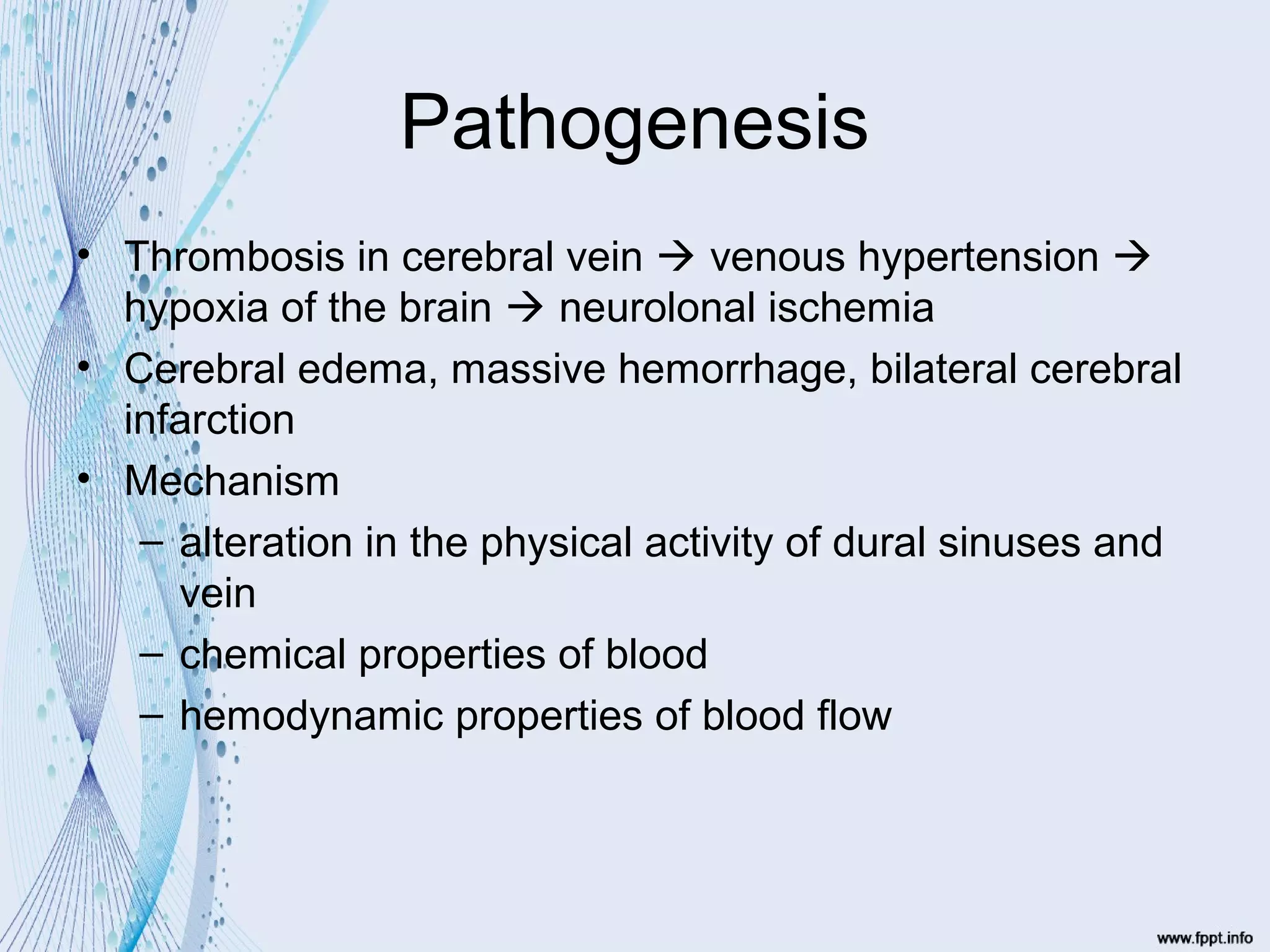


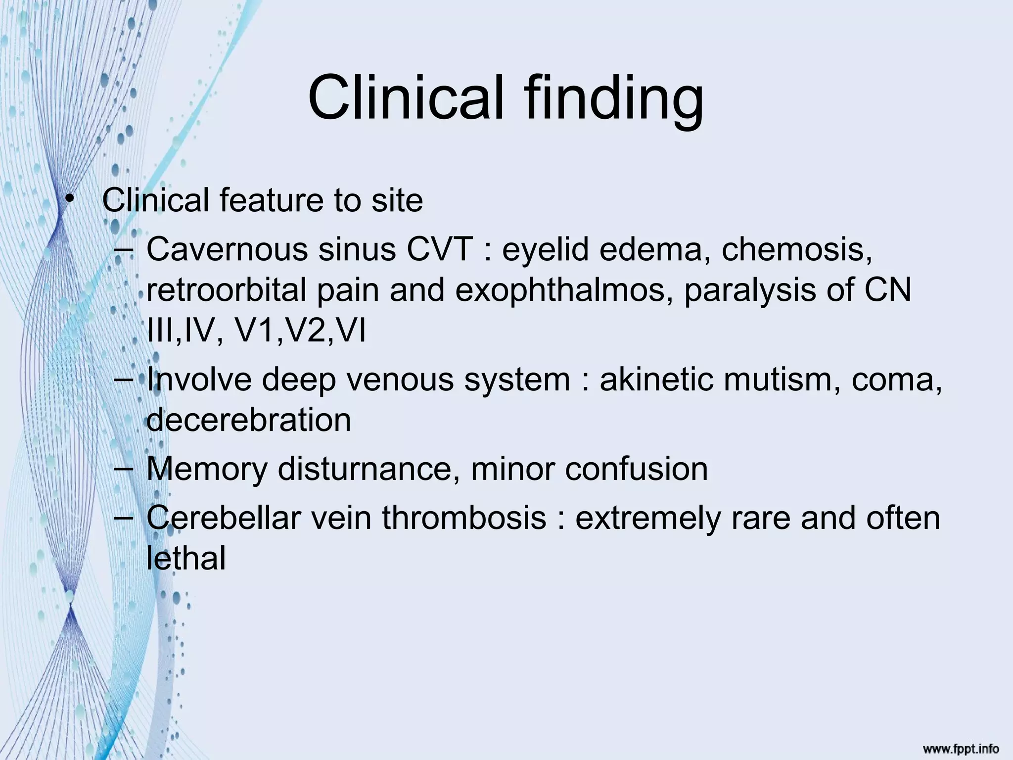
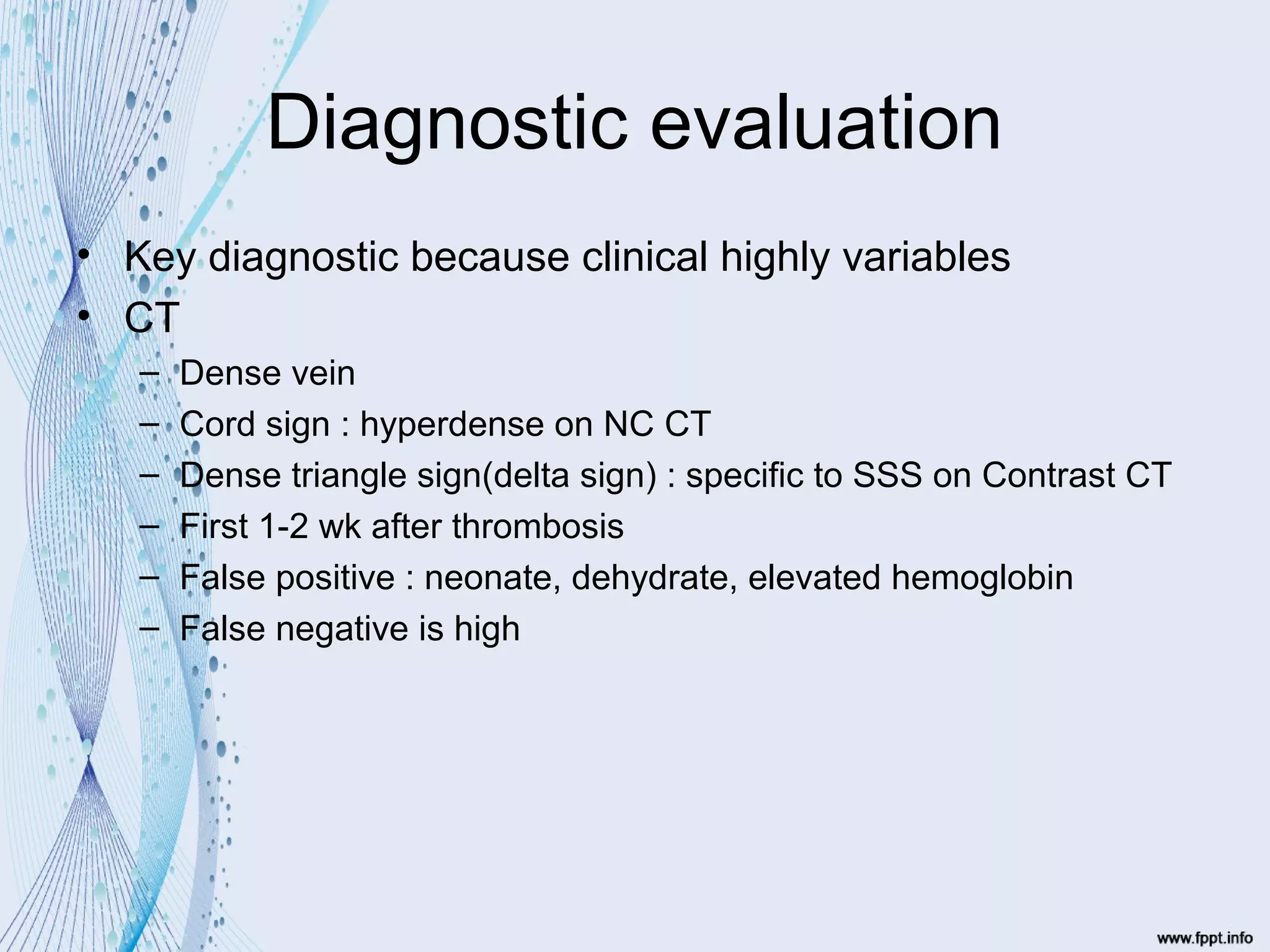


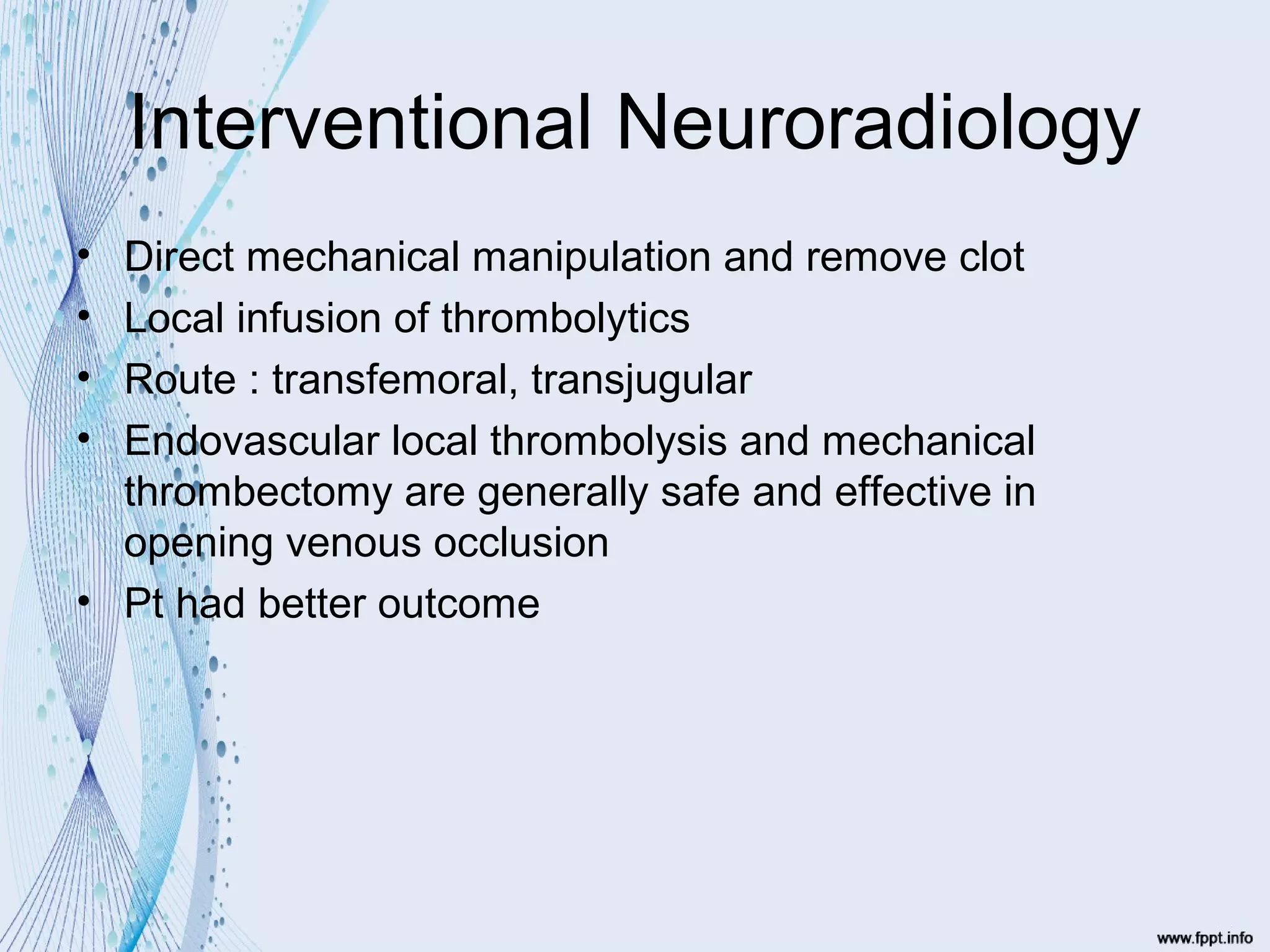


This document summarizes cerebral venous and sinus thrombosis. It discusses the pathogenesis, which can involve thrombosis in cerebral veins leading to venous hypertension and hypoxia. Risk factors include trauma, hypercoagulable states, infections, oral contraceptive use, and idiopathic causes. Clinical findings often include headache, seizures, neurological deficits, and features depending on the site of thrombosis. Diagnosis involves CT, MRI, MRA and MRV to detect thrombus. Treatment involves anticoagulants like heparin and warfarin, thrombolytics through interventional radiology, and occasionally surgery for complications. Outcomes depend on factors like coma, site of thrombosis, and degree of intracranial pressure.


