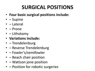
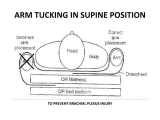
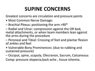
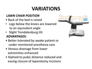
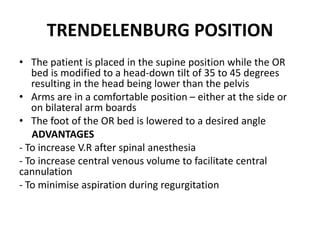
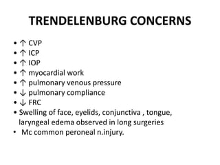
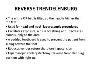
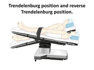
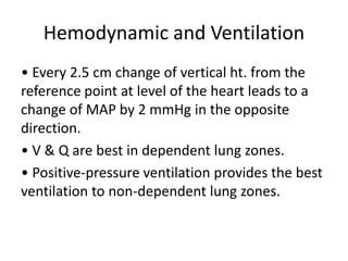
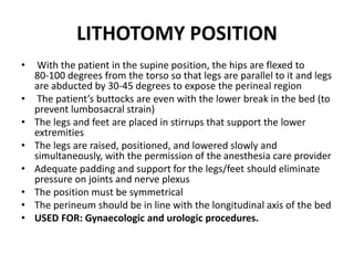
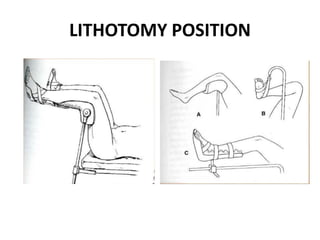
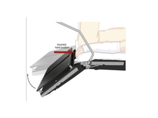
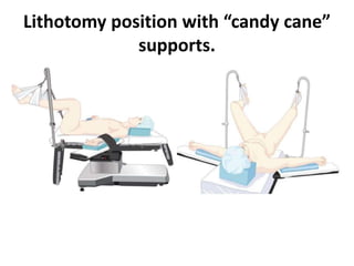
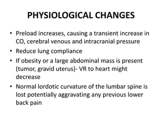
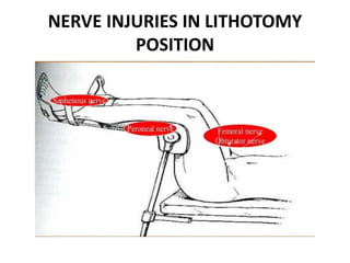
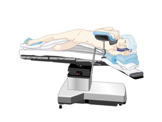
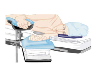
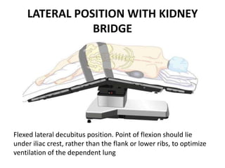
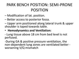
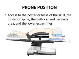
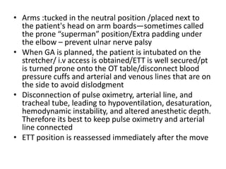
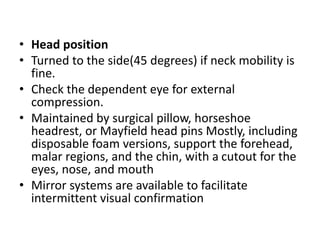
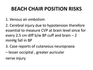
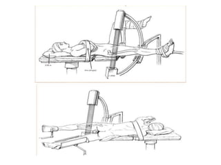
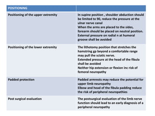
PATIENT POSITIONING
AND ANAESTHESIA
INTRODUCTION
• Positioning is the joint responsibility of the
surgeon & anesthesiologist.
• Ideal pt. positioning involves balancing
surgical comfort, against the risks related to
the pt. position.
• Pt. positioning & postural limitation should
be considered during the PAC.
OVERVIEW
• One must be aware of the anatomic and physiologic changes
associated with anesthesia, patient positioning, and the
procedure.
• The following criteria should be met to prevent injury from
pressure, obstruction or stretching:
– No interference with respiration
– No interference with circulation
– No pressure on peripheral nerves
– Minimal skin pressure
– Accessibility to operative site
– Accessibility for anesthetic administration
– No undue musculoskeletal discomfort
– Maintenance of individual requirements
ASSESSMENT
• The team should assess the following prior to
positioning of the patient:
– Procedure length
– Surgeon’s preference of position
– Required position for procedure
– Anesthesia to be administered
– Patient’s risk factors (age, weight, skin condition,
mobility/limitations, pre ‐ existing conditions,
airway etc.)
– Patient’s privacy and medical needs
GENERAL PHYSIOLOGICAL CONCERNS
•
PULMONARY CONCERNS
• Any position which limits movements of abdomen ,
chest wall or diaphragm increase atelectasis and
intrapulmonary shunt
• Change from standing to supine ‐ decrease FRC due
to cephalad displacement of the diaphragm
SURGICAL POSITIONS
• Four basic surgical positions include:
• – Supine
• – Lateral
• – Prone
• – Lithotomy
• Variations include:
• – Trendelenburg
• – Reverse Trendelenburg
• – Fowler’s/semifowler
• – Beach chair position
• – Wattson jone position
• – Position for robotic surgeries
SUPINE
• Most common with the least amount of harm
• Placed on back with legs extended and uncrossed at the
ankles
• Arms either on arm boards abducted <90* with palms up or
tucked (not touching metal or constricted)
• Spinal column should be in alignment with legs parallel to the
bed
– Head in line with the spine and the face is upward
– Hips are parallel to the spine
• Padding is placed under the head, arms, and heels with a
pillow placed under the knees
• Safety belt placed 2” above the knees while not impeding
circulation
ARM TUCKING IN SUPINE POSITION
TO PREVENT BRACHIAL PLEXUS INJURY
SUPINE CONCERNS
Greatest concerns are circulation and pressure points
• Most Common Nerve Damage:
– Brachial Plexus: positioning the arm >90*
– Radial and Ulnar: compression against the OR bed,
metal attachments, or when team members lean against
the arms during the procedure
– Peroneal and Tibial: Crossing of feet and plantar flexion
of ankles and feet
• Vulnerable Bony Prominences: (due to rubbing and
sustained pressure)
– Occiput, spine, scapula, Olecranon, Sacrum, Calcaneous
Comp: pressure alopecia,back ache , tissue ishemia.
VARIATIONS
LAWN CHAIR POSITION
• Back of the bed is raised
• Legs below the knees are lowered
to an equivalent angle
• Slight Trendelenburg tilt
ADVANTAGES:
• Better tolerated by awake patient or
under monitored anesthesia care
• Venous drainage from lower
extremities enhanced
• Xiphoid to pubic distance reduced and
easing closure of laparotomy incisions
TRENDELENBURG POSITION
• The patient is placed in the supine position while the OR
bed is modified to a head‐down tilt of 35 to 45 degrees
resulting in the head being lower than the pelvis
• Arms are in a comfortable position – either at the side or
on bilateral arm boards
• The foot of the OR bed is lowered to a desired angle
ADVANTAGES
- To increase V.R after spinal anesthesia
- To increase central venous volume to facilitate central
cannulation
- To minimise aspiration during regurgitation
TRENDELENBURG CONCERNS
• ↑ CVP
• ↑ ICP
• ↑ IOP
• ↑ myocardial work
• ↑ pulmonary venous pressure
• ↓ pulmonary compliance
• ↓ FRC
• Swelling of face, eyelids, conjunctiva , tongue,
laryngeal edema observed in long surgeries
• Mc common peroneal n.injury.
REVERSE TRENDELENBURG
• The entire OR bed is tilted so the head is higher than
the feet
• Used for head and neck, laparoscopic procedures
• Facilitates exposure, aids in breathing and decreases
blood supply to the area
• A padded footboard is used to prevent the patient from
sliding toward the foot
• Reduces venous return therefore hypotension
• Laparoscopic cholecystectomy : reverse trendelenburg
position with right up
Trendelenburg position and reverse
Trendelenburg position.
Hemodynamic and Ventilation
• Every 2.5 cm change of vertical ht. from the
reference point at level of the heart leads to a
change of MAP by 2 mmHg in the opposite
direction.
• V & Q are best in dependent lung zones.
• Positive‐pressure ventilation provides the best
ventilation to non‐dependent lung zones.
LITHOTOMY POSITION
• With the patient in the supine position, the hips are flexed to
80‐100 degrees from the torso so that legs are parallel to it and legs
are abducted by 30‐45 degrees to expose the perineal region
• The patient’s buttocks are even with the lower break in the bed (to
prevent lumbosacral strain)
• The legs and feet are placed in stirrups that support the lower
extremities
• The legs are raised, positioned, and lowered slowly and
simultaneously, with the permission of the anesthesia care provider
• Adequate padding and support for the legs/feet should eliminate
pressure on joints and nerve plexus
• The position must be symmetrical
• The perineum should be in line with the longitudinal axis of the bed
• USED FOR: Gynaecologic and urologic procedures.
LITHOTOMY POSITION
Lithotomy position with “candy cane”
supports.
PHYSIOLOGICAL CHANGES
• Preload increases, causing a transient increase in
CO, cerebral venous and intracranial pressure
• Reduce lung compliance
• If obesity or a large abdominal mass is present
(tumor, gravid uterus)‐ VR to heart might
decrease
• Normal lordotic curvature of the lumbar spine is
lost potentially aggravating any previous lower
back pain
NERVE INJURIES IN LITHOTOMY
POSITION
COMPARTMENT SYNDROME
• Rare but serious (mc seen in lithotomy 1:9000
whereas in supine 1:92000)
• Dorsiflexion of the ankle
• Excessive pressure of leg straps
• Surgeons leaning on suspended legs for long
durations
LATERAL POSITION
• Anesthetized supine prior to turning
• Shoulder & hips turned simultaneously to prevent torsion of the
spine & great vessels
• Lower leg is flexed at the hip; upper leg is straight
• Head must be in cervical alignment with the spine
• Breasts and genitalia to be free from torsion and pressure
• Axillary roll placed caudal to axilla of the downside arm (to protect
brachial plexus)
• Padding placed under lower leg, to ankle and foot of upper leg, and
to lower arm (palm up) and upper arm
• Pillow placed lengthwise between legs and between arms (if lateral
arm holder is not used)
• Stabilize patient with safety strap and silk tape, if needed
• USED FOR: Surgical access to the hemithorax, kidney,
retroperitoneal space.
LATERAL POSITION WITH KIDNEY
BRIDGE
Flexed lateral decubitus position. Point of flexion should lie
under iliac crest, rather than the flank or lower ribs, to optimize
ventilation of the dependent lung
• Pulse should be monitored in the dependent
arm for early detection of compression to
axillary neurovascular structures.
• Low saturation reading in pulse oximetry may be
an early warning of compromised circulation.
• When a kidney rest is used, it must be properly
placed under the dependent iliac crest to prevent
inadvertent compression of the inferior vena cava
PARK BENCH POSITION: SEMI‐PRONE
POSITION
– Modification of lat. position.
– Better access to posterior fossa.
– Upper arm positioned along lateral trunk & upper
shoulder is taped towards table.
• Hemodynamics and Ventilation:
- Lung tissue above 18 cm from bed level is not
perfused.
-During GA & positive pressure ventilation, the
non‐dependent lung zones are ventilated better ‐
worsening V/Q mismatch
PRONE POSITION
• Access to the posterior fossa of the skull, the
posterior spine, the buttocks and perirectal
area, and the lower extremities
• Arms :tucked in the neutral position /placed next to
the patient's head on arm boards—sometimes called
the prone “superman” position/Extra padding under
the elbow – prevent ulnar nerve palsy
• When GA is planned, the patient is intubated on the
stretcher/ i.v access is obtained/ETT is well secured/pt
is turned prone onto the OT table/disconnect blood
pressure cuffs and arterial and venous lines that are on
the side to avoid dislodgment
• Disconnection of pulse oximetry, arterial line, and
tracheal tube, leading to hypoventilation, desaturation,
hemodynamic instability, and altered anesthetic depth.
Therefore its best to keep pulse oximetry and arterial
line connected
• ETT position is reassessed immediately after the move
• Head position
• Turned to the side(45 degrees) if neck mobility is
fine.
• Check the dependent eye for external
compression.
• Maintained by surgical pillow, horseshoe
headrest, or Mayfield head pins Mostly, including
disposable foam versions, support the forehead,
malar regions, and the chin, with a cutout for the
eyes, nose, and mouth
• Mirror systems are available to facilitate
intermittent visual confirmation
Head support devices used in
prone position
• 1. MIRROR SYSTEM
• 2. HORSE SHOE REST
• Increased intra‐abdominal pressure decreases FRC,
compliance and increased PAP and transmits elevated VP to
the abdominal and spine vessels‐increase bleeding risk.
• Its imp that the abdomen hangs free and moves with
respiration‐ space of atleast 6 cms!
• Thorax: firm rolls or bolsters placed each side from the
clavicle to the iliac crest ( wilson frame, jackson table,
relton frame)
• Pendulous structures (e.g., Male genitalia and female
breasts) should be clear of compression and placed medial
to bolsters.
• It is essential to check the ETT position at a required degree
of flexion
Hemodynamics and Ventilation
• increases intraabdominal pressure, decreases VR to the
heart, and increases systemic and pulmonary vascular
resistance‐HYPOTENSION
• Oxygenation and oxygen delivery, however, may
improve as
1) Perfusion of the entire lungs improves
2) Increase in intraabdominal pressure decreases chest
wall compliance, which under PPV, improves ventilation
of the dependent zones of the lung, and
3) Previously atelectatic dorsal zones of lungs may open.
Fowler’s Position & semi-fowler ‘s
position
• Patient begins in the supine position
• Foot of the OR bed is lowered slightly, flexing the knees, while the
body section is raised to 35 – 45 degrees, thereby becoming a
backrest
• The entire OR bed is tilted slightly with the head end downward
(preventing the patient from sliding)
• Feet rest against a padded footboard
• Arms are crossed loosely over the abdomen and taped or placed on
a pillow on the patient’s lap
• A pillow is placed under the knees.
• For cranial procedures, the head is supported in a head rest and/or
with sterile tongs
• This position can be used for shoulder
procedures ( BEACH CHAIR POSITION)
BEACH CHAIR POSITION RISKS
1. Venous air embolism
2. Cerebral injury due to hypotension therefore
essential to measure CVP at brain level since for
every 2.5 cm diff b/w BP cuff and brain – 2
mmHg fall in BP
3. Case reports of cutaneous neuropraxia
– lesser occipital , greater auricular
nerve injury
SITTING POSITION
This is actually a modified recumbent position as the legs are
kept as high as possible to promote venous return.
• Arms must be supported to prevent shoulder traction.
• Head holder support is preferably attached to the back
section of the table.
ADVANTAGES:
• Excellent surgical exposure
• Reduced perioperative blood loss
• Superior access to the airway
• Reduced facial swelling
• Improved ventilation, particularly in obese
patients
• Modern monitoring‐ early indication of air
embolism
RELATIVE CONTRAINDICATIONS OF
SITTING POSITION
• VP shunt
• Cerebral ischemia upright awake
• Patent foramen ovale(detected preop by
contrast ECHO)
• Cardiac instability /extreme ages
PROBLEMS
• Venous air embolism
• Hypotension (prevented by stockings)
• Arms if not supported well‐ brachial plexus
injury
SPECIAL CONCERNS IN
NEUROSURGICAL PATIENTS
• Head Positioning
• Ideal position of head for Craniotomies & spine
procedures is based on the 2 principles:
1) An imaginary trajectory from the highest point at
skull surface to area of interest in brain should be
the shortest distance between the 2 points.
2) The exposed surface of the skull & an imaginary
perimeter of craniotomy should be parallel to the
floor.
Types of Craniotomies
• A. Ant. Parasagittal
• B. Frontosphenotemporal
• C. Sub‐temporal
• D. Lat Sub‐occipital
• E. Midline Sub‐occipital
• F. Post. Parasagittal
Application of a skeletal fixation pins
• Tachycardia and hypertension
• Rupture of untreated cerebral aneurysms
Local infiltration
iv anesthetic agent (propofol 0.5‐1 mg/kg)
Inhalational anesthetic
• Benefits : Immobility, surgical comfort.
• Risks : Bleeding, air embolism, scalp and eye
laceration, pressure alopecia.
Head and Neck positioning: Rotation,
Hyperflexion, Hyperextension
• Brain stem & cervical spine ischemia.
• Quadriparesis, quadriplegia &
cerebral infarction.
• At risk :
– Osteophytes
– Arthritis
– Vascular atherosclerosis
– Obesity
– Hemodynamically unstable
• Head safely rotated b/w 0‐45°.
• For more rotation, a roll/pillow place under the
opposite shoulder.
• Maintaining 2‐3 finger breadths thyromental
distance during neck flexion.
Orthopaedic surgery position
• Orthopedic fracture table – Wattson‐Jone’s
• Body section to support head & thorax
• Sacral plate for pelvis
• Perineal post
• Adjustable foot plates
• Table maintains traction of the extremity
• Allows surgical & fluroscopic access
• Anesthesia induced & then the patients are
positioned on this table . Arm on side
Problems with Jones position
• Brachial plexus injury due to > than 90*
extension of the upper limb
• Lower extremity compartment syndrome due
to long surgeries & compression
• Pudendal nerve injury Due to pressure of the
perineal post
POSITIONING IN ROBOTIC SURGERIES
• Robot‐assisted laparoscopic surgery(RALS) ‐ referred to
as da Vinci surgery‐used for gynecologic, urological,
and gynecologic oncology procedures.
• Pt is placed in dorsal lithotomy and steep
trendelenburg position.(30‐45 degrees table tilt)
• Steps to avoid slipping of patient are:
1. Place an surgical sheet made of antiskid material on
the OT bed.
2. Placing surgical gel pads against patient’s bare skin.
3. Use of the Bean Bag Positioner
POSITIONING IN ROBOTIC SURGERIES
• The risks are same as in Trendelenburg position
including:
• Rhabdomyolysis
• Corneal abrasions
• Prolonged surgery
• Difficult immediate access to the airway and iv
• Fixed position of the robot can cause injury if
position is changed
• Monitoring is difficult
PERIPHERAL NERVE INJURIES
Most frequent site of injury:
• Ulnar nerve (28%)
• Brachial plexus (20%)
• Lumbosacral nerve root (16%)
• Spinal cord (13%)
• Either : stretch or compression type
Brachial plexus is vulnerable to stretch and compression
• ASA GUIDELINES TO PREVENT
PERIOPERATIVE PERIPHERAL
NEUROPATHY
POSITIONING
Positioning of the upper extremity In supine position , shoulder abduction should
be limited to 90, reduce the pressure at the
ulnar nerve canal
When the arms are placed to the sides,
forearm should be placed on neutral position.
External pressure on radial n at humeral
groove shall be avoided
Positioning of the lower extremity The lithotomy position that stretches the
hamstring gp beyond a comfortable range
may pull the sciatic nerve.
Extended pressure at the head of the fibula
shall be avoided
Neither hip extension or flexion inc risk of
femoral neuropathy
Padded protection Padded armrests may reduce the potential for
upper limb neuropathy
Elbow and head of the fibula padding reduce
the risk of peripheral neuropathies
Post surgical evaluation The postsurgical evaluation of the limb nerve
function should lead to an early diagnosis of a
peripheral neuropathy
Eye injuries
• Rare (0.05%).
• Mc- Corneal abrasions
• Blindness rare,0.1% in prone surgery.(risk
factors –prolonged surgery, hypotension ,
blood loss ,anemia and pressure sec to prone
positioning)
ASA GUIDELINES FOR PREVENTION OF
PERIOPERATIVE VISUAL
LOSS‐POSITIONING
• There is no pathophysiologic mechanism by which facial
edema can cause perioperative ION.
• There is no evidence that ocular compression causes
isolated perioperative anterior ION or posterior ION.
– However, direct pressure on the eye should be avoided to
prevent central retinal artery occlusion (CRAO).
• The high‐risk patient should be positioned so that the head
is level with or higher than the heart when possible.
• The high‐risk patient's head should be maintained in a
neutral forward position (e.g., without significant neck
flexion, extension, lateral flexion, or rotation) when
possible.
Thank you!!!!

PATIENT POSITIONING AND ANAESTHESIA ppt

  • 1.
  • 2.
    INTRODUCTION • Positioning isthe joint responsibility of the surgeon & anesthesiologist. • Ideal pt. positioning involves balancing surgical comfort, against the risks related to the pt. position. • Pt. positioning & postural limitation should be considered during the PAC.
  • 3.
    OVERVIEW • One mustbe aware of the anatomic and physiologic changes associated with anesthesia, patient positioning, and the procedure. • The following criteria should be met to prevent injury from pressure, obstruction or stretching: – No interference with respiration – No interference with circulation – No pressure on peripheral nerves – Minimal skin pressure – Accessibility to operative site – Accessibility for anesthetic administration – No undue musculoskeletal discomfort – Maintenance of individual requirements
  • 4.
    ASSESSMENT • The teamshould assess the following prior to positioning of the patient: – Procedure length – Surgeon’s preference of position – Required position for procedure – Anesthesia to be administered – Patient’s risk factors (age, weight, skin condition, mobility/limitations, pre ‐ existing conditions, airway etc.) – Patient’s privacy and medical needs
  • 5.
    GENERAL PHYSIOLOGICAL CONCERNS • PULMONARYCONCERNS • Any position which limits movements of abdomen , chest wall or diaphragm increase atelectasis and intrapulmonary shunt • Change from standing to supine ‐ decrease FRC due to cephalad displacement of the diaphragm
  • 6.
    SURGICAL POSITIONS • Fourbasic surgical positions include: • – Supine • – Lateral • – Prone • – Lithotomy • Variations include: • – Trendelenburg • – Reverse Trendelenburg • – Fowler’s/semifowler • – Beach chair position • – Wattson jone position • – Position for robotic surgeries
  • 7.
    SUPINE • Most commonwith the least amount of harm • Placed on back with legs extended and uncrossed at the ankles • Arms either on arm boards abducted <90* with palms up or tucked (not touching metal or constricted) • Spinal column should be in alignment with legs parallel to the bed – Head in line with the spine and the face is upward – Hips are parallel to the spine • Padding is placed under the head, arms, and heels with a pillow placed under the knees • Safety belt placed 2” above the knees while not impeding circulation
  • 9.
    ARM TUCKING INSUPINE POSITION TO PREVENT BRACHIAL PLEXUS INJURY
  • 10.
    SUPINE CONCERNS Greatest concernsare circulation and pressure points • Most Common Nerve Damage: – Brachial Plexus: positioning the arm >90* – Radial and Ulnar: compression against the OR bed, metal attachments, or when team members lean against the arms during the procedure – Peroneal and Tibial: Crossing of feet and plantar flexion of ankles and feet • Vulnerable Bony Prominences: (due to rubbing and sustained pressure) – Occiput, spine, scapula, Olecranon, Sacrum, Calcaneous Comp: pressure alopecia,back ache , tissue ishemia.
  • 11.
    VARIATIONS LAWN CHAIR POSITION •Back of the bed is raised • Legs below the knees are lowered to an equivalent angle • Slight Trendelenburg tilt ADVANTAGES: • Better tolerated by awake patient or under monitored anesthesia care • Venous drainage from lower extremities enhanced • Xiphoid to pubic distance reduced and easing closure of laparotomy incisions
  • 12.
    TRENDELENBURG POSITION • Thepatient is placed in the supine position while the OR bed is modified to a head‐down tilt of 35 to 45 degrees resulting in the head being lower than the pelvis • Arms are in a comfortable position – either at the side or on bilateral arm boards • The foot of the OR bed is lowered to a desired angle ADVANTAGES - To increase V.R after spinal anesthesia - To increase central venous volume to facilitate central cannulation - To minimise aspiration during regurgitation
  • 13.
    TRENDELENBURG CONCERNS • ↑CVP • ↑ ICP • ↑ IOP • ↑ myocardial work • ↑ pulmonary venous pressure • ↓ pulmonary compliance • ↓ FRC • Swelling of face, eyelids, conjunctiva , tongue, laryngeal edema observed in long surgeries • Mc common peroneal n.injury.
  • 14.
    REVERSE TRENDELENBURG • Theentire OR bed is tilted so the head is higher than the feet • Used for head and neck, laparoscopic procedures • Facilitates exposure, aids in breathing and decreases blood supply to the area • A padded footboard is used to prevent the patient from sliding toward the foot • Reduces venous return therefore hypotension • Laparoscopic cholecystectomy : reverse trendelenburg position with right up
  • 15.
    Trendelenburg position andreverse Trendelenburg position.
  • 16.
    Hemodynamic and Ventilation •Every 2.5 cm change of vertical ht. from the reference point at level of the heart leads to a change of MAP by 2 mmHg in the opposite direction. • V & Q are best in dependent lung zones. • Positive‐pressure ventilation provides the best ventilation to non‐dependent lung zones.
  • 17.
    LITHOTOMY POSITION • Withthe patient in the supine position, the hips are flexed to 80‐100 degrees from the torso so that legs are parallel to it and legs are abducted by 30‐45 degrees to expose the perineal region • The patient’s buttocks are even with the lower break in the bed (to prevent lumbosacral strain) • The legs and feet are placed in stirrups that support the lower extremities • The legs are raised, positioned, and lowered slowly and simultaneously, with the permission of the anesthesia care provider • Adequate padding and support for the legs/feet should eliminate pressure on joints and nerve plexus • The position must be symmetrical • The perineum should be in line with the longitudinal axis of the bed • USED FOR: Gynaecologic and urologic procedures.
  • 18.
  • 20.
    Lithotomy position with“candy cane” supports.
  • 21.
    PHYSIOLOGICAL CHANGES • Preloadincreases, causing a transient increase in CO, cerebral venous and intracranial pressure • Reduce lung compliance • If obesity or a large abdominal mass is present (tumor, gravid uterus)‐ VR to heart might decrease • Normal lordotic curvature of the lumbar spine is lost potentially aggravating any previous lower back pain
  • 22.
    NERVE INJURIES INLITHOTOMY POSITION
  • 23.
    COMPARTMENT SYNDROME • Rarebut serious (mc seen in lithotomy 1:9000 whereas in supine 1:92000) • Dorsiflexion of the ankle • Excessive pressure of leg straps • Surgeons leaning on suspended legs for long durations
  • 24.
    LATERAL POSITION • Anesthetizedsupine prior to turning • Shoulder & hips turned simultaneously to prevent torsion of the spine & great vessels • Lower leg is flexed at the hip; upper leg is straight • Head must be in cervical alignment with the spine • Breasts and genitalia to be free from torsion and pressure • Axillary roll placed caudal to axilla of the downside arm (to protect brachial plexus) • Padding placed under lower leg, to ankle and foot of upper leg, and to lower arm (palm up) and upper arm • Pillow placed lengthwise between legs and between arms (if lateral arm holder is not used) • Stabilize patient with safety strap and silk tape, if needed • USED FOR: Surgical access to the hemithorax, kidney, retroperitoneal space.
  • 27.
    LATERAL POSITION WITHKIDNEY BRIDGE Flexed lateral decubitus position. Point of flexion should lie under iliac crest, rather than the flank or lower ribs, to optimize ventilation of the dependent lung
  • 28.
    • Pulse shouldbe monitored in the dependent arm for early detection of compression to axillary neurovascular structures. • Low saturation reading in pulse oximetry may be an early warning of compromised circulation. • When a kidney rest is used, it must be properly placed under the dependent iliac crest to prevent inadvertent compression of the inferior vena cava
  • 29.
    PARK BENCH POSITION:SEMI‐PRONE POSITION – Modification of lat. position. – Better access to posterior fossa. – Upper arm positioned along lateral trunk & upper shoulder is taped towards table. • Hemodynamics and Ventilation: - Lung tissue above 18 cm from bed level is not perfused. -During GA & positive pressure ventilation, the non‐dependent lung zones are ventilated better ‐ worsening V/Q mismatch
  • 30.
    PRONE POSITION • Accessto the posterior fossa of the skull, the posterior spine, the buttocks and perirectal area, and the lower extremities
  • 31.
    • Arms :tuckedin the neutral position /placed next to the patient's head on arm boards—sometimes called the prone “superman” position/Extra padding under the elbow – prevent ulnar nerve palsy • When GA is planned, the patient is intubated on the stretcher/ i.v access is obtained/ETT is well secured/pt is turned prone onto the OT table/disconnect blood pressure cuffs and arterial and venous lines that are on the side to avoid dislodgment • Disconnection of pulse oximetry, arterial line, and tracheal tube, leading to hypoventilation, desaturation, hemodynamic instability, and altered anesthetic depth. Therefore its best to keep pulse oximetry and arterial line connected • ETT position is reassessed immediately after the move
  • 32.
    • Head position •Turned to the side(45 degrees) if neck mobility is fine. • Check the dependent eye for external compression. • Maintained by surgical pillow, horseshoe headrest, or Mayfield head pins Mostly, including disposable foam versions, support the forehead, malar regions, and the chin, with a cutout for the eyes, nose, and mouth • Mirror systems are available to facilitate intermittent visual confirmation
  • 33.
    Head support devicesused in prone position • 1. MIRROR SYSTEM • 2. HORSE SHOE REST
  • 34.
    • Increased intra‐abdominalpressure decreases FRC, compliance and increased PAP and transmits elevated VP to the abdominal and spine vessels‐increase bleeding risk. • Its imp that the abdomen hangs free and moves with respiration‐ space of atleast 6 cms! • Thorax: firm rolls or bolsters placed each side from the clavicle to the iliac crest ( wilson frame, jackson table, relton frame) • Pendulous structures (e.g., Male genitalia and female breasts) should be clear of compression and placed medial to bolsters. • It is essential to check the ETT position at a required degree of flexion
  • 35.
    Hemodynamics and Ventilation •increases intraabdominal pressure, decreases VR to the heart, and increases systemic and pulmonary vascular resistance‐HYPOTENSION • Oxygenation and oxygen delivery, however, may improve as 1) Perfusion of the entire lungs improves 2) Increase in intraabdominal pressure decreases chest wall compliance, which under PPV, improves ventilation of the dependent zones of the lung, and 3) Previously atelectatic dorsal zones of lungs may open.
  • 36.
    Fowler’s Position &semi-fowler ‘s position • Patient begins in the supine position • Foot of the OR bed is lowered slightly, flexing the knees, while the body section is raised to 35 – 45 degrees, thereby becoming a backrest • The entire OR bed is tilted slightly with the head end downward (preventing the patient from sliding) • Feet rest against a padded footboard • Arms are crossed loosely over the abdomen and taped or placed on a pillow on the patient’s lap • A pillow is placed under the knees. • For cranial procedures, the head is supported in a head rest and/or with sterile tongs • This position can be used for shoulder procedures ( BEACH CHAIR POSITION)
  • 37.
    BEACH CHAIR POSITIONRISKS 1. Venous air embolism 2. Cerebral injury due to hypotension therefore essential to measure CVP at brain level since for every 2.5 cm diff b/w BP cuff and brain – 2 mmHg fall in BP 3. Case reports of cutaneous neuropraxia – lesser occipital , greater auricular nerve injury
  • 39.
    SITTING POSITION This isactually a modified recumbent position as the legs are kept as high as possible to promote venous return. • Arms must be supported to prevent shoulder traction. • Head holder support is preferably attached to the back section of the table.
  • 40.
    ADVANTAGES: • Excellent surgicalexposure • Reduced perioperative blood loss • Superior access to the airway • Reduced facial swelling • Improved ventilation, particularly in obese patients • Modern monitoring‐ early indication of air embolism
  • 41.
    RELATIVE CONTRAINDICATIONS OF SITTINGPOSITION • VP shunt • Cerebral ischemia upright awake • Patent foramen ovale(detected preop by contrast ECHO) • Cardiac instability /extreme ages
  • 42.
    PROBLEMS • Venous airembolism • Hypotension (prevented by stockings) • Arms if not supported well‐ brachial plexus injury
  • 43.
    SPECIAL CONCERNS IN NEUROSURGICALPATIENTS • Head Positioning • Ideal position of head for Craniotomies & spine procedures is based on the 2 principles: 1) An imaginary trajectory from the highest point at skull surface to area of interest in brain should be the shortest distance between the 2 points. 2) The exposed surface of the skull & an imaginary perimeter of craniotomy should be parallel to the floor.
  • 44.
    Types of Craniotomies •A. Ant. Parasagittal • B. Frontosphenotemporal • C. Sub‐temporal • D. Lat Sub‐occipital • E. Midline Sub‐occipital • F. Post. Parasagittal
  • 45.
    Application of askeletal fixation pins • Tachycardia and hypertension • Rupture of untreated cerebral aneurysms Local infiltration iv anesthetic agent (propofol 0.5‐1 mg/kg) Inhalational anesthetic • Benefits : Immobility, surgical comfort. • Risks : Bleeding, air embolism, scalp and eye laceration, pressure alopecia.
  • 46.
    Head and Neckpositioning: Rotation, Hyperflexion, Hyperextension • Brain stem & cervical spine ischemia. • Quadriparesis, quadriplegia & cerebral infarction.
  • 47.
    • At risk: – Osteophytes – Arthritis – Vascular atherosclerosis – Obesity – Hemodynamically unstable • Head safely rotated b/w 0‐45°. • For more rotation, a roll/pillow place under the opposite shoulder. • Maintaining 2‐3 finger breadths thyromental distance during neck flexion.
  • 48.
    Orthopaedic surgery position •Orthopedic fracture table – Wattson‐Jone’s • Body section to support head & thorax • Sacral plate for pelvis • Perineal post • Adjustable foot plates • Table maintains traction of the extremity • Allows surgical & fluroscopic access • Anesthesia induced & then the patients are positioned on this table . Arm on side
  • 50.
    Problems with Jonesposition • Brachial plexus injury due to > than 90* extension of the upper limb • Lower extremity compartment syndrome due to long surgeries & compression • Pudendal nerve injury Due to pressure of the perineal post
  • 51.
    POSITIONING IN ROBOTICSURGERIES • Robot‐assisted laparoscopic surgery(RALS) ‐ referred to as da Vinci surgery‐used for gynecologic, urological, and gynecologic oncology procedures. • Pt is placed in dorsal lithotomy and steep trendelenburg position.(30‐45 degrees table tilt) • Steps to avoid slipping of patient are: 1. Place an surgical sheet made of antiskid material on the OT bed. 2. Placing surgical gel pads against patient’s bare skin. 3. Use of the Bean Bag Positioner
  • 52.
  • 53.
    POSITIONING IN ROBOTICSURGERIES • The risks are same as in Trendelenburg position including: • Rhabdomyolysis • Corneal abrasions • Prolonged surgery • Difficult immediate access to the airway and iv • Fixed position of the robot can cause injury if position is changed • Monitoring is difficult
  • 54.
    PERIPHERAL NERVE INJURIES Mostfrequent site of injury: • Ulnar nerve (28%) • Brachial plexus (20%) • Lumbosacral nerve root (16%) • Spinal cord (13%) • Either : stretch or compression type
  • 55.
    Brachial plexus isvulnerable to stretch and compression
  • 56.
    • ASA GUIDELINESTO PREVENT PERIOPERATIVE PERIPHERAL NEUROPATHY
  • 57.
    POSITIONING Positioning of theupper extremity In supine position , shoulder abduction should be limited to 90, reduce the pressure at the ulnar nerve canal When the arms are placed to the sides, forearm should be placed on neutral position. External pressure on radial n at humeral groove shall be avoided Positioning of the lower extremity The lithotomy position that stretches the hamstring gp beyond a comfortable range may pull the sciatic nerve. Extended pressure at the head of the fibula shall be avoided Neither hip extension or flexion inc risk of femoral neuropathy Padded protection Padded armrests may reduce the potential for upper limb neuropathy Elbow and head of the fibula padding reduce the risk of peripheral neuropathies Post surgical evaluation The postsurgical evaluation of the limb nerve function should lead to an early diagnosis of a peripheral neuropathy
  • 58.
    Eye injuries • Rare(0.05%). • Mc- Corneal abrasions • Blindness rare,0.1% in prone surgery.(risk factors –prolonged surgery, hypotension , blood loss ,anemia and pressure sec to prone positioning)
  • 59.
    ASA GUIDELINES FORPREVENTION OF PERIOPERATIVE VISUAL LOSS‐POSITIONING • There is no pathophysiologic mechanism by which facial edema can cause perioperative ION. • There is no evidence that ocular compression causes isolated perioperative anterior ION or posterior ION. – However, direct pressure on the eye should be avoided to prevent central retinal artery occlusion (CRAO). • The high‐risk patient should be positioned so that the head is level with or higher than the heart when possible. • The high‐risk patient's head should be maintained in a neutral forward position (e.g., without significant neck flexion, extension, lateral flexion, or rotation) when possible.
  • 60.