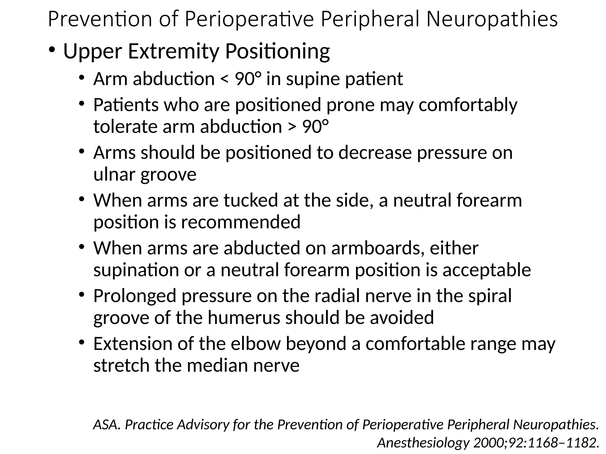
Basics of Head Fixation and
Positioning
Dhaval Shukla
Additional Professor of Neurosurgery
NIMHANS, Bangalore.
Basic Principles of Head Fixation
Falling into the pins
• Firm placement of the two-
point swivel arm before
engaging a contralateral single
pin
• Spatial positioning of the pins
relative to the cranial equator
and gravity
• Ensure that two of the three pins
are below the cranial equator
• Insuring that the cranium will be
held safely even if the pins were
to be less tight than optimal
• Tightness of the pins
• Pressure for adults 60 - 80 lbs
• Pressure for children 30 - 40 lbs
Coppa ND, et al. AANS 2010.
Basic Principles of Head Fixation
Avoid
• Critical neurovascular structures
• superficial temporal artery
• supraorbital artery and nerve
• occipital artery
• Areas of thin bone
• Squamous temporal
• Regions with underlying venous
structures
• Air sinuses
• Pins in children less than 3 years of
age
Coppa ND, et al. AANS 2010.
Bifrontal Craniotomy
• All 3 pins below equator
• Approximate position of
ears
• Double pin
• One on mastoid process
• Other superiorly and
toward midline
• Single pin superior and
posterior to the pinna of
the contralateral ear
Coppa ND, et al. AANS 2010.
Pterional Craniotomy
• Pterion facing upward
• Two pins at or below
equator
• Double pins
• One pin just superior and
lateral to the occipital
protuberance, below
equator
• Other pin on mastoid
process ipsilateral to the
side of surgical approach
• Single pin superior to the
temporal fossa
(contralateral to surgical
approach) Coppa ND, et al. AANS 2010.
Middle Fossa Craniotomy
• Double pins
• Superior to occipital
protuberance, which
indicates the, and on
either side of superior
sagittal sinus and superior
to transverse sinuses
• Single pin
• Should be located at or
near midline several
centimeters above nasion
Coppa ND, et al. AANS 2010.
Prone Craniotomy
• Double pins
• One pin is posterior and
superior to the pinna
• Other pin is anterior in
relation to the temporal
fossa, and should be at or
near margin of this fossa
• Single pin
• Superior to the pinna of
the contralateral ear at
the margin of the
temporal fossa
• Place single pin on right
side to reduce risk to
language area
Coppa ND, et al. AANS 2010.
Positioning in Neurosurgery
Garg N, Indira Devi B, Shukla D. Positioning in Neurosurgery.
Ramamurthi and Tandon’s Textbook of Neurosurgery (3rd ed.). 2012. pp 2299-2305.
Proper positioning
To provide an adequate exposure of the operative site
without excessive retraction of the brain.
• Operative site should be easily accessible
• Operative field should be least dependent
• Area should also have a direct line of sight
• Reduce venous congestion and oozing, thus providing a
bloodless field
• The operative field should be at a higher position than the heart
• No pressure on the neck and no compression of the abdomen while
the patient is in the prone position
• Making use of gravity to help in retracting the brain
Positioning in Neurosurgery
Garg N, Indira Devi B, Shukla D. Positioning in Neurosurgery.
Ramamurthi and Tandon’s Textbook of Neurosurgery (3rd ed.). 2012. pp 2299-2305.
Proper positioning
• To make surgery ergonomical and less tiring for
surgeon.
• To ensure patient safety and comfort, so as to
reduce the post-operative discomfort of the patient.
Avoid undue pressure/ traction of limbs
Avoid undue pressure on vital tissues
• To permit free access to the anaesthetist.
• To permit extension of the operative field if
required.
• Preoperative Assessment
• When judged appropriate, it is helpful to ascertain that
patients can comfortably tolerate the anticipated
operative position.
• Protective Padding
• Padded armboards may decrease the risk of upper
extremity neuropathy.
• The use of chest rolls in laterally positioned patients may
decrease the risk of upper extremity neuropathies.
• Padding at the elbow and at the fibular head may
decrease the risk of upper and lower extremity
neuropathies, respectively.
• Equipment
• Shoulder braces in steep head-down positions may
increase the risk of brachial plexus neuropathies.
Prevention of Perioperative Peripheral Neuropathies
ASA. Practice Advisory for the Prevention of Perioperative Peripheral Neuropathies.
Anesthesiology 2000;92:1168–1182.
• Upper Extremity Positioning
• Arm abduction < 90° in supine patient
• Patients who are positioned prone may comfortably
tolerate arm abduction > 90°
• Arms should be positioned to decrease pressure on
ulnar groove
• When arms are tucked at the side, a neutral forearm
position is recommended
• When arms are abducted on armboards, either
supination or a neutral forearm position is acceptable
• Prolonged pressure on the radial nerve in the spiral
groove of the humerus should be avoided
• Extension of the elbow beyond a comfortable range may
stretch the median nerve
Prevention of Perioperative Peripheral Neuropathies
ASA. Practice Advisory for the Prevention of Perioperative Peripheral Neuropathies.
Anesthesiology 2000;92:1168–1182.
• Lower Extremity Positioning
• Prolonged pressure on the peroneal nerve at the fibular
head should be avoided.
• Neither extension nor flexion of the hip increases the
risk of femoral neuropathy
• Postoperative Assessment
• A simple postoperative assessment of extremity nerve
function may lead to early recognition of peripheral
neuropathies.
• Documentation
• Charting specific positioning actions during the care of
patients may result in improvements of care
Prevention of Perioperative Peripheral Neuropathies
ASA. Practice Advisory for the Prevention of Perioperative Peripheral Neuropathies.
Anesthesiology 2000;92:1168–1182.
Take Home Message
• Positioning is the joint responsibility of the surgeon
and anesthesiologist.
• Each member of the team has to use knowledge of
anatomy and physiology, as well as experience in
using various positioning aids and accessories, to
the safe positioning of patients.

Head Fixation and Positioning in Neurosurgery.pptx

  • 1.
    Basics of HeadFixation and Positioning Dhaval Shukla Additional Professor of Neurosurgery NIMHANS, Bangalore.
  • 2.
    Basic Principles ofHead Fixation
  • 4.
    Basic Principles ofHead Fixation Falling into the pins • Firm placement of the two- point swivel arm before engaging a contralateral single pin • Spatial positioning of the pins relative to the cranial equator and gravity • Ensure that two of the three pins are below the cranial equator • Insuring that the cranium will be held safely even if the pins were to be less tight than optimal • Tightness of the pins • Pressure for adults 60 - 80 lbs • Pressure for children 30 - 40 lbs Coppa ND, et al. AANS 2010.
  • 5.
    Basic Principles ofHead Fixation Avoid • Critical neurovascular structures • superficial temporal artery • supraorbital artery and nerve • occipital artery • Areas of thin bone • Squamous temporal • Regions with underlying venous structures • Air sinuses • Pins in children less than 3 years of age Coppa ND, et al. AANS 2010.
  • 6.
    Bifrontal Craniotomy • All3 pins below equator • Approximate position of ears • Double pin • One on mastoid process • Other superiorly and toward midline • Single pin superior and posterior to the pinna of the contralateral ear Coppa ND, et al. AANS 2010.
  • 7.
    Pterional Craniotomy • Pterionfacing upward • Two pins at or below equator • Double pins • One pin just superior and lateral to the occipital protuberance, below equator • Other pin on mastoid process ipsilateral to the side of surgical approach • Single pin superior to the temporal fossa (contralateral to surgical approach) Coppa ND, et al. AANS 2010.
  • 8.
    Middle Fossa Craniotomy •Double pins • Superior to occipital protuberance, which indicates the, and on either side of superior sagittal sinus and superior to transverse sinuses • Single pin • Should be located at or near midline several centimeters above nasion Coppa ND, et al. AANS 2010.
  • 9.
    Prone Craniotomy • Doublepins • One pin is posterior and superior to the pinna • Other pin is anterior in relation to the temporal fossa, and should be at or near margin of this fossa • Single pin • Superior to the pinna of the contralateral ear at the margin of the temporal fossa • Place single pin on right side to reduce risk to language area Coppa ND, et al. AANS 2010.
  • 10.
  • 11.
    Positioning in Neurosurgery GargN, Indira Devi B, Shukla D. Positioning in Neurosurgery. Ramamurthi and Tandon’s Textbook of Neurosurgery (3rd ed.). 2012. pp 2299-2305. Proper positioning To provide an adequate exposure of the operative site without excessive retraction of the brain. • Operative site should be easily accessible • Operative field should be least dependent • Area should also have a direct line of sight • Reduce venous congestion and oozing, thus providing a bloodless field • The operative field should be at a higher position than the heart • No pressure on the neck and no compression of the abdomen while the patient is in the prone position • Making use of gravity to help in retracting the brain
  • 12.
    Positioning in Neurosurgery GargN, Indira Devi B, Shukla D. Positioning in Neurosurgery. Ramamurthi and Tandon’s Textbook of Neurosurgery (3rd ed.). 2012. pp 2299-2305. Proper positioning • To make surgery ergonomical and less tiring for surgeon. • To ensure patient safety and comfort, so as to reduce the post-operative discomfort of the patient. Avoid undue pressure/ traction of limbs Avoid undue pressure on vital tissues • To permit free access to the anaesthetist. • To permit extension of the operative field if required.
  • 20.
    • Preoperative Assessment •When judged appropriate, it is helpful to ascertain that patients can comfortably tolerate the anticipated operative position. • Protective Padding • Padded armboards may decrease the risk of upper extremity neuropathy. • The use of chest rolls in laterally positioned patients may decrease the risk of upper extremity neuropathies. • Padding at the elbow and at the fibular head may decrease the risk of upper and lower extremity neuropathies, respectively. • Equipment • Shoulder braces in steep head-down positions may increase the risk of brachial plexus neuropathies. Prevention of Perioperative Peripheral Neuropathies ASA. Practice Advisory for the Prevention of Perioperative Peripheral Neuropathies. Anesthesiology 2000;92:1168–1182.
  • 21.
    • Upper ExtremityPositioning • Arm abduction < 90° in supine patient • Patients who are positioned prone may comfortably tolerate arm abduction > 90° • Arms should be positioned to decrease pressure on ulnar groove • When arms are tucked at the side, a neutral forearm position is recommended • When arms are abducted on armboards, either supination or a neutral forearm position is acceptable • Prolonged pressure on the radial nerve in the spiral groove of the humerus should be avoided • Extension of the elbow beyond a comfortable range may stretch the median nerve Prevention of Perioperative Peripheral Neuropathies ASA. Practice Advisory for the Prevention of Perioperative Peripheral Neuropathies. Anesthesiology 2000;92:1168–1182.
  • 22.
    • Lower ExtremityPositioning • Prolonged pressure on the peroneal nerve at the fibular head should be avoided. • Neither extension nor flexion of the hip increases the risk of femoral neuropathy • Postoperative Assessment • A simple postoperative assessment of extremity nerve function may lead to early recognition of peripheral neuropathies. • Documentation • Charting specific positioning actions during the care of patients may result in improvements of care Prevention of Perioperative Peripheral Neuropathies ASA. Practice Advisory for the Prevention of Perioperative Peripheral Neuropathies. Anesthesiology 2000;92:1168–1182.
  • 23.
    Take Home Message •Positioning is the joint responsibility of the surgeon and anesthesiologist. • Each member of the team has to use knowledge of anatomy and physiology, as well as experience in using various positioning aids and accessories, to the safe positioning of patients.

Editor's Notes

  • #6 Bifrontal Craniotomyall three pins are placed below the cranial equatorapproximated by position of the earsOne double pin is placed on the mastoid process while the other is placed superiorly and toward midlinesingle pin is placed superior and posterior to the pinna of the contralateral ear
  • #7 Pterional Craniotomy
  • #8 For the middle fossa approach (Figure 4), pins are placed such that the double pins are superior the occipital protuberance, which indicates the torcular herophili, and on either side of the superior sagittal sinus and superior to the transverse sinuses. The single pin should be located at or near midline several centimeters above the nasion. This avoids the bilateral supraorbital neurovascular structures. By placing the pins in these locations, the surgeon is sure to place two pins at or below the cranial equator and thus the head will fall into the pins. Secondly, these pin locations avoid hazard zones that include the supraorbital neurovascular structures and the venous sinuses posteriorly.
  • #9 Prone Craniotomyalways place the single pin on the right side to reduce this riskone pin is posterior and superior to the pinna of the ear while the other pin is anterior in relation to the temporal fossa, and should be at or near the margin of this fossasuperior to the pinna of the contralateral ear at the margin of the temporal fossaSingle pin