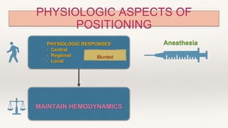
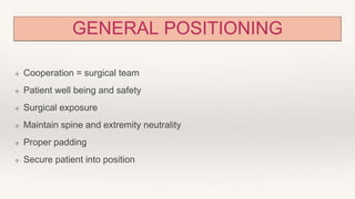
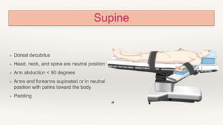
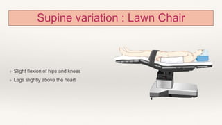
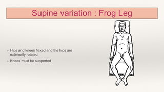
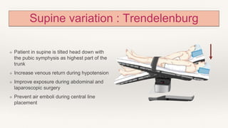
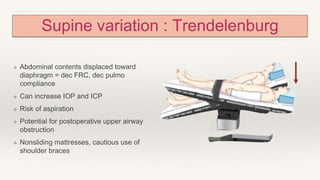
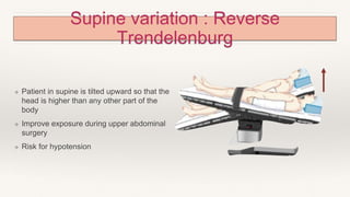
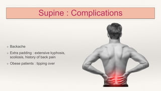
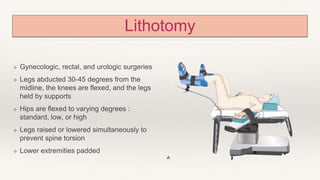
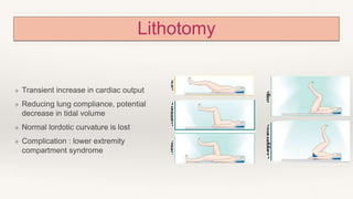
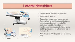
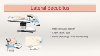
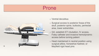
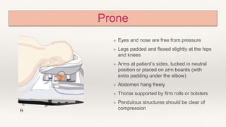
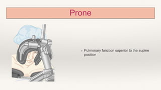
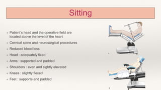
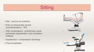
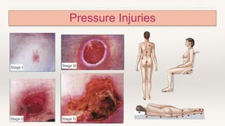
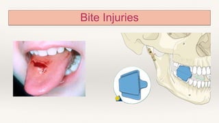
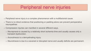
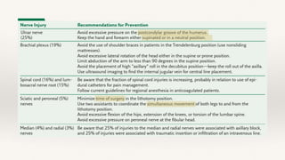
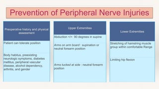
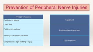
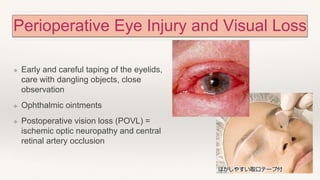
PATIENT
POSITIONING AND
ASSOCIATED RISKS
Presented by
PHYSIOLOGIC ASPECTS OF
POSITIONING
PHYSIOLOGIC RESPONSES
• Central
• Regional
• Local
MAINTAIN HEMODYNAMICS
Anesthesia
Blunted
PHYSIOLOGIC ASPECTS OF
POSITIONING
PULMONARY PHYSIOLOGY
⬇️ FUNCTIONAL RESIDUAL
CAPACITY
⬇️
⬆️ V/Q MISMATCH HYPOXEMIA
Anesthesia
GENERAL POSITIONING
❖ Cooperation = surgical team
❖ Patient well being and safety
❖ Surgical exposure
❖ Maintain spine and extremity neutrality
❖ Proper padding
❖ Secure patient into position
Supine
❖ Dorsal decubitus
❖ Head, neck, and spine are neutral position
❖ Arm abduction < 90 degrees
❖ Arms and forearms supinated or in neutral
position with palms toward the body
❖ Padding
Supine variation : Lawn Chair
❖ Slight flexion of hips and knees
❖ Legs slightly above the heart
Supine variation : Frog Leg
❖ Hips and knees flexed and the hips are
externally rotated
❖ Knees must be supported
Supine variation : Trendelenburg
❖ Patient in supine is tilted head down with
the pubic symphysis as highest part of the
trunk
❖ Increase venous return during hypotension
❖ Improve exposure during abdominal and
laparoscopic surgery
❖ Prevent air emboli during central line
placement
Supine variation : Trendelenburg
❖ Abdominal contents displaced toward
diaphragm = dec FRC, dec pulmo
compliance
❖ Can increase IOP and ICP
❖ Risk of aspiration
❖ Potential for postoperative upper airway
obstruction
❖ Nonsliding mattresses, cautious use of
shoulder braces
Supine variation : Reverse
Trendelenburg
❖ Patient in supine is tilted upward so that the
head is higher than any other part of the
body
❖ Improve exposure during upper abdominal
surgery
❖ Risk for hypotension
Supine : Complications
❖ Backache
❖ Extra padding : extensive kyphosis,
scoliosis, history of back pain
❖ Obese patients : tipping over
Lithotomy
❖ Gynecologic, rectal, and urologic surgeries
❖ Legs abducted 30-45 degrees from the
midline, the knees are flexed, and the legs
held by supports
❖ Hips are flexed to varying degrees :
standard, low, or high
❖ Legs raised or lowered simultaneously to
prevent spine torsion
❖ Lower extremities padded
Lithotomy
❖ Transient increase in cardiac output
❖ Reducing lung compliance, potential
decrease in tidal volume
❖ Normal lordotic curvature is lost
❖ Complication : lower extremity
compartment syndrome
Lateral decubitus
❖ Patient lies on the nonoperative side
❖ Must be well secured
❖ Extremities : dependent leg somewhat
flexed, pillow or padding placed between
knees, dependent arm in front of the
patient on a padded arm board,
nondependent arm supported over folded
bedding or suspended with an arm rest or
foam cradle
❖ Arm abducted <90 degrees, use of axillary
rolls
Lateral decubitus
❖ Head in neutral position
❖ Check : ears, eyes
❖ Pulmo physiology : V/Q mismatching
Prone
❖ Ventral decubitus
❖ Surgical access to posterior fossa of the
skull, posterior spine, buttocks, perirectal
area, lower extremities
❖ GA: establish ET intubation, IV access,
Foley catheter and invasive hemodynamic
access before turning patient
❖ Head maintained in neutral position using
surgical pillow, horseshoe hadrest, or
Mayfield rigid head pins
Prone
❖ Eyes and nose are free from pressure
❖ Legs padded and flexed slightly at the hips
and knees
❖ Arms at patient’s sides, tucked in neutral
position or placed on arm boards (with
extra padding under the elbow)
❖ Abdomen hang freely
❖ Thorax supported by firm rolls or bolsters
❖ Pendulous structures should be clear of
compression
Prone
❖ Pulmonary function superior to the supine
position
Sitting
❖ Patient’s head and the operative field are
located above the level of the heart
❖ Cervical spine and neurosurgical procedures
❖ Reduced blood loss
❖ Head : adequately fixed
❖ Arms : supported and padded
❖ Shoulders : even and sightly elevated
❖ Knees : slightly flexed
❖ Feet : supporte and padded
Sitting
❖ Risk : venous air embolism
❖ Rule out intracardiac shunts
(contraindication) : TEE
❖ VAE complications : arrhythmias, acute
pulmonary hypertension, and circulatory
collapse
❖ Hypotension = compression stockings
❖ Pneumocephalus
Robotic Surgery : Steep Trendelenburg
❖ Supine tilted head down at 30 - 45 degrees
and lithotomy with arms tucked in neutral
position
❖ Nonslip mattress, chest straps in X
configuration, shoulder braces (monitor for
stretching at the neck)
❖ Perform test prior to docking robot
❖ Hemodynamic and respiratory changes
❖ Laryngeal edema and optic neuropathy
Pressure Injuries
Stage I
Stage II
Stage III
Stage IV
Bite Injuries
Peripheral nerve injuries
❖ Peripheral nerve injury is a complex phenomenon with a multifactorial cause.
❖ There is no direct evidence that positioning or padding alone can prevent perioperative
neuropathies
❖ Compression injuries can manifest in several different ways.
❖ Neurapraxia is caused by a relatively short ischemia time and usually causes only a
transient dysfunction.
❖ Axonotmesis is a demyelinating injury.
❖ Neurotmesis is due to a severed or disrupted nerve and usually deficits are permanent.
Prevention of Peripheral Nerve Injuries
Preoperative history and physical
assessment
Patient can tolerate position
Body habitus, preexisting
neurologic symptoms, diabetes
mellitus, peripheral vascular
disease, alcohol dependency,
arthritis, and gender
Upper Extremities
Abduction </= 90 degrees in supine
Arms on arm board : supination or
neutral forearm position
Arms tucked at side : neutral forearm
position
Lower Extremities
Stretching of hamstring muscle
group within comfortable Range
Limiting hip flexion
Prevention of Peripheral Nerve Injuries
Protective Padding
Padded arm boards
Chest rolls
Padding at the elbow
Padding to protect fibular nerve
Complications : tight padding = injury
Equipment
Postoperative Assessment
Documentation
Evaluation and Treatment of Perioperative
Neuropathies
❖ Correlate and document the extent of sensory or motor deficits with the
preoperative exam as well as any intraoperative events
❖ Neurologic consultation
❖ Proper diagnosis and management = most injuries resolve
❖ Sensory neuropathies = mostly transient
❖ Motor neuropathies = 4-6 weeks recovery; interim PT
❖ Electrophysiologic evaluation after 4 weeks = more definitive info
Perioperative Eye Injury and Visual Loss
❖ Corneal abrasions are most common
❖ GA: no lid reflex, decreased tear
production
❖ FB sensation, photophobia, blurry
vision, erythema
❖ Risk factors : increased age, long
surgery, prone and Trendelenburg
position, supplemental oxygen
Perioperative Eye Injury and Visual Loss
❖ Early and careful taping of the eyelids,
care with dangling objects, close
observation
❖ Ophthalmic ointments
❖ Postoperative vision loss (POVL) =
ischemic optic neuropathy and central
retinal artery occlusion
Anesthesia outside the Operating Room
❖ GI endoscopy
❖ Cardiac catheterization
❖ Interventional radiology
❖ Neuroradiology
❖ MRI / CT tomography
❖ Office-based procedures
In summary…
Positioning of patients = essential aspect of
intraoperative care
Operative team works together when positioning to
ensure patient safety and comfort in addition to
desired surgical exposure
Final position should appear natural
Thank you!

Report on Patient Positioning for Anesthesia

  • 1.
  • 3.
    PHYSIOLOGIC ASPECTS OF POSITIONING PHYSIOLOGICRESPONSES • Central • Regional • Local MAINTAIN HEMODYNAMICS Anesthesia Blunted
  • 4.
    PHYSIOLOGIC ASPECTS OF POSITIONING PULMONARYPHYSIOLOGY ⬇️ FUNCTIONAL RESIDUAL CAPACITY ⬇️ ⬆️ V/Q MISMATCH HYPOXEMIA Anesthesia
  • 6.
    GENERAL POSITIONING ❖ Cooperation= surgical team ❖ Patient well being and safety ❖ Surgical exposure ❖ Maintain spine and extremity neutrality ❖ Proper padding ❖ Secure patient into position
  • 7.
    Supine ❖ Dorsal decubitus ❖Head, neck, and spine are neutral position ❖ Arm abduction < 90 degrees ❖ Arms and forearms supinated or in neutral position with palms toward the body ❖ Padding
  • 8.
    Supine variation :Lawn Chair ❖ Slight flexion of hips and knees ❖ Legs slightly above the heart
  • 9.
    Supine variation :Frog Leg ❖ Hips and knees flexed and the hips are externally rotated ❖ Knees must be supported
  • 10.
    Supine variation :Trendelenburg ❖ Patient in supine is tilted head down with the pubic symphysis as highest part of the trunk ❖ Increase venous return during hypotension ❖ Improve exposure during abdominal and laparoscopic surgery ❖ Prevent air emboli during central line placement
  • 11.
    Supine variation :Trendelenburg ❖ Abdominal contents displaced toward diaphragm = dec FRC, dec pulmo compliance ❖ Can increase IOP and ICP ❖ Risk of aspiration ❖ Potential for postoperative upper airway obstruction ❖ Nonsliding mattresses, cautious use of shoulder braces
  • 12.
    Supine variation :Reverse Trendelenburg ❖ Patient in supine is tilted upward so that the head is higher than any other part of the body ❖ Improve exposure during upper abdominal surgery ❖ Risk for hypotension
  • 13.
    Supine : Complications ❖Backache ❖ Extra padding : extensive kyphosis, scoliosis, history of back pain ❖ Obese patients : tipping over
  • 14.
    Lithotomy ❖ Gynecologic, rectal,and urologic surgeries ❖ Legs abducted 30-45 degrees from the midline, the knees are flexed, and the legs held by supports ❖ Hips are flexed to varying degrees : standard, low, or high ❖ Legs raised or lowered simultaneously to prevent spine torsion ❖ Lower extremities padded
  • 15.
    Lithotomy ❖ Transient increasein cardiac output ❖ Reducing lung compliance, potential decrease in tidal volume ❖ Normal lordotic curvature is lost ❖ Complication : lower extremity compartment syndrome
  • 16.
    Lateral decubitus ❖ Patientlies on the nonoperative side ❖ Must be well secured ❖ Extremities : dependent leg somewhat flexed, pillow or padding placed between knees, dependent arm in front of the patient on a padded arm board, nondependent arm supported over folded bedding or suspended with an arm rest or foam cradle ❖ Arm abducted <90 degrees, use of axillary rolls
  • 17.
    Lateral decubitus ❖ Headin neutral position ❖ Check : ears, eyes ❖ Pulmo physiology : V/Q mismatching
  • 18.
    Prone ❖ Ventral decubitus ❖Surgical access to posterior fossa of the skull, posterior spine, buttocks, perirectal area, lower extremities ❖ GA: establish ET intubation, IV access, Foley catheter and invasive hemodynamic access before turning patient ❖ Head maintained in neutral position using surgical pillow, horseshoe hadrest, or Mayfield rigid head pins
  • 19.
    Prone ❖ Eyes andnose are free from pressure ❖ Legs padded and flexed slightly at the hips and knees ❖ Arms at patient’s sides, tucked in neutral position or placed on arm boards (with extra padding under the elbow) ❖ Abdomen hang freely ❖ Thorax supported by firm rolls or bolsters ❖ Pendulous structures should be clear of compression
  • 20.
    Prone ❖ Pulmonary functionsuperior to the supine position
  • 21.
    Sitting ❖ Patient’s headand the operative field are located above the level of the heart ❖ Cervical spine and neurosurgical procedures ❖ Reduced blood loss ❖ Head : adequately fixed ❖ Arms : supported and padded ❖ Shoulders : even and sightly elevated ❖ Knees : slightly flexed ❖ Feet : supporte and padded
  • 22.
    Sitting ❖ Risk :venous air embolism ❖ Rule out intracardiac shunts (contraindication) : TEE ❖ VAE complications : arrhythmias, acute pulmonary hypertension, and circulatory collapse ❖ Hypotension = compression stockings ❖ Pneumocephalus
  • 24.
    Robotic Surgery :Steep Trendelenburg ❖ Supine tilted head down at 30 - 45 degrees and lithotomy with arms tucked in neutral position ❖ Nonslip mattress, chest straps in X configuration, shoulder braces (monitor for stretching at the neck) ❖ Perform test prior to docking robot ❖ Hemodynamic and respiratory changes ❖ Laryngeal edema and optic neuropathy
  • 26.
    Pressure Injuries Stage I StageII Stage III Stage IV
  • 28.
  • 30.
    Peripheral nerve injuries ❖Peripheral nerve injury is a complex phenomenon with a multifactorial cause. ❖ There is no direct evidence that positioning or padding alone can prevent perioperative neuropathies ❖ Compression injuries can manifest in several different ways. ❖ Neurapraxia is caused by a relatively short ischemia time and usually causes only a transient dysfunction. ❖ Axonotmesis is a demyelinating injury. ❖ Neurotmesis is due to a severed or disrupted nerve and usually deficits are permanent.
  • 32.
    Prevention of PeripheralNerve Injuries Preoperative history and physical assessment Patient can tolerate position Body habitus, preexisting neurologic symptoms, diabetes mellitus, peripheral vascular disease, alcohol dependency, arthritis, and gender Upper Extremities Abduction </= 90 degrees in supine Arms on arm board : supination or neutral forearm position Arms tucked at side : neutral forearm position Lower Extremities Stretching of hamstring muscle group within comfortable Range Limiting hip flexion
  • 33.
    Prevention of PeripheralNerve Injuries Protective Padding Padded arm boards Chest rolls Padding at the elbow Padding to protect fibular nerve Complications : tight padding = injury Equipment Postoperative Assessment Documentation
  • 35.
    Evaluation and Treatmentof Perioperative Neuropathies ❖ Correlate and document the extent of sensory or motor deficits with the preoperative exam as well as any intraoperative events ❖ Neurologic consultation ❖ Proper diagnosis and management = most injuries resolve ❖ Sensory neuropathies = mostly transient ❖ Motor neuropathies = 4-6 weeks recovery; interim PT ❖ Electrophysiologic evaluation after 4 weeks = more definitive info
  • 37.
    Perioperative Eye Injuryand Visual Loss ❖ Corneal abrasions are most common ❖ GA: no lid reflex, decreased tear production ❖ FB sensation, photophobia, blurry vision, erythema ❖ Risk factors : increased age, long surgery, prone and Trendelenburg position, supplemental oxygen
  • 38.
    Perioperative Eye Injuryand Visual Loss ❖ Early and careful taping of the eyelids, care with dangling objects, close observation ❖ Ophthalmic ointments ❖ Postoperative vision loss (POVL) = ischemic optic neuropathy and central retinal artery occlusion
  • 40.
    Anesthesia outside theOperating Room ❖ GI endoscopy ❖ Cardiac catheterization ❖ Interventional radiology ❖ Neuroradiology ❖ MRI / CT tomography ❖ Office-based procedures
  • 41.
  • 42.
    Positioning of patients= essential aspect of intraoperative care Operative team works together when positioning to ensure patient safety and comfort in addition to desired surgical exposure Final position should appear natural
  • 43.

Editor's Notes

  • #2 Patient positioning in the operating room facilitates surgical procedures; however, positioning can be a source of patient injury and can alter intraoperative physiology.
  • #4 Physiologic responses play an essential role in blunting hemodynamic changes that would otherwise occur from positional changes in our day-to-day lives. Central, regional, and local mechanisms are involved. Upright to supine = increased venous return, increased preload, stroke vol, cardiac output = brief inc BP = activates baroreceptors, stimulating parasympathetic response = maintain BP
  • #5 Pulmonary physiology is also altered by positional changes, which are further exaggerated during anesthesia. For example, when nonanesthetized people lie down, their functional residual capacity (volume of air present in the lungs at the end of passive expiration) decreases as a result of the diaphragm shifting upward. In anesthetized patients, the decrease in FRC is more dramatic, and often closing capacity (point during expiration when small airways begin to close) exceeds FRC (in young individuals, it is half the FRC), leading to increases inventilation-perfusion(V/̇Q) mismatching and hypoxemia.
  • #7 Maintaining neutrality of the patient’s spine and extremities prevents undue stretch. Tissues overlying all bony prominences, such as the heels and sacrum, must be padded to prevent soft tissue ischemia due to pressure. Ideally, patients are placed in a surgical position that they can tolerate when awake. The duration of more extreme positions, when necessary, should be limited as much as possible.
  • #8 the most common position for surgery One or both arms can be abducted or adducted alongside the patient Arm abduction should be limited to less than 90 degrees in order to prevent brachial plexus injury from the head of the humerus pushing into the axilla Hands and forearms are either supinated or kept in a neutral position with the palm toward the body to reduce external pressure on the ulnar nerve pad all bony prominences as well as stopcocks or IV lines that may exert pressure on the skin
  • #9 often better tolerated by patients who are awake or undergoing monitored anesthesia care facilitates venous drainage from the lower extremities. Furthermore, the xiphoid to pubic distance is decreased, reducing tension on the abdominal musculature
  • #10 facilitates procedures to the perineum, medial thighs, genitalia, and rectum. The knees must be supported in order to minimize stress or disloca- tion of the hips.
  • #11 Trendelenburg positioning is commonly used today to increase venous return during hypotension, improve exposure during abdominal and laparoscopic surgery, and prevent air emboli during central line placement. Initially, placement of the patient head-down causes an autotransfusion from the legs with about a 9% from baseline increase in cardiac output in 1 minute. However, these changes are not sustained and within 10 minutes many hemodynamic variables, including cardiac output, return to baseline values. still part of the initial resuscitative efforts to treat hypovolemia
  • #12 decreases FRC and can also decrease pulmonary compliance necessitating higher airway pressures during mechanical ventilation In patients with increased ICP and impaired cerebral autoregula- tion, Trendelenburg positioning should be avoided. For patients receiving general anesthesia who will be placed in the Trendelenburg position, endotracheal intubation is strongly recommended over supraglottic d/t risk of aspiration Prolonged head-down position can lead to swelling of the face, conjunctivae, larynx, and tongue with an increased potential for postoperative upper airway obstruction. An air leak should be verified around the endotracheal tube or the larynx visualized prior to extubation measures should be taken to ensure the patient does not slide or shift
  • #14 Backache may occur in the supine position as the nor- mal lumbar lordotic curvature is lost during general anesthesia with muscle relaxation or a neuraxial block- ade. Consequently, patients with extensive kyphosis, scoliosis, or a previous history of back pain may require extra padding of the spine or slight flexion at the hip and knee Operating room table weight limits should be strictly observed; they differ substantially with regard to normal and reverse positioning.
  • #15 The common peroneal nerve wraps around the head of the fibula on the lateral leg and is at significant risk of injury if insufficiently padded. The foot section of the operating room table is low- ered or taken away
  • #16 Lower extremity compartment syndrome is a rare but devastating complication associated with the lithotomy posi- tion. It occurs when perfusion to an extremity is inadequate because of either restricted arterial flow (from leg elevation) or obstructed venous outflow (direct limb compression or excessive hip flexion). This results in ischemia, edema, and rhabdomyolysis from increased tissue pressure within a fas- cial compartment it is recom- mended to periodically lower the legs to the level of the body if surgery extends beyond several hours
  • #17 facilitate surgery in the thorax, retroperitoneum, or hip axillary roll prevents compression injury to the dependent brachial plexus and dependent axillary vascular structures
  • #18 The patient’s head must be kept in a neutral position to prevent excessive lateral rotation of the neck and stretch injuries to the brachial plexus. This positioning may require additional head support The depen- dent ear should be checked to avoid folding and undue pressure The eyes should be securely taped before repo- sitioning if the patient is asleep. The dependent eye must be checked frequently for external compression In a patient who is mechanically ventilated, the combination of the lateral weight of the mediastinum and disproportionate cephalad pressure of abdominal contents on the dependent diaphragm decreases compli- ance of the dependent lung and favors ventilation of the nondependent lung. Simultaneously pulmonary blood flow to the dependent lung increases because of the effect of gravity. This causes ventilation-perfusion mismatching and can affect alveolar ventilation and gas exchange
  • #19 Turning the patient from supine to prone requires coor- dination of all operating room providers. The anesthe- sia provider is primarily responsible for coordinating the move and for the repositioning of the head. An exception is in cases in which the head is placed in rigid pin fixa- tion and the surgeon holds the pin frame. During the turn to prone, the head, neck, and spine are maintained in a neutral position strokes apparently can occur from presumed carotid and vertebral injury during turning disconnect the endotracheal tube during movement and to reconnect immediately upon prone positioning
  • #20 Turning the patient from supine to prone requires coor- dination of all operating room providers. The anesthe- sia provider is primarily responsible for coordinating the move and for the repositioning of the head. An exception is in cases in which the head is placed in rigid pin fixa- tion and the surgeon holds the pin frame. During the turn to prone, the head, neck, and spine are maintained in a neutral position strokes apparently can occur from presumed carotid and vertebral injury during turning disconnect the endotracheal tube during movement and to reconnect immediately upon prone positioning The abdomen should hang relatively freely for patients in the prone position. This alleviates external pressure on the abdomen, which can otherwise cause problems with ventilation and hypotension by compressing the inferior vena cava and reducing venous return
  • #21 The prone position has been utilized to improve respiratory function and mortality rate in patients with adult respiratory distress syndrome
  • #22 The anesthesia provider should ensure that the shoulders are even or very mildly elevated in order to avoid stretch injury between the neck and shoulders he knees are usually slightly flexed for balance and to reduce stretching of the sciatic nerve, and the feet are also sup- ported and padded
  • #23 During intracranial procedures, a significant amount of air can be entrained through the open dural venous sinuses. Low venous pressure in the operative field creates a gradi- ent for air entry into the venous system, similar to the risk of venous air entry during central line placement Patients undergoing planned surgery in the sitting position should be first evaluated to rule-out anatomic intracardiac shunts. If an intracardiac shunt is present, even small amounts of entrained venous air may result in a stroke or myocardial infarction Currently TEE is the gold standard for detec- tion of intracardiac shunts Pneumocephalus occurs in almost all patients under- going cervical spine or posterior fossa surgery in the sitting position. occurs because of the lower pressure of cerebrospinal fluid in the sitting position.
  • #25 Robotic surgery came into use around 1999 and has quickly become the norm for many urologic operations as well as in gynecologic surgery where it is also dra- matically increasing Other complications from robotic laparoscopic surger- ies include laryngeal edema and optic neuropathy
  • #27 Pressure injuries are due to prolonged pressure that inhibits capillary blood flow over a bony prominence. In animal models, damage has been shown to start within 2 hours with 70 mm Hg force Stages range from intact, nonblanchable erythema (stage 1) to full- thickness tissue loss (stage 4). Muscle damage occurs before skin and subcutaneous tissue damage and is likely due to increased oxygen requirements of muscle In the supine position, areas most at risk include the sacrum, heels, and occiput prone position, the chest and knees are at the most frequent risk for pressure injury and in the sitting position, the ischial tuberosities are at the most frequent risk Most intraoperative pressure injuries (>80%) are dis- covered within 72 hours of surgery and occur most often in operations lasting more than 3 hours pressure injuries in the lips, tongue, and nasal alae can occur from endotracheal tubes, nasogastric tubes, and other medical devices
  • #29 Transcranial motor-evoked potentials (Tc-MEPs) are becoming more commonly used for both spine surgi- cal procedures and neurosurgical procedures. Tc-MEPs involve contraction of the temporalis and masseter muscle, which has been implicated in tongue, lip, and even tooth injuries because of biting motion.
  • #31 Axonotmesis - 2-3 weeks
  • #41 Vigilance is particu- larly important outside the operating room to maintain patient safety because of the less familiar environment, relative lack of positioning equipment, and variability in staff and nursing training with regard to patient posi- tioning