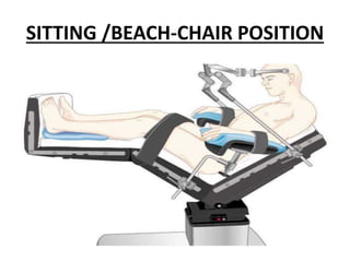
Patient Positioning For
Surgery and its Anaesthestic
Consideration
MODERATOR: DR.PROF VIKRAM VARDHAN
PRESENTER: DR NIDHI DUBEY
Introduction
• Different patient positions are required to
provide access for different surgical procedures.
• Each position has implications for ventilation and
haemodynamics, as well as exposing patients to
possible complications such as nerve injury and
pressure sores.
• The anaesthetist plays an important role in
minimising the risks associated with these
positions
• After induction, when positioning the patient
on operation table for the proposed surgery,
the anesthetist should take into account
1. Surgical access
2. Patient safety
3. anesthetic technique
4. monitoring
5. positioning of peripheral lines
Continued…
Each position may have adverse effect in
terms of
1. Airway
2. Skeletal ,
3. Neurological,
4. Ventilator
5. Circulatory effect .
Patient position required most commonly in
surgery are:
1. Supine
2. Prone
3. Sitting /beach –chair-position
4. Lateral
5. Lithotomy
6. Trendelenburg
7. Reverse Trendelenburg
8. …..and etc.
Supine position
Used in majority of surgical procedures.
Patient Placed on his back with legs
extended and uncrossed at the ankles.
• Spinal column in alignment with legs
parallel to the OT bed
 Head in line with the spine and the
face is upward
 Hips are parallel to the spine
Changes
•Supine position has an effect on ventilaton by
leading to a significant reduction in FRC in
anesthetised patient.
•As the FRC decrease there is potential for the
closing capacity to exceed the FRC, which leads
to V/Q mis -match and subsequent to
hypoxaemia.
Continued..
• Certain patient group are more at risk of this
occurring.,
• These include
1. The elderly who have higher closing
capacities
2. The obese or pregnant patient who already
have reduced FRC.
This effect can be mitigated with application of
positive end expiratory pressure.
Complication
• Neuropathies
• Pressure area edema
• Reduction in perfusion leading to ischemia ,
and subsequent tissue breakdown.
• Greatest concerns are circulation and
pressure points.
Complication
• Most Common Nerve Damage:
 Brachial Plexus: positioning the arm
>90*
 Radial and Ulnar:
compression against the OT bed,
metal attachments, or when team
members lean against the arms
during the procedure.
 Peroneal and Tibial:
Crossing of feet and plantar flexion
of ankles and feet
Pressure point in supine position
CONTINUED…
Pressure alopecia:
• Lumps, such as those caused by
monitoring cable connectors, should not
be placed under head padding because
they may create focal areas of pressure.)
Backache :
• as the normal lumbar lordotic curvature,
particularly the tone of the paraspinous
musculature is lost during general
anesthesia with muscle relaxation or a
neuraxial block.
Concern and Prevention of complication
• The head should be maintained in the
neutral position ,if possible, or turned
towards the abducted arm with
endotracheal tube fixed and tapped
properly.
• Proper head ring (donut, gel-pad) to be
used.
• Tissues overlying all bony prominences,
such as the heels and sacrum, must be
padded to prevent soft tissue ischemia
owing to pressure, especially during
prolonged surgery.
Continued..
• External rotation of the arm and posterior
displacement should be avoided.
• ASA practice Advisory recommends limiting
arm abduction in supine patient to less than
90 degrees at the shoulder with the hand
and forearm either supinated or kept in
neutral position.
• The elbows and any protruding objects such
as intravenous line and arterial line should
be padded.
PRONE (VENTRAL DECUBITUS)
INDICATION
• Posterior fossa of the skull
• Spine surgeries
Changes
• Ventilation improves with prone due to increase
in FRC.
• Can be hampered if pressure is exerted on the
abdomen leading to raised intra abdominal
pressure and causing decrease lung compliances.
• Any pressure on the abdomen causes
compression on IVC and decreasing venous
return.
continued...
• Causes decrease in cardiac output , effects on
arterial filling and decrease left ventricular
compliances due to higher intra thoracic
pressure.
• During spinal surgeries, compression of the
IVC may also lead to surgical difficulties.
Continued..
• Blood unable to return to the heart via the IVC
will alternatively be shunted through the
vertebral column venous plexus and increase
blood in the surgical field.
Caution
• The airway is very difficult to access after a
patient has been positioned prone and therefore
care and attention must be spent securing it .
• Tapes or ties are appropriate , but consider the
pressure that a tie may exert on the face when
the patient is turned.
• Take care when turning the patient ,as the tube s
vulnerable to movement and tube position
should be re checked clinicaly after turing .
Continued…
• Access to the patient is limited once the
patient is positoned .
• Consider this when securing i.v access and
avoid i.v cannulae in the antecubital fossa, as
these are likely to become kinked while prone.
• Disconnect non essential lines when turning
the patient to minimise the risk of inadvertent
removal.
Contiuned…
• Patient should be supported on bony area with
supports placed across the chest (just below the
clavicle)and the plevis, allowing the abdomen to
remain free of pressure.
• CPR is problematic in the prone position and
positioning of defibrillator pads is very diffcult.
• In high risk cases , consider application prior to
turning the patient prone.
Complication
• Injury to brachial plexus
• Spinal nerve inury
• Reduced blood supply to carotid ,vertebral
artery due to excessive movement of head
and neck.
• Post –operative visual loss
(ischemic optic neuropathy associated
with prone positioning and spinal surgery)
Management
Recommendation
• Maintaing head in neutral position
• Avoiding trendelenburg position
• Maintaining arterial blood pressure.
• Proper padding at pressure points.
It is important that patients remain
anesthesised until they have been
repositioned supine.
SITTING /BEACH-CHAIR POSITION
INDICATION
• Posterior fossa surgery
The main advantages of the sitting position
over the prone position for neurosurgical and
cervical spine surgeries are;
excellent surgical exposure
decreased blood in the operative field
reduced peri-operative blood loss.
superior access to the airway, reduced facial
swelling, and improved ventilation,
particularly in obese patients.(to the
anesthesiologist)
Caution
• Limited access to airway once surgical draping is
done , so it is very important to ensure
endoracheal tube to be well secured.
• Head elevated to 15*-30*, and fixed with 3 point
holder with neck flexed….excessive flexion or
rotation of the neck impedes Jugular Venous
Drainage and can increase ICP.
Can also cause swelling of upper airway
• Back is elevated to 60*, and the legs are elevated
with knee flexed.
Continued…
• Arms remain at the sides with the hands
resting on lap.
• Careful positioning and padding of pressure
point avoid injuries.
• Proper care for iv lines with extensions,
arterial cannula must be take in note.
Complication
• Hypotension may result after sitting the
patient up due to less active baroreceptor
reflexes in anesthetised patient.
t/t: sit up patient slowly and treat
hypotension with volume resuscitation and
vasopressors.
• If hypotension is unable to be effectively
treated ,lay the patient in supine.
Continued…
• Pneumocephalus:
features include : delayed recovery,neurological
deficit,headache ,confusion ,agitation or
convulsion.
t/t:
high flow oxygen.,
Burr hole aspiration of air.
Continued..
Macroglossia:
• Due to execcseive flexion of neck during
prolonged surgeries causes obstruction of
venous and lymphatics drainage.
• It may cause post-op respiratory obstruction
,particularly in children.
Continued….
Quadriplegia:
• Due to prolonged focal pressure on the spinal
cord secondary to the acute flexion of the head
in sitting position.
• Proper positioning and avoidance of hypotension
during surgery can help avoid this complication.
Continued…
Venous air embolism
• Negative venous pressure may occur at the
surgical site,as the veins are held open by dura
and bone .
• Air embolism effect depends on sizes
small <10 ml detected by TEE
moderate 10-15 ml noted clinically with
decrease in
ETCO2,tachycardia,hypertensionfrom
sympathetic response.
Continued.…
• Large >50 ml can be catastrophic ,leading to
tachycardia ,arrhythmias ,hypotension , right
ventricular failure and cardiac arrest.
T/t:
• Surgical field fluid irrigation.
• Increase oxygen concentration to
100%,manage hypotension with fluid
resuscitations and vasopressors , treat
arrhythmias.
• If possible ,place patient to left lateral
tredelenburg position.
• Attempt to aspiration of air via central venous
catheter can be attempted.
LATERAL DECUBITUS
Indication
• Thoracic
• Hip
• Shoulder surgeries.
Effects
Respiratory system :
• Ventilation of the lower lung is decreased ,
while perfusion is increased leading to V/Q
mismatch.
• Decrease in thoracic compliances , tidal
volume , vital capacity and FRC.
Continued….
• Neuropathies of the cervical plexus , brachial
plexus and common peroneal nerve may
occur.
Prevention of complications….
• Support the dependent arm.
• Pad all pressure points.
• Axillary roll placed under dependent chest .
• Avoid too much tension on shoulder(brachial
plexus).
• Considerable rotation & flexion of the neck
(kinking of ETT,IJV) …(use of flexo-metallic
ETT).
LITHOTOMY
INDICATIONS
• Gynaecological
• Urological surgery.
Effects
• Placing the legs in lithotomy position decreases the blood volume in
the leg veins and redistributes this blood volume centrally,
increasing venous return to the heart and therefore cardiac output.
• In susceptible patients, this increase in central blood volume can
lead to pulmonary oedema. On returning the legs to the supine
position at the end of the procedure, blood will again fill the venous
system of the legs. Venous return will decrease, leading to a fall in
cardiac output.
• Hypotension may result until baroreceptor reflexes are activated.
Blood pressure must be monitored closely during these periods and
treated accordingly.
Complications
• Peripheral neuropathies have been associated with use
of the lithotomy position. Certain nerves are
particularly vulnerable in this position and include the
sciatic, common peroneal and saphenous nerves.
• As the common peroneal nerve runs superficially over
the fibular head and pressure from leg supports may
lead to nerve injury, it is important to pad the area and
avoid any pressure on the nerve.
• The saphenous nerve may also be damaged by
pressure from leg supports as it passes over the medial
condyle of the tibia. Flexion and external rotation at
the hip can stretch and damage the sciatic nerve.
• When positioning during anesthesia, always consider the
normal range of movement for the patient and limit the
positioning to this. It is important to monitor patient
positioning throughout the procedure, as the leg supports
may be moved once the patient has been draped, making
assessment of the movement at the hip joint more difficult.
• Both legs should be positioned in stirrups simultaneously to
avoid inadvertent musculoskeletal injury.
• The lithotomy position is also associated with the
uncommon complication of compartment syndrome of the
lower leg.
Prevention
• Anesthetists need to be aware of the potential for this complication
and consider intermittently lowering the legs during prolonged
procedures in lithotomy.
• Obstruction to venous drainage also predisposes patients to
development of venous thrombosis. Therefore, prophylaxis with
compression stockings or sequential compression devices should be
considered in all cases.
• Special care must be taken with the patient hands, which when
placed by the patient’s side may be injured when the table position
is altered. Hands must be adequately protected and then
monitored with any movement of the table in order to avoid crush
injuries.
TRENDELENBURG Position
• Trendelenburg is the term used when the patient is tilted
15 degrees or greater head down.
• Prolonged Trendelenburg positioning can lead to facial and
laryngeal oedema.
• Minimising the amount of intravenous fluid administered
during the case may help lessen the incidence of this
complication.
• The endotracheal tube tip may move caudad during
positioning, leading to endobronchial intubation.
• The Trendelenburg position leads to a further reduction in
FRC from the supine position, due to further cephalad
movement of the diaphragm.
• Trendelenburg predisposes to atelectasis and causes
decreased respiratory compliance, so patients may need
higher airway pressures to achieve adequate tidal volumes.
• Barotrauma may result from high peak inspiratory pressure.
• With steep Trendelenburg positioning, the patient may
slide down the table and care must be taken to secure the
patient prior to tilting the bed.
• Arms must be secured to prevent falling from arm boards,
which can lead to brachial plexus injury.
• Trendelenburg position will lead to increases in intracranial
and intraocular pressure and should be avoided in patients
who cannot tolerate this.
REVERSE TRENDELENBURG
• FRC is increased in the reverse Trendelenburg
position relative to supine. Lung compliance also
increases and therefore care must be taken with
lung volumes during positive pressure
ventilation.
• Hypotension may result from positioning in
reverse Trendelenburg and the anesthetist
should account for the hydrostatic gradient
between the blood pressure cuff and the brain,
to prevent cerebral hypoperfusion.
TAKE AWAY HOME MESSAGE
• As with any change in position, care must be
taken to ensure that there is no dislodgement
or movement of the endotracheal tube with
any positional changes.
• Particular care needs to be taken to ensure
pressure areas are padded and limbs are
positioned anatomically to minimise the risk
of nerve injury.
• Patient position can affect the patient’s
airway, ventilation and haemodynamics.
•Nerve injury and pressure areas can occur
under anesthesia in any position and special
care needs to be taken to minimise their
occurrence.
THANK -YOU

Patient Positioning For.pptx

  • 1.
    Patient Positioning For Surgeryand its Anaesthestic Consideration MODERATOR: DR.PROF VIKRAM VARDHAN PRESENTER: DR NIDHI DUBEY
  • 2.
    Introduction • Different patientpositions are required to provide access for different surgical procedures. • Each position has implications for ventilation and haemodynamics, as well as exposing patients to possible complications such as nerve injury and pressure sores. • The anaesthetist plays an important role in minimising the risks associated with these positions
  • 3.
    • After induction,when positioning the patient on operation table for the proposed surgery, the anesthetist should take into account 1. Surgical access 2. Patient safety 3. anesthetic technique 4. monitoring 5. positioning of peripheral lines
  • 4.
    Continued… Each position mayhave adverse effect in terms of 1. Airway 2. Skeletal , 3. Neurological, 4. Ventilator 5. Circulatory effect .
  • 5.
    Patient position requiredmost commonly in surgery are: 1. Supine 2. Prone 3. Sitting /beach –chair-position 4. Lateral 5. Lithotomy 6. Trendelenburg 7. Reverse Trendelenburg 8. …..and etc.
  • 6.
  • 7.
    Used in majorityof surgical procedures. Patient Placed on his back with legs extended and uncrossed at the ankles. • Spinal column in alignment with legs parallel to the OT bed  Head in line with the spine and the face is upward  Hips are parallel to the spine
  • 8.
    Changes •Supine position hasan effect on ventilaton by leading to a significant reduction in FRC in anesthetised patient. •As the FRC decrease there is potential for the closing capacity to exceed the FRC, which leads to V/Q mis -match and subsequent to hypoxaemia.
  • 9.
    Continued.. • Certain patientgroup are more at risk of this occurring., • These include 1. The elderly who have higher closing capacities 2. The obese or pregnant patient who already have reduced FRC. This effect can be mitigated with application of positive end expiratory pressure.
  • 10.
    Complication • Neuropathies • Pressurearea edema • Reduction in perfusion leading to ischemia , and subsequent tissue breakdown. • Greatest concerns are circulation and pressure points.
  • 11.
    Complication • Most CommonNerve Damage:  Brachial Plexus: positioning the arm >90*  Radial and Ulnar: compression against the OT bed, metal attachments, or when team members lean against the arms during the procedure.  Peroneal and Tibial: Crossing of feet and plantar flexion of ankles and feet
  • 12.
    Pressure point insupine position
  • 13.
    CONTINUED… Pressure alopecia: • Lumps,such as those caused by monitoring cable connectors, should not be placed under head padding because they may create focal areas of pressure.) Backache : • as the normal lumbar lordotic curvature, particularly the tone of the paraspinous musculature is lost during general anesthesia with muscle relaxation or a neuraxial block.
  • 14.
    Concern and Preventionof complication • The head should be maintained in the neutral position ,if possible, or turned towards the abducted arm with endotracheal tube fixed and tapped properly. • Proper head ring (donut, gel-pad) to be used. • Tissues overlying all bony prominences, such as the heels and sacrum, must be padded to prevent soft tissue ischemia owing to pressure, especially during prolonged surgery.
  • 15.
    Continued.. • External rotationof the arm and posterior displacement should be avoided. • ASA practice Advisory recommends limiting arm abduction in supine patient to less than 90 degrees at the shoulder with the hand and forearm either supinated or kept in neutral position. • The elbows and any protruding objects such as intravenous line and arterial line should be padded.
  • 17.
  • 18.
    INDICATION • Posterior fossaof the skull • Spine surgeries
  • 19.
    Changes • Ventilation improveswith prone due to increase in FRC. • Can be hampered if pressure is exerted on the abdomen leading to raised intra abdominal pressure and causing decrease lung compliances. • Any pressure on the abdomen causes compression on IVC and decreasing venous return.
  • 20.
    continued... • Causes decreasein cardiac output , effects on arterial filling and decrease left ventricular compliances due to higher intra thoracic pressure. • During spinal surgeries, compression of the IVC may also lead to surgical difficulties.
  • 21.
    Continued.. • Blood unableto return to the heart via the IVC will alternatively be shunted through the vertebral column venous plexus and increase blood in the surgical field.
  • 22.
    Caution • The airwayis very difficult to access after a patient has been positioned prone and therefore care and attention must be spent securing it . • Tapes or ties are appropriate , but consider the pressure that a tie may exert on the face when the patient is turned. • Take care when turning the patient ,as the tube s vulnerable to movement and tube position should be re checked clinicaly after turing .
  • 23.
    Continued… • Access tothe patient is limited once the patient is positoned . • Consider this when securing i.v access and avoid i.v cannulae in the antecubital fossa, as these are likely to become kinked while prone. • Disconnect non essential lines when turning the patient to minimise the risk of inadvertent removal.
  • 24.
    Contiuned… • Patient shouldbe supported on bony area with supports placed across the chest (just below the clavicle)and the plevis, allowing the abdomen to remain free of pressure. • CPR is problematic in the prone position and positioning of defibrillator pads is very diffcult. • In high risk cases , consider application prior to turning the patient prone.
  • 25.
    Complication • Injury tobrachial plexus • Spinal nerve inury • Reduced blood supply to carotid ,vertebral artery due to excessive movement of head and neck. • Post –operative visual loss (ischemic optic neuropathy associated with prone positioning and spinal surgery)
  • 26.
    Management Recommendation • Maintaing headin neutral position • Avoiding trendelenburg position • Maintaining arterial blood pressure. • Proper padding at pressure points. It is important that patients remain anesthesised until they have been repositioned supine.
  • 27.
  • 28.
  • 29.
    The main advantagesof the sitting position over the prone position for neurosurgical and cervical spine surgeries are; excellent surgical exposure decreased blood in the operative field reduced peri-operative blood loss. superior access to the airway, reduced facial swelling, and improved ventilation, particularly in obese patients.(to the anesthesiologist)
  • 30.
    Caution • Limited accessto airway once surgical draping is done , so it is very important to ensure endoracheal tube to be well secured. • Head elevated to 15*-30*, and fixed with 3 point holder with neck flexed….excessive flexion or rotation of the neck impedes Jugular Venous Drainage and can increase ICP. Can also cause swelling of upper airway • Back is elevated to 60*, and the legs are elevated with knee flexed.
  • 31.
    Continued… • Arms remainat the sides with the hands resting on lap. • Careful positioning and padding of pressure point avoid injuries. • Proper care for iv lines with extensions, arterial cannula must be take in note.
  • 32.
    Complication • Hypotension mayresult after sitting the patient up due to less active baroreceptor reflexes in anesthetised patient. t/t: sit up patient slowly and treat hypotension with volume resuscitation and vasopressors. • If hypotension is unable to be effectively treated ,lay the patient in supine.
  • 33.
    Continued… • Pneumocephalus: features include: delayed recovery,neurological deficit,headache ,confusion ,agitation or convulsion. t/t: high flow oxygen., Burr hole aspiration of air.
  • 34.
    Continued.. Macroglossia: • Due toexeccseive flexion of neck during prolonged surgeries causes obstruction of venous and lymphatics drainage. • It may cause post-op respiratory obstruction ,particularly in children.
  • 35.
    Continued…. Quadriplegia: • Due toprolonged focal pressure on the spinal cord secondary to the acute flexion of the head in sitting position. • Proper positioning and avoidance of hypotension during surgery can help avoid this complication.
  • 36.
    Continued… Venous air embolism •Negative venous pressure may occur at the surgical site,as the veins are held open by dura and bone . • Air embolism effect depends on sizes small <10 ml detected by TEE moderate 10-15 ml noted clinically with decrease in ETCO2,tachycardia,hypertensionfrom sympathetic response.
  • 37.
    Continued.… • Large >50ml can be catastrophic ,leading to tachycardia ,arrhythmias ,hypotension , right ventricular failure and cardiac arrest. T/t: • Surgical field fluid irrigation. • Increase oxygen concentration to 100%,manage hypotension with fluid resuscitations and vasopressors , treat arrhythmias.
  • 38.
    • If possible,place patient to left lateral tredelenburg position. • Attempt to aspiration of air via central venous catheter can be attempted.
  • 39.
  • 40.
  • 41.
    Effects Respiratory system : •Ventilation of the lower lung is decreased , while perfusion is increased leading to V/Q mismatch. • Decrease in thoracic compliances , tidal volume , vital capacity and FRC.
  • 42.
    Continued…. • Neuropathies ofthe cervical plexus , brachial plexus and common peroneal nerve may occur.
  • 43.
    Prevention of complications…. •Support the dependent arm. • Pad all pressure points. • Axillary roll placed under dependent chest . • Avoid too much tension on shoulder(brachial plexus). • Considerable rotation & flexion of the neck (kinking of ETT,IJV) …(use of flexo-metallic ETT).
  • 44.
  • 45.
  • 46.
    Effects • Placing thelegs in lithotomy position decreases the blood volume in the leg veins and redistributes this blood volume centrally, increasing venous return to the heart and therefore cardiac output. • In susceptible patients, this increase in central blood volume can lead to pulmonary oedema. On returning the legs to the supine position at the end of the procedure, blood will again fill the venous system of the legs. Venous return will decrease, leading to a fall in cardiac output. • Hypotension may result until baroreceptor reflexes are activated. Blood pressure must be monitored closely during these periods and treated accordingly.
  • 47.
    Complications • Peripheral neuropathieshave been associated with use of the lithotomy position. Certain nerves are particularly vulnerable in this position and include the sciatic, common peroneal and saphenous nerves. • As the common peroneal nerve runs superficially over the fibular head and pressure from leg supports may lead to nerve injury, it is important to pad the area and avoid any pressure on the nerve. • The saphenous nerve may also be damaged by pressure from leg supports as it passes over the medial condyle of the tibia. Flexion and external rotation at the hip can stretch and damage the sciatic nerve.
  • 48.
    • When positioningduring anesthesia, always consider the normal range of movement for the patient and limit the positioning to this. It is important to monitor patient positioning throughout the procedure, as the leg supports may be moved once the patient has been draped, making assessment of the movement at the hip joint more difficult. • Both legs should be positioned in stirrups simultaneously to avoid inadvertent musculoskeletal injury. • The lithotomy position is also associated with the uncommon complication of compartment syndrome of the lower leg.
  • 49.
    Prevention • Anesthetists needto be aware of the potential for this complication and consider intermittently lowering the legs during prolonged procedures in lithotomy. • Obstruction to venous drainage also predisposes patients to development of venous thrombosis. Therefore, prophylaxis with compression stockings or sequential compression devices should be considered in all cases. • Special care must be taken with the patient hands, which when placed by the patient’s side may be injured when the table position is altered. Hands must be adequately protected and then monitored with any movement of the table in order to avoid crush injuries.
  • 50.
  • 51.
    • Trendelenburg isthe term used when the patient is tilted 15 degrees or greater head down. • Prolonged Trendelenburg positioning can lead to facial and laryngeal oedema. • Minimising the amount of intravenous fluid administered during the case may help lessen the incidence of this complication. • The endotracheal tube tip may move caudad during positioning, leading to endobronchial intubation. • The Trendelenburg position leads to a further reduction in FRC from the supine position, due to further cephalad movement of the diaphragm.
  • 52.
    • Trendelenburg predisposesto atelectasis and causes decreased respiratory compliance, so patients may need higher airway pressures to achieve adequate tidal volumes. • Barotrauma may result from high peak inspiratory pressure. • With steep Trendelenburg positioning, the patient may slide down the table and care must be taken to secure the patient prior to tilting the bed. • Arms must be secured to prevent falling from arm boards, which can lead to brachial plexus injury. • Trendelenburg position will lead to increases in intracranial and intraocular pressure and should be avoided in patients who cannot tolerate this.
  • 53.
  • 54.
    • FRC isincreased in the reverse Trendelenburg position relative to supine. Lung compliance also increases and therefore care must be taken with lung volumes during positive pressure ventilation. • Hypotension may result from positioning in reverse Trendelenburg and the anesthetist should account for the hydrostatic gradient between the blood pressure cuff and the brain, to prevent cerebral hypoperfusion.
  • 55.
    TAKE AWAY HOMEMESSAGE • As with any change in position, care must be taken to ensure that there is no dislodgement or movement of the endotracheal tube with any positional changes. • Particular care needs to be taken to ensure pressure areas are padded and limbs are positioned anatomically to minimise the risk of nerve injury.
  • 56.
    • Patient positioncan affect the patient’s airway, ventilation and haemodynamics. •Nerve injury and pressure areas can occur under anesthesia in any position and special care needs to be taken to minimise their occurrence.
  • 57.