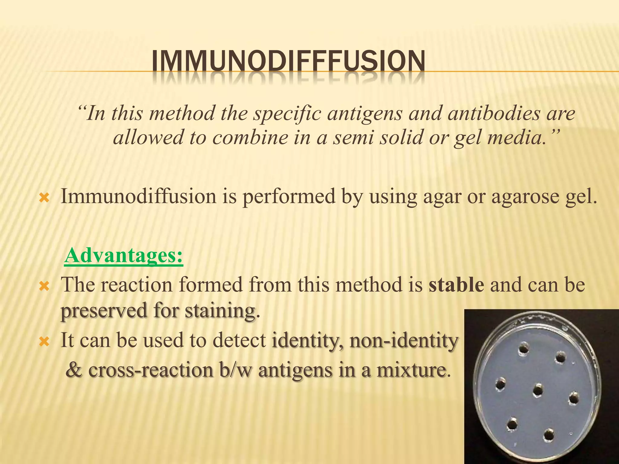
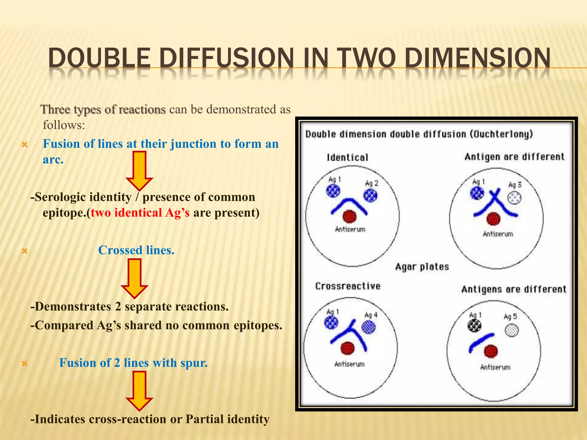
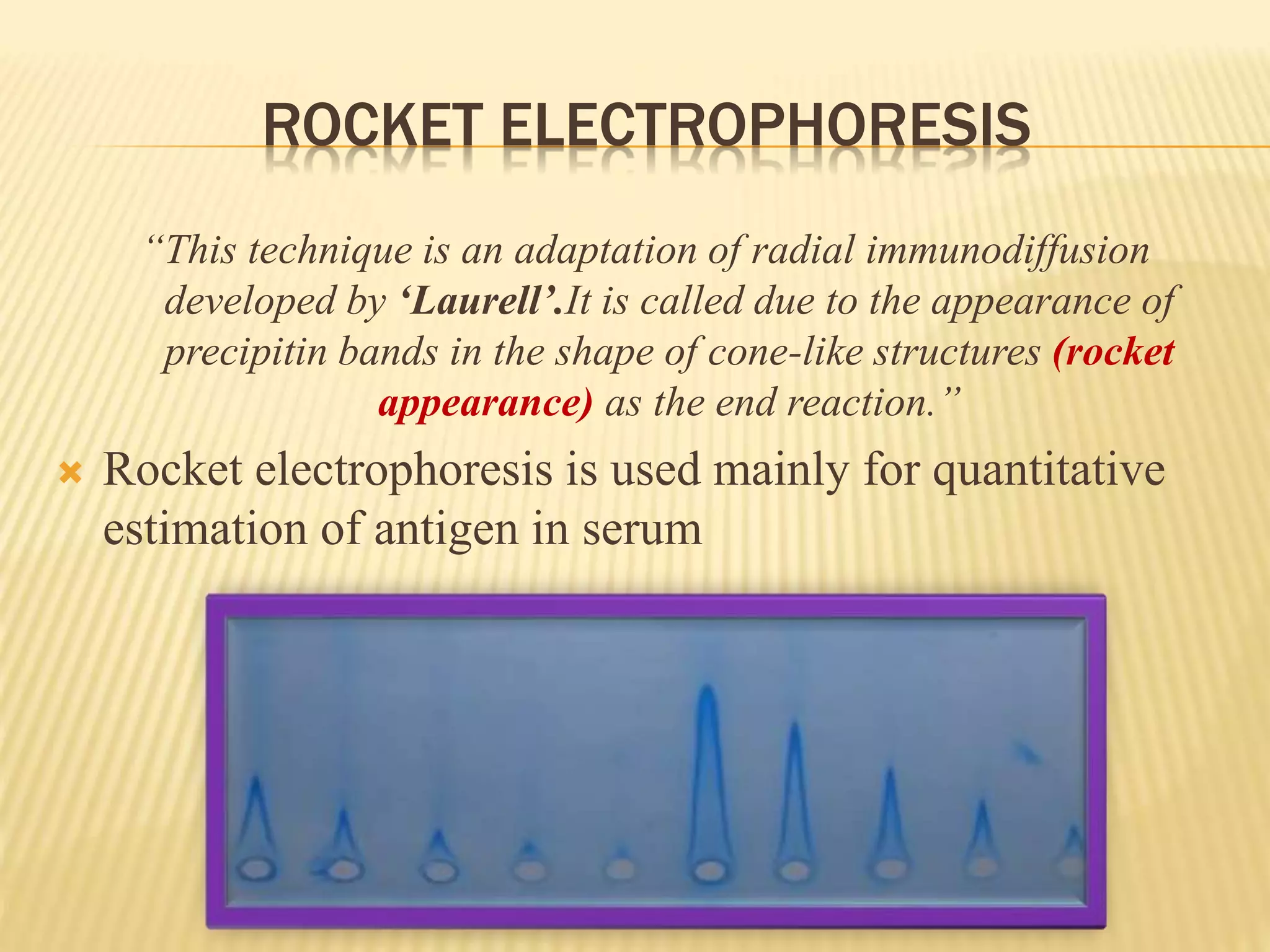
Part :02
TYPES OF ANTIGEN – ANTIBODY REACTION
The types of antigen – antibody reactions are:
 Precipitation Reaction.
 Agglutination Reaction.
 Complement Fixation.
 Immunodiffusion.
 Immuno-electrophoresis
COMPLEMENT FIXATION TEST (CFT)
 The complement fixation test (CFT) was extensively used in syphilis
serology (wassermann test) after being introduced by Wasserman in
1909.
 Lysis of RBC or bacteria requires some non-specific unstable
components of fresh serum which are called “complement.”
 Complement is a protein (globulin) present in normal serum.
 This complement system comprises of 11 proteins and are present
in every individual. They bind to Fc component of Ab involved in
Ag-Ab complex.
 This ability of the Ag-Ab complex to fix complement is used in
complement Fixation tests.
PRINCIPLE OF CFT
 The CFT is a technique that has been used over many years to detect and
quantify antibody that Serology does not agglutinate or precipitate when reacted
with its antigen, but can be demonstrated by its use or fixation of complement.
 Complement takes part in many of the immunological reactions. It gets
absorbed during the combination of antigens and antibody.
 This property of Ag-Ab complex to fix the complement is used in complement
fixation test for the identification of specific antibodies.
 The hemolytic system containing sheep erythrocytes (RBC) and its
corresponding antibody (amboceptor) is used as an indicator which
shows the utilization or availability of the complement.
 If the complement is fixed then there will be no lysis of sheep erythrocytes,
thus denoting a positive test.
 If the complement is available then there will be haemolysis which is a property
of complement, denoting a negative test.
COMPONENTS OF CFT
Test System
 Antigen: It may be soluble or particulate.
 Antibody: Human serum (May or may not contain Antibody towards
specific Antigen)
 Complement: It is pooled serum obtained from 4 to 5 guinea pigs. It
should be fresh or specially preserved as the complement activity is heat
labile (stored at -30 °C in small fractions). The complement activity
should be initially standardized before using in the test.
Indicator System (Hemolytic system)
 Erythrocytes: Sheep RBC
 Amboceptor (Hemolysins): Rabbit antibody to sheep red cells prepared
by inoculating sheep erythrocytes into rabbit under standard
immunization protocol.
The test requires five reagents and is carried out in two steps.
Complement
fixation stage
Indicator
stage
Complement fixation
No Complement fixation
Complement proteins are heat labile
and are destroyed by heating at 56°C
for 20 – 30 minutes.
A B
Heamolysis(-ve test)
No Heamolysis(+ve test)
COMPLEMENT FIXATION TEST (CFT)
Advantages of CFT:
 Ability to screen against a large number of viral and bacterial
infections at the same time.
 Economically Cheap.
Disadvantages of CFT:
 Not sensitive - cannot be used for immunity screening.
 Time consuming and labor intensive.
 Often non-specific e.g. cross-reactivity between HSV .
“PRECIPITATION IN AGAR”
IMMUNODIFFFUSION
“In this method the specific antigens and antibodies are
allowed to combine in a semi solid or gel media.”
 Immunodiffusion is performed by using agar or agarose gel.
Advantages:
 The reaction formed from this method is stable and can be
preserved for staining.
 It can be used to detect identity, non-identity
& cross-reaction b/w antigens in a mixture.
IMMUNODIFFUSION
 Immunodiffusion reactions are classified on the basis of:
(a) number of reactants diffusing.
(b) direaction of diffusion.
Single
diffusion in
one
dimension
Double
diffusion in
one
dimension
Single
diffusion in
two
dimension
Double
diffusion in
two
dimension.
1. SINGLE DIFFUSION IN ONE DIMENSION
“Single diffusion of antigen in agar in one
dimension called single diffusion in one
dimension.”
 It is also called oudin procedure, because this technique
was introduced by oudin. Who for the first time used
gels for precipitation reaction.
SINGLE DIFFUSION IN ONE DIMENSION
Oudin Procedure:
 The antibody is incorporated in a
agar gel in a test tube and then
the antigen is layered over it.
 The antigen diffuses downwards
through the gel, forming a line
of precipitation.
 Precipitate dissolves as the
concentration of the antigen
increases.
 The number of bands indicates
the number of different antigens
2. SINGLE DIFFUSION IN TWO DIMENSION
“Single diffusion in two dimension is also called
Radial immunodiffusion(Mancini test)”
 RID has been used for the
quantitative estimation of
antibodies and antigens in
the serum.
3.DOUBLE DIFFUSION IN ONE DIMENSION
This method is also called “Oakley-
fulthorpe procedure”. In this method
plain agar is also used.
Oakley-fulthorpe procedure.
 Ab is incorporated in a gel, above
which a column of plain agar
 The antigen is layered on top of the
agar medium.
 The Ag and Ab move towards each
other through the intervening column
of plain agar.
 This will result in the formation of
precipitate when they meet at optimum
proportion.
4. DOUBLE DIFFUSION IN TWO DIMENSION
“This method is also called
‘Ouchterlony’s procedure’.(also
known as agar gel immunodiffusion
or passive double immunodiffusion)
“In this method both Ag & Ab diffuse
independently through agar gel in 2
dimensions, horizontally & vertically.”
 The technique is named for Örjan
Ouchterlony, the Swedish physician
who invented the test in 1948.
Ouchterlony’s procedure:
 The test is performed by cutting
wells in the agar gel poured on a
glass slide or in a petri-dish.
 The antibodies is placed in a center
well and different antigens are added
to the well surrounding the center
well.
 After an incubation period of 12-48
hours in a moisture chamber, the
lines precipitins are formed at the site
of the combination of antigen and
antibody.
Precipitin line
DOUBLE DIFFUSION IN TWO DIMENSION
Three types of reactions can be demonstrated as
follows:
 Fusion of lines at their junction to form an
arc.
-Serologic identity / presence of common
epitope.(two identical Ag’s are present)
 Crossed lines.
-Demonstrates 2 separate reactions.
-Compared Ag’s shared no common epitopes.
 Fusion of 2 lines with spur.
-Indicates cross-reaction or Partial identity
DOUBLE DIFFUSION IN TWO DIMENSION
Uses:
 It is an immunological technique used in the detection,
identification and quantification of antibodies and
antigens, such as immunoglobulins and extractable
nuclear antigens.
 Demonstration of Antibodies in the serodiagnosis of
small pox.
 This technique is also used for the Identification of
fungal antigens.
“PRECIPITATION IN AGAR WITH AN ELECTRIC FIELD”
IMMUNO-ELECTROPHORESIS
“It is a method in which different antigens in serum are
separated according to their charge under an electric
field.”
 It’s a process of combination of immunodiffusion and
electrophoresis.
Advantages:
 The main advantage of immunoelectrophoresis is that a no. of
antigens can be identified in serum.
PROCEDURE:
 In this method, a drop of Ag is
placed into a well in agar on glass
slide.
 An electric current is then passed
through the agar.
 During electrophoresis, Antigens
move in the electric field according
to their charge & size.
 A trough is cut into agar and is
filled with the antibody and
diffusion is allowed to occur.
 As the Ag and Ab diffuse towards
each other, they form a series of
lines of precipitation.
COUNTER CURRENT IMMUNO-ELECTROPHORESIS
“It depends on movement of antigen towards the anode
(positive charge) & antibody towards the cathode (negative
charge) through the agar in the electric field.”
Advanteges:
 This method is a highly specific for the detection of both antigen
& antibodies in the serum, CSF and other body fluids in diagnosis
of many infectious diseases including bacterial, fungal, viral &
parasitic.
 This method is commonly used for HBsAg
PROCEDURE:
 The test is performed on a glass slide with agarose in which pair
of wells are punched out.
 One well is filled with Ag & the other with Ab.
 Electric current is then passed through the gel.
 The migration of Ag and Ab is greatly facilitated under electric
field, & the line of precipitation is made visible in 30-60 mins.
ROCKET ELECTROPHORESIS
“This technique is an adaptation of radial immunodiffusion
developed by ‘Laurell’.It is called due to the appearance of
precipitin bands in the shape of cone-like structures (rocket
appearance) as the end reaction.”
 Rocket electrophoresis is used mainly for quantitative
estimation of antigen in serum
PROCEDURE:
 In this method Ab is incorporated in the gel, & antigens are placed in wells cut
in gel.
 Electric current is then passed through the gel, which facilitated the migration
of Ag into the agar.
 This results in formation of a precipitin line(conical in shape), resembling a
rocket.
 The height of rocket is directly proportional to concentration of antigen in the
sample.
Antigen antibody reaction (part: 2)

Antigen antibody reaction (part: 2)

  • 1.
  • 2.
    TYPES OF ANTIGEN– ANTIBODY REACTION The types of antigen – antibody reactions are:  Precipitation Reaction.  Agglutination Reaction.  Complement Fixation.  Immunodiffusion.  Immuno-electrophoresis
  • 4.
    COMPLEMENT FIXATION TEST(CFT)  The complement fixation test (CFT) was extensively used in syphilis serology (wassermann test) after being introduced by Wasserman in 1909.  Lysis of RBC or bacteria requires some non-specific unstable components of fresh serum which are called “complement.”  Complement is a protein (globulin) present in normal serum.  This complement system comprises of 11 proteins and are present in every individual. They bind to Fc component of Ab involved in Ag-Ab complex.  This ability of the Ag-Ab complex to fix complement is used in complement Fixation tests.
  • 5.
    PRINCIPLE OF CFT The CFT is a technique that has been used over many years to detect and quantify antibody that Serology does not agglutinate or precipitate when reacted with its antigen, but can be demonstrated by its use or fixation of complement.  Complement takes part in many of the immunological reactions. It gets absorbed during the combination of antigens and antibody.  This property of Ag-Ab complex to fix the complement is used in complement fixation test for the identification of specific antibodies.  The hemolytic system containing sheep erythrocytes (RBC) and its corresponding antibody (amboceptor) is used as an indicator which shows the utilization or availability of the complement.  If the complement is fixed then there will be no lysis of sheep erythrocytes, thus denoting a positive test.  If the complement is available then there will be haemolysis which is a property of complement, denoting a negative test.
  • 6.
    COMPONENTS OF CFT TestSystem  Antigen: It may be soluble or particulate.  Antibody: Human serum (May or may not contain Antibody towards specific Antigen)  Complement: It is pooled serum obtained from 4 to 5 guinea pigs. It should be fresh or specially preserved as the complement activity is heat labile (stored at -30 °C in small fractions). The complement activity should be initially standardized before using in the test. Indicator System (Hemolytic system)  Erythrocytes: Sheep RBC  Amboceptor (Hemolysins): Rabbit antibody to sheep red cells prepared by inoculating sheep erythrocytes into rabbit under standard immunization protocol. The test requires five reagents and is carried out in two steps.
  • 7.
    Complement fixation stage Indicator stage Complement fixation NoComplement fixation Complement proteins are heat labile and are destroyed by heating at 56°C for 20 – 30 minutes. A B Heamolysis(-ve test) No Heamolysis(+ve test)
  • 8.
    COMPLEMENT FIXATION TEST(CFT) Advantages of CFT:  Ability to screen against a large number of viral and bacterial infections at the same time.  Economically Cheap. Disadvantages of CFT:  Not sensitive - cannot be used for immunity screening.  Time consuming and labor intensive.  Often non-specific e.g. cross-reactivity between HSV .
  • 9.
  • 10.
    IMMUNODIFFFUSION “In this methodthe specific antigens and antibodies are allowed to combine in a semi solid or gel media.”  Immunodiffusion is performed by using agar or agarose gel. Advantages:  The reaction formed from this method is stable and can be preserved for staining.  It can be used to detect identity, non-identity & cross-reaction b/w antigens in a mixture.
  • 11.
    IMMUNODIFFUSION  Immunodiffusion reactionsare classified on the basis of: (a) number of reactants diffusing. (b) direaction of diffusion. Single diffusion in one dimension Double diffusion in one dimension Single diffusion in two dimension Double diffusion in two dimension.
  • 12.
    1. SINGLE DIFFUSIONIN ONE DIMENSION “Single diffusion of antigen in agar in one dimension called single diffusion in one dimension.”  It is also called oudin procedure, because this technique was introduced by oudin. Who for the first time used gels for precipitation reaction.
  • 13.
    SINGLE DIFFUSION INONE DIMENSION Oudin Procedure:  The antibody is incorporated in a agar gel in a test tube and then the antigen is layered over it.  The antigen diffuses downwards through the gel, forming a line of precipitation.  Precipitate dissolves as the concentration of the antigen increases.  The number of bands indicates the number of different antigens
  • 14.
    2. SINGLE DIFFUSIONIN TWO DIMENSION “Single diffusion in two dimension is also called Radial immunodiffusion(Mancini test)”  RID has been used for the quantitative estimation of antibodies and antigens in the serum.
  • 15.
    3.DOUBLE DIFFUSION INONE DIMENSION This method is also called “Oakley- fulthorpe procedure”. In this method plain agar is also used. Oakley-fulthorpe procedure.  Ab is incorporated in a gel, above which a column of plain agar  The antigen is layered on top of the agar medium.  The Ag and Ab move towards each other through the intervening column of plain agar.  This will result in the formation of precipitate when they meet at optimum proportion.
  • 16.
    4. DOUBLE DIFFUSIONIN TWO DIMENSION “This method is also called ‘Ouchterlony’s procedure’.(also known as agar gel immunodiffusion or passive double immunodiffusion) “In this method both Ag & Ab diffuse independently through agar gel in 2 dimensions, horizontally & vertically.”  The technique is named for Örjan Ouchterlony, the Swedish physician who invented the test in 1948. Ouchterlony’s procedure:  The test is performed by cutting wells in the agar gel poured on a glass slide or in a petri-dish.  The antibodies is placed in a center well and different antigens are added to the well surrounding the center well.  After an incubation period of 12-48 hours in a moisture chamber, the lines precipitins are formed at the site of the combination of antigen and antibody. Precipitin line
  • 17.
    DOUBLE DIFFUSION INTWO DIMENSION Three types of reactions can be demonstrated as follows:  Fusion of lines at their junction to form an arc. -Serologic identity / presence of common epitope.(two identical Ag’s are present)  Crossed lines. -Demonstrates 2 separate reactions. -Compared Ag’s shared no common epitopes.  Fusion of 2 lines with spur. -Indicates cross-reaction or Partial identity
  • 19.
    DOUBLE DIFFUSION INTWO DIMENSION Uses:  It is an immunological technique used in the detection, identification and quantification of antibodies and antigens, such as immunoglobulins and extractable nuclear antigens.  Demonstration of Antibodies in the serodiagnosis of small pox.  This technique is also used for the Identification of fungal antigens.
  • 20.
    “PRECIPITATION IN AGARWITH AN ELECTRIC FIELD”
  • 21.
    IMMUNO-ELECTROPHORESIS “It is amethod in which different antigens in serum are separated according to their charge under an electric field.”  It’s a process of combination of immunodiffusion and electrophoresis. Advantages:  The main advantage of immunoelectrophoresis is that a no. of antigens can be identified in serum.
  • 22.
    PROCEDURE:  In thismethod, a drop of Ag is placed into a well in agar on glass slide.  An electric current is then passed through the agar.  During electrophoresis, Antigens move in the electric field according to their charge & size.  A trough is cut into agar and is filled with the antibody and diffusion is allowed to occur.  As the Ag and Ab diffuse towards each other, they form a series of lines of precipitation.
  • 23.
    COUNTER CURRENT IMMUNO-ELECTROPHORESIS “Itdepends on movement of antigen towards the anode (positive charge) & antibody towards the cathode (negative charge) through the agar in the electric field.” Advanteges:  This method is a highly specific for the detection of both antigen & antibodies in the serum, CSF and other body fluids in diagnosis of many infectious diseases including bacterial, fungal, viral & parasitic.  This method is commonly used for HBsAg
  • 24.
    PROCEDURE:  The testis performed on a glass slide with agarose in which pair of wells are punched out.  One well is filled with Ag & the other with Ab.  Electric current is then passed through the gel.  The migration of Ag and Ab is greatly facilitated under electric field, & the line of precipitation is made visible in 30-60 mins.
  • 25.
    ROCKET ELECTROPHORESIS “This techniqueis an adaptation of radial immunodiffusion developed by ‘Laurell’.It is called due to the appearance of precipitin bands in the shape of cone-like structures (rocket appearance) as the end reaction.”  Rocket electrophoresis is used mainly for quantitative estimation of antigen in serum
  • 26.
    PROCEDURE:  In thismethod Ab is incorporated in the gel, & antigens are placed in wells cut in gel.  Electric current is then passed through the gel, which facilitated the migration of Ag into the agar.  This results in formation of a precipitin line(conical in shape), resembling a rocket.  The height of rocket is directly proportional to concentration of antigen in the sample.