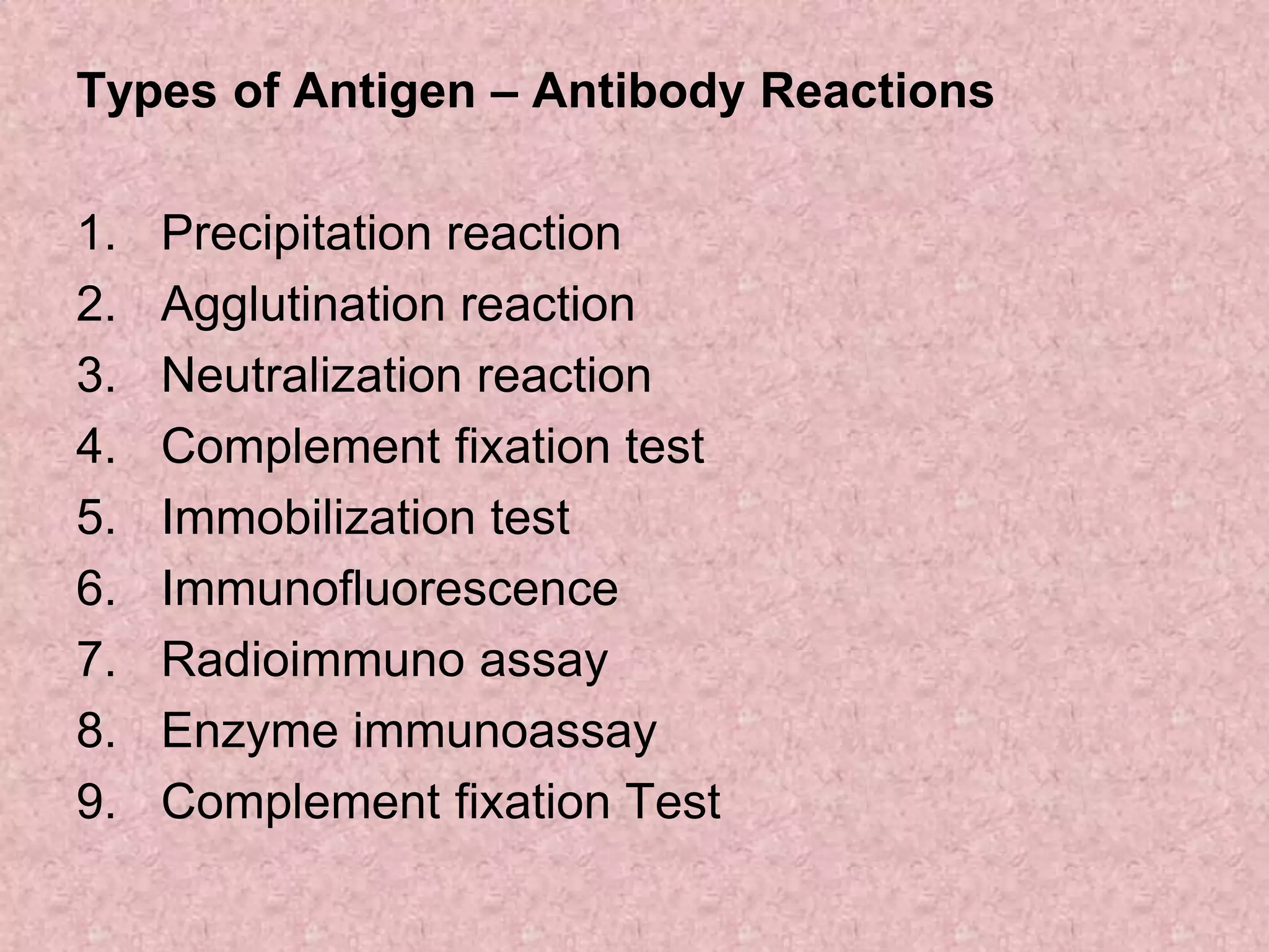
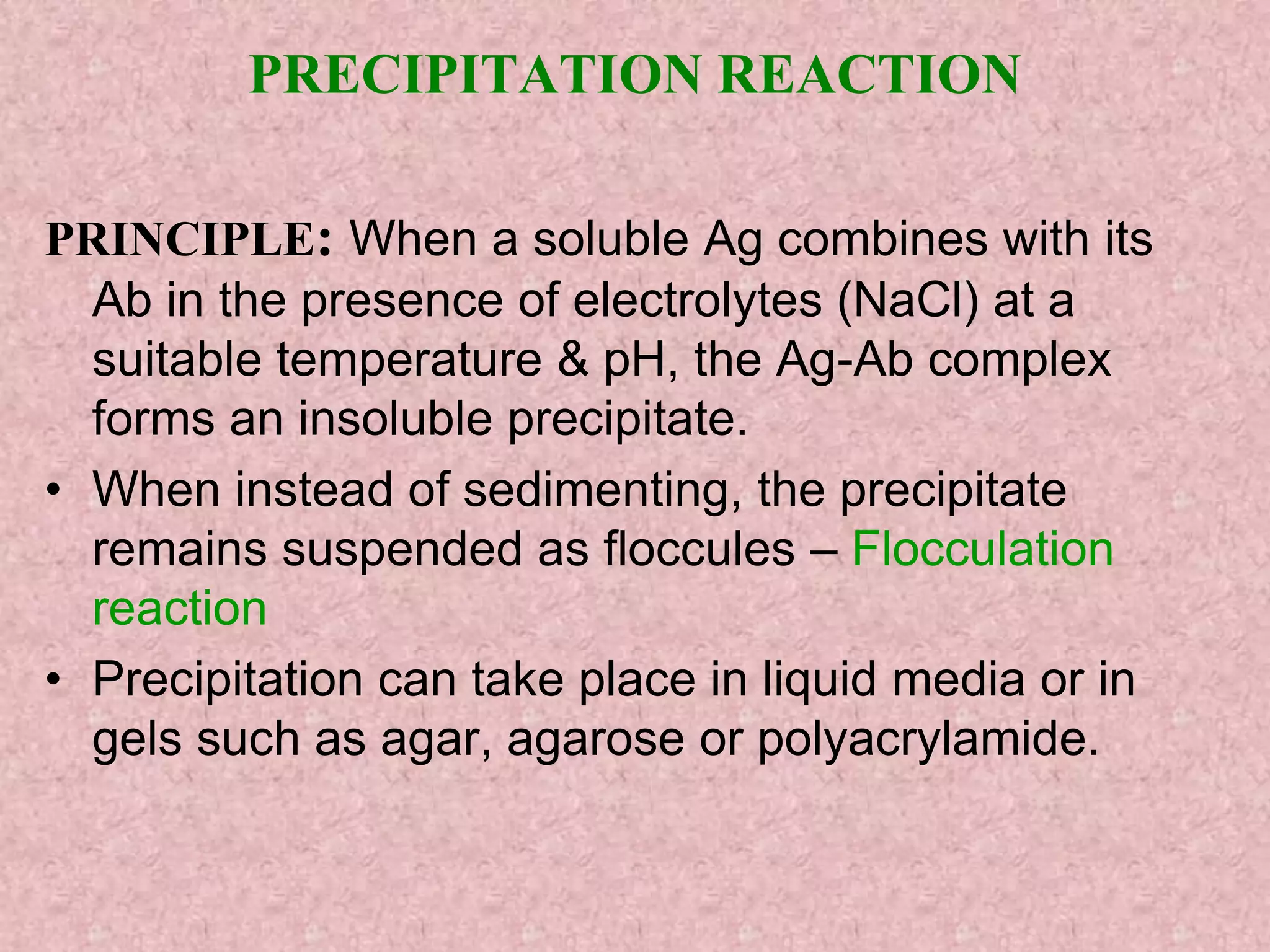
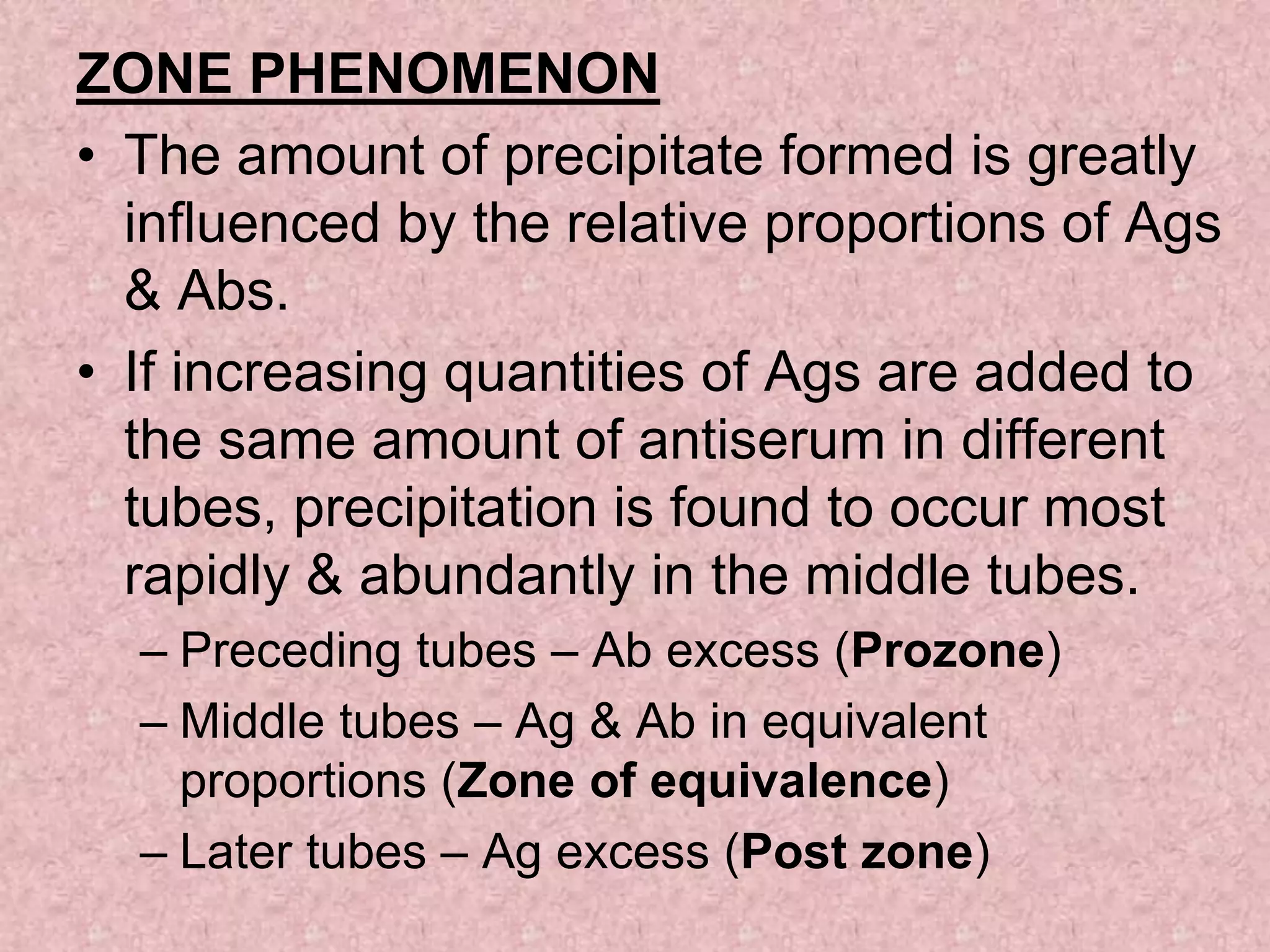
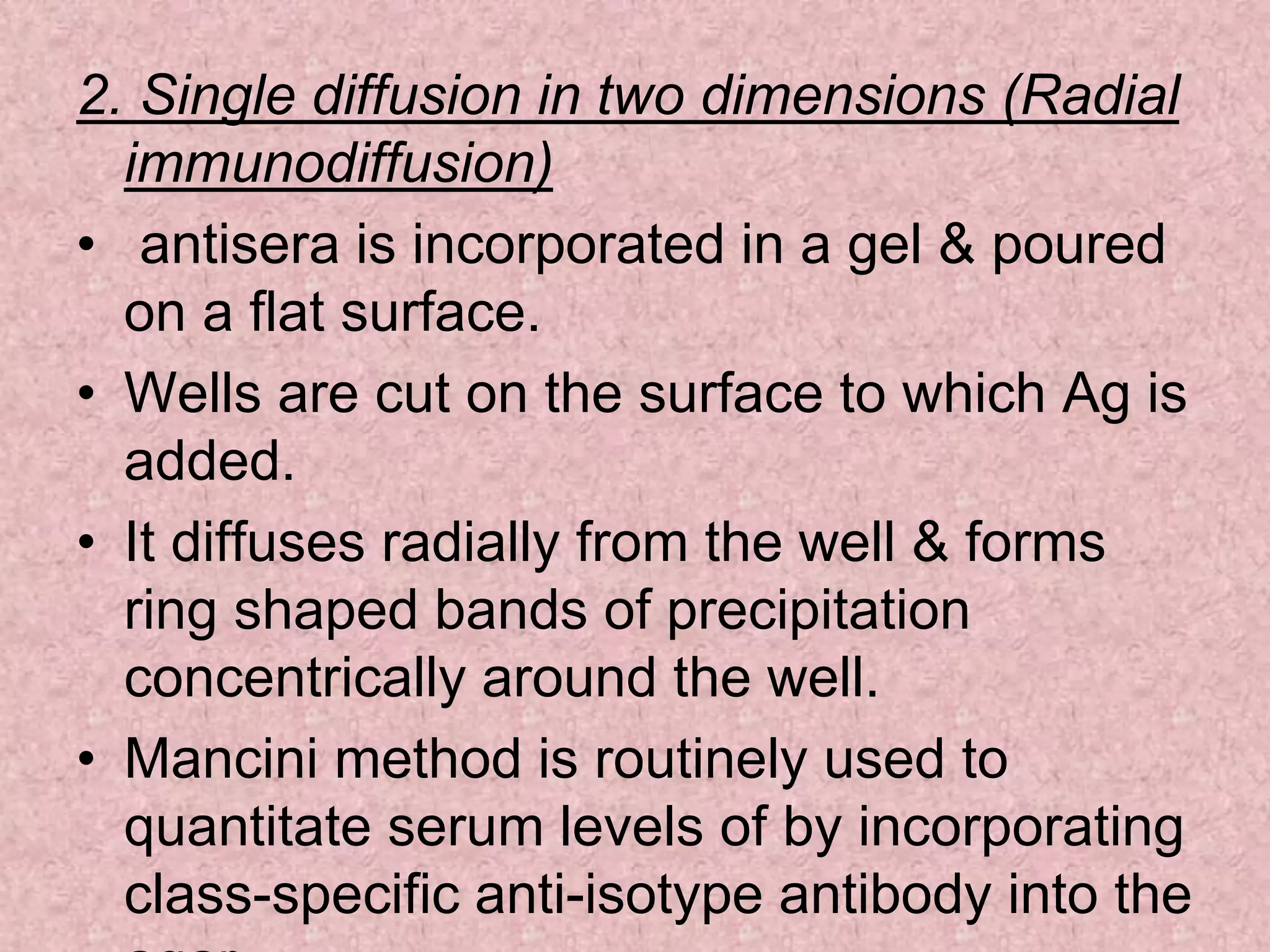
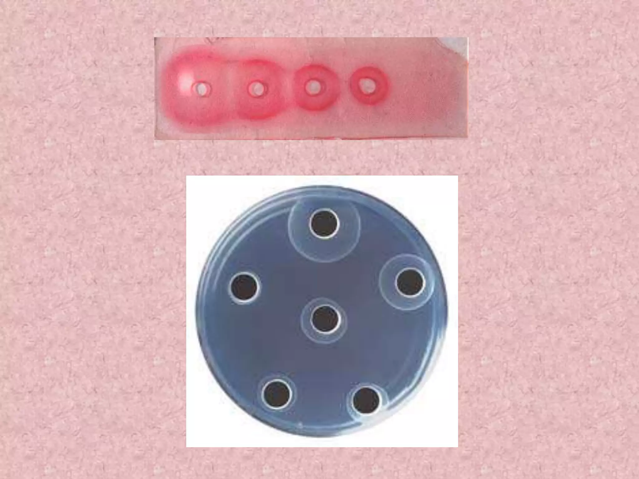
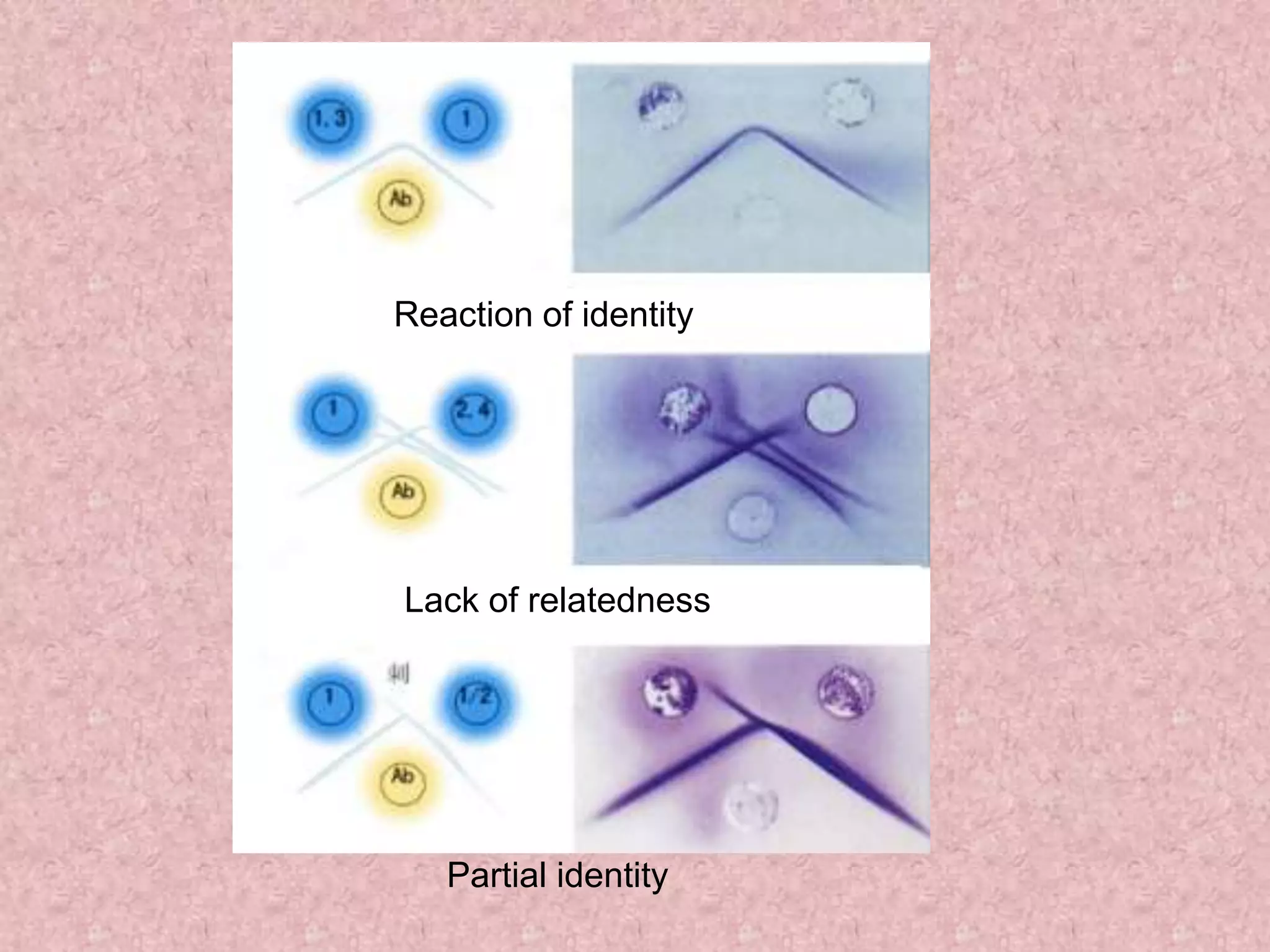
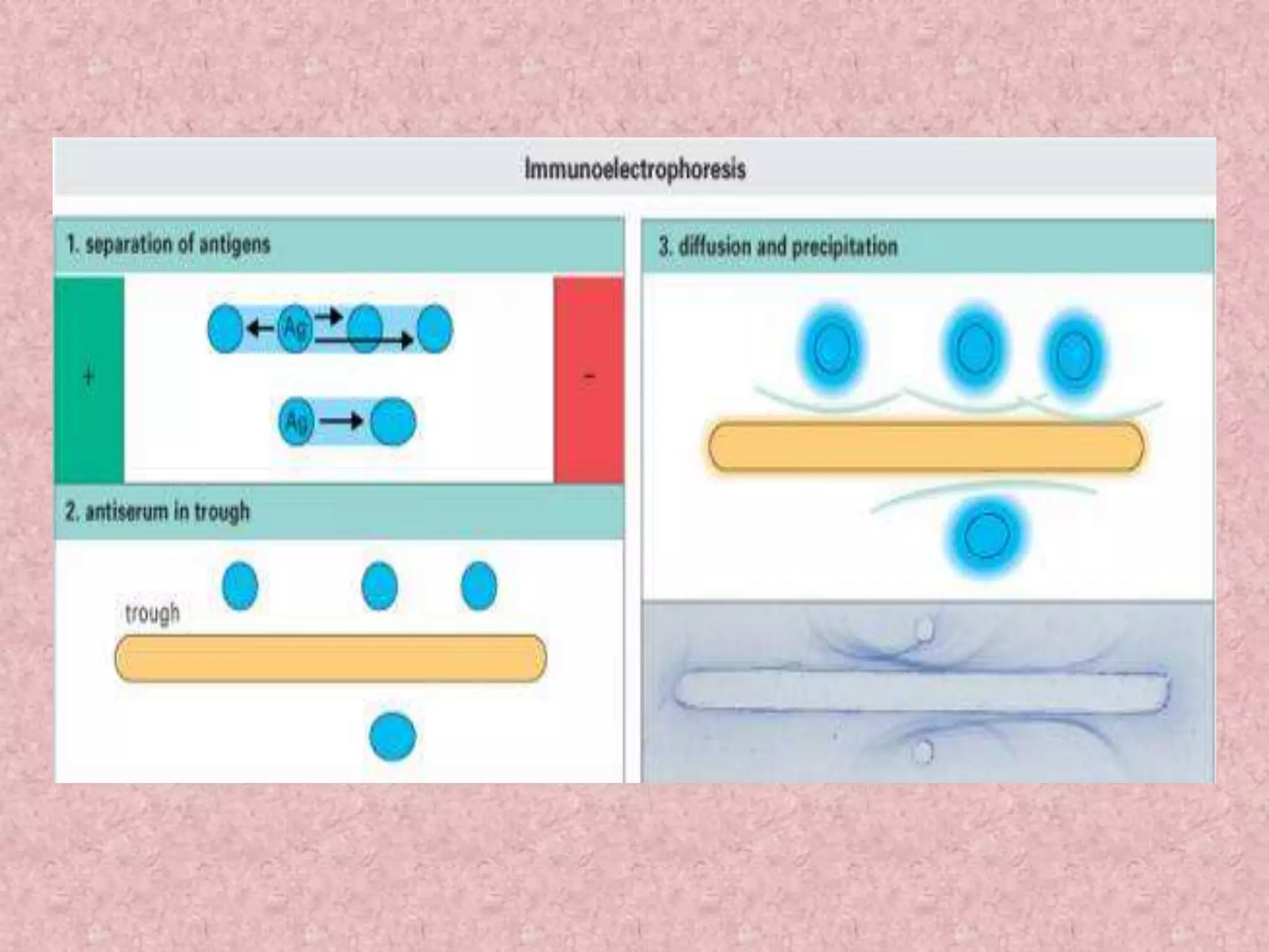
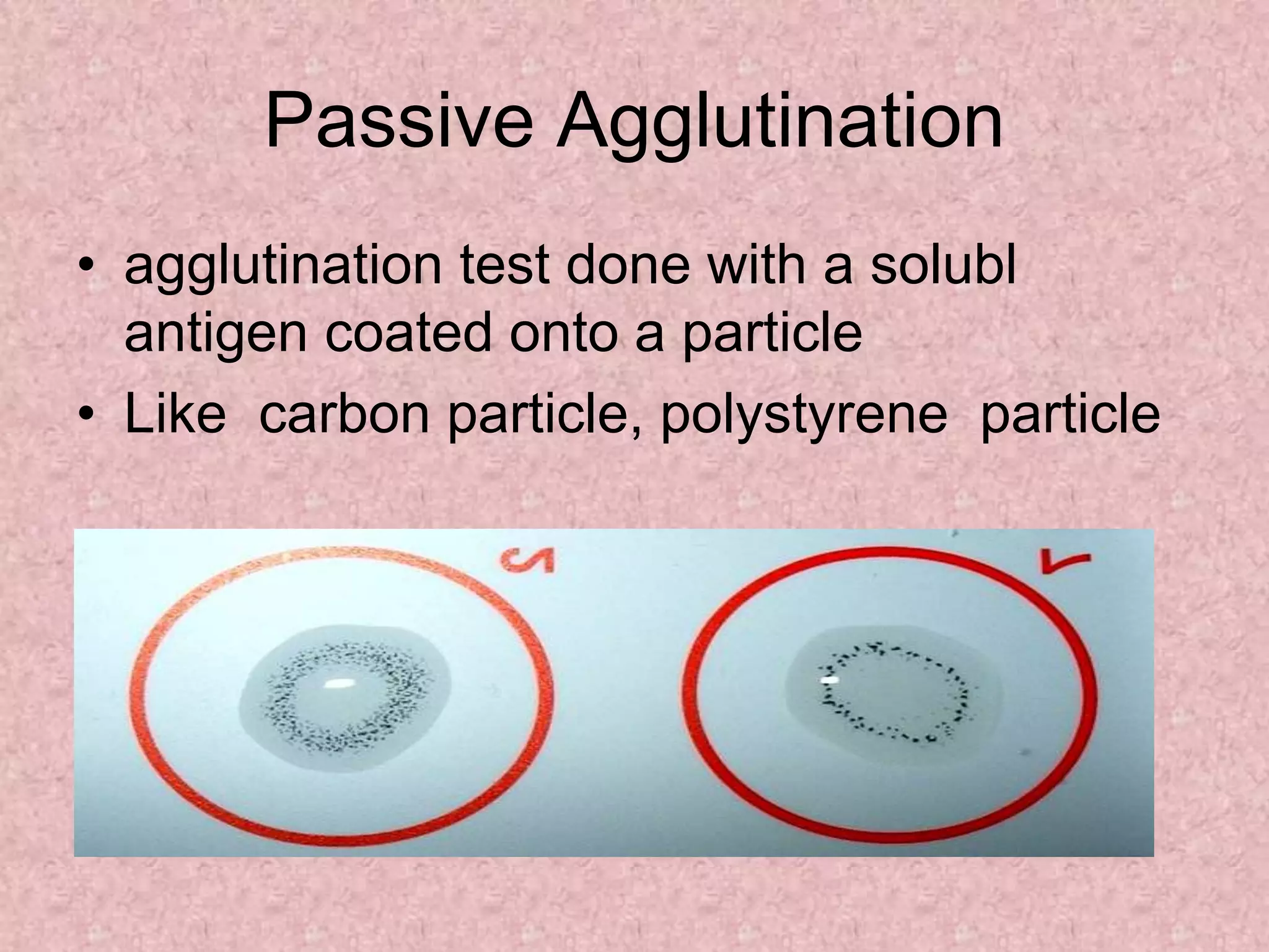
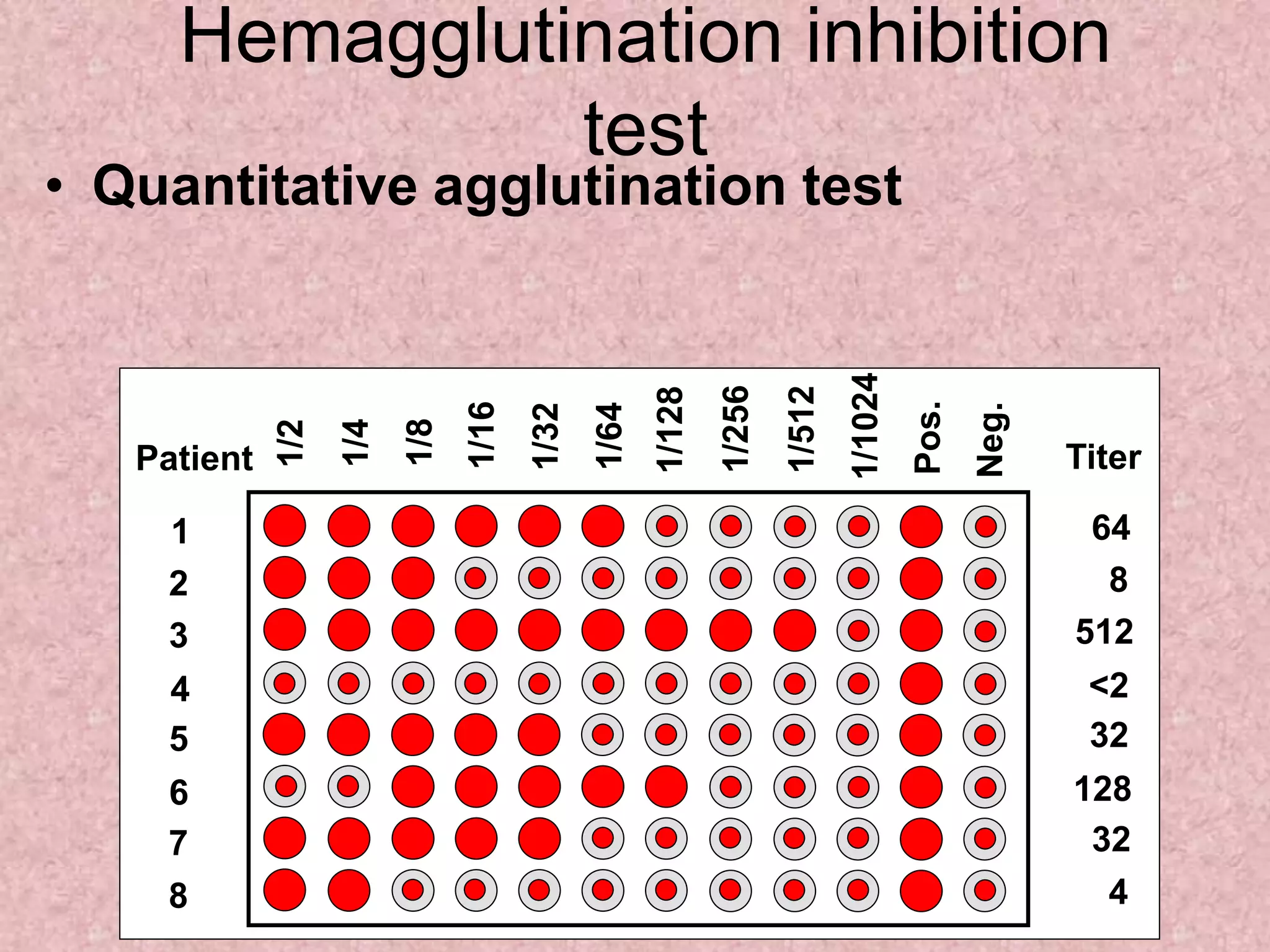
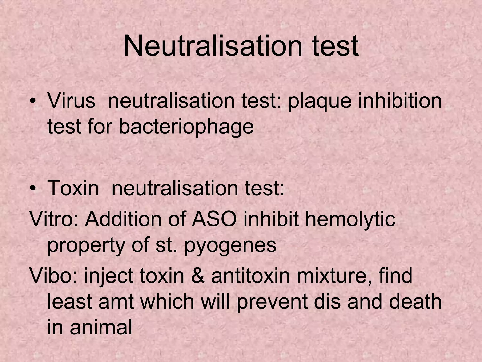
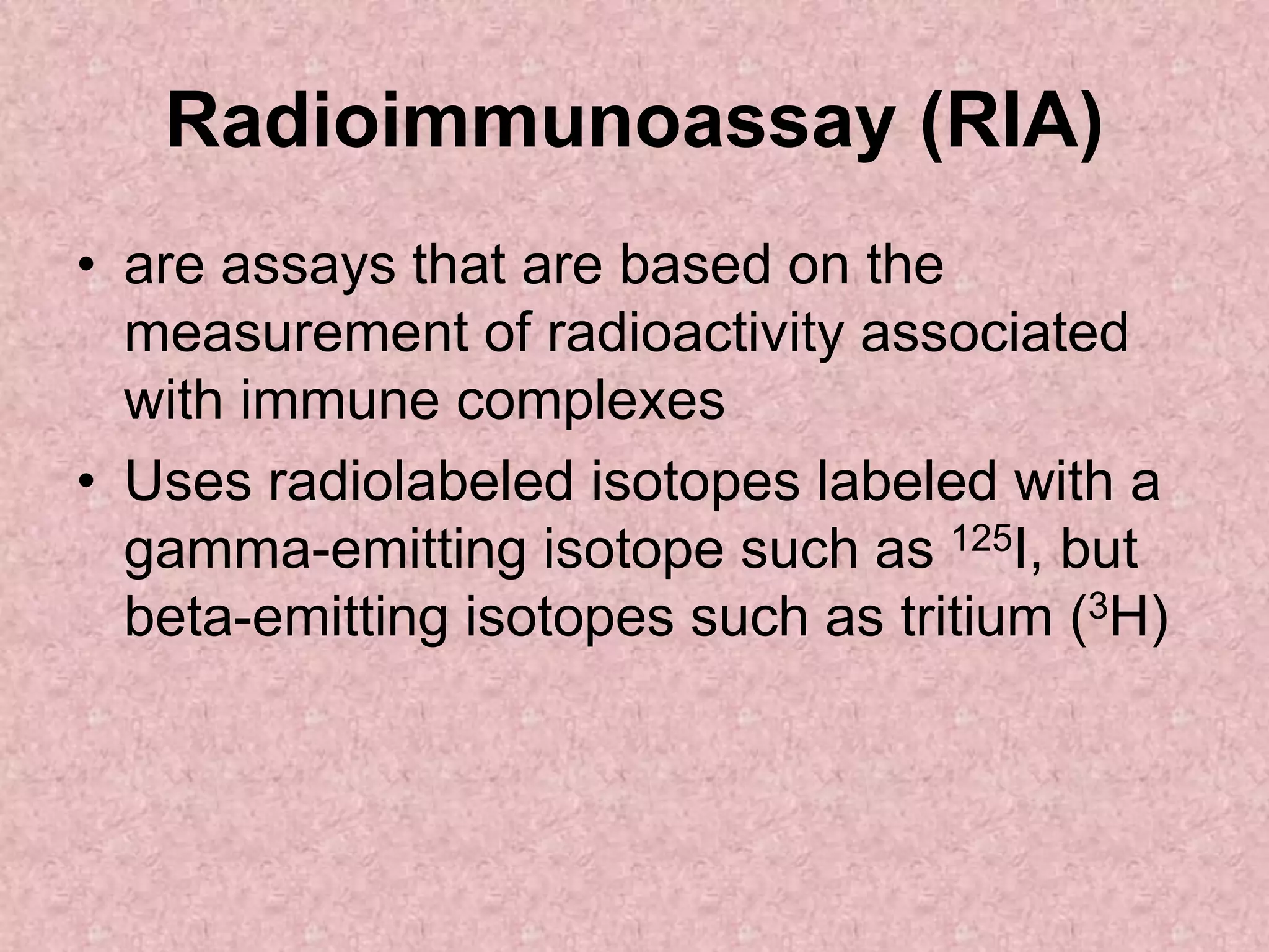
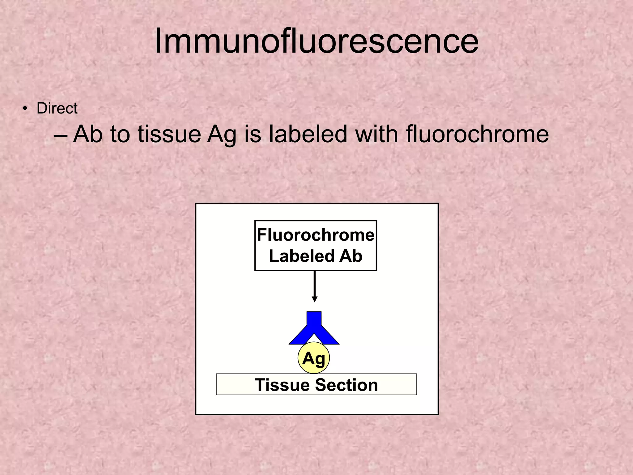
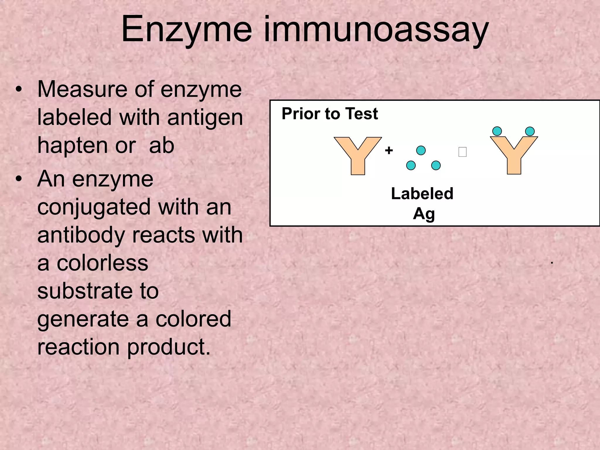
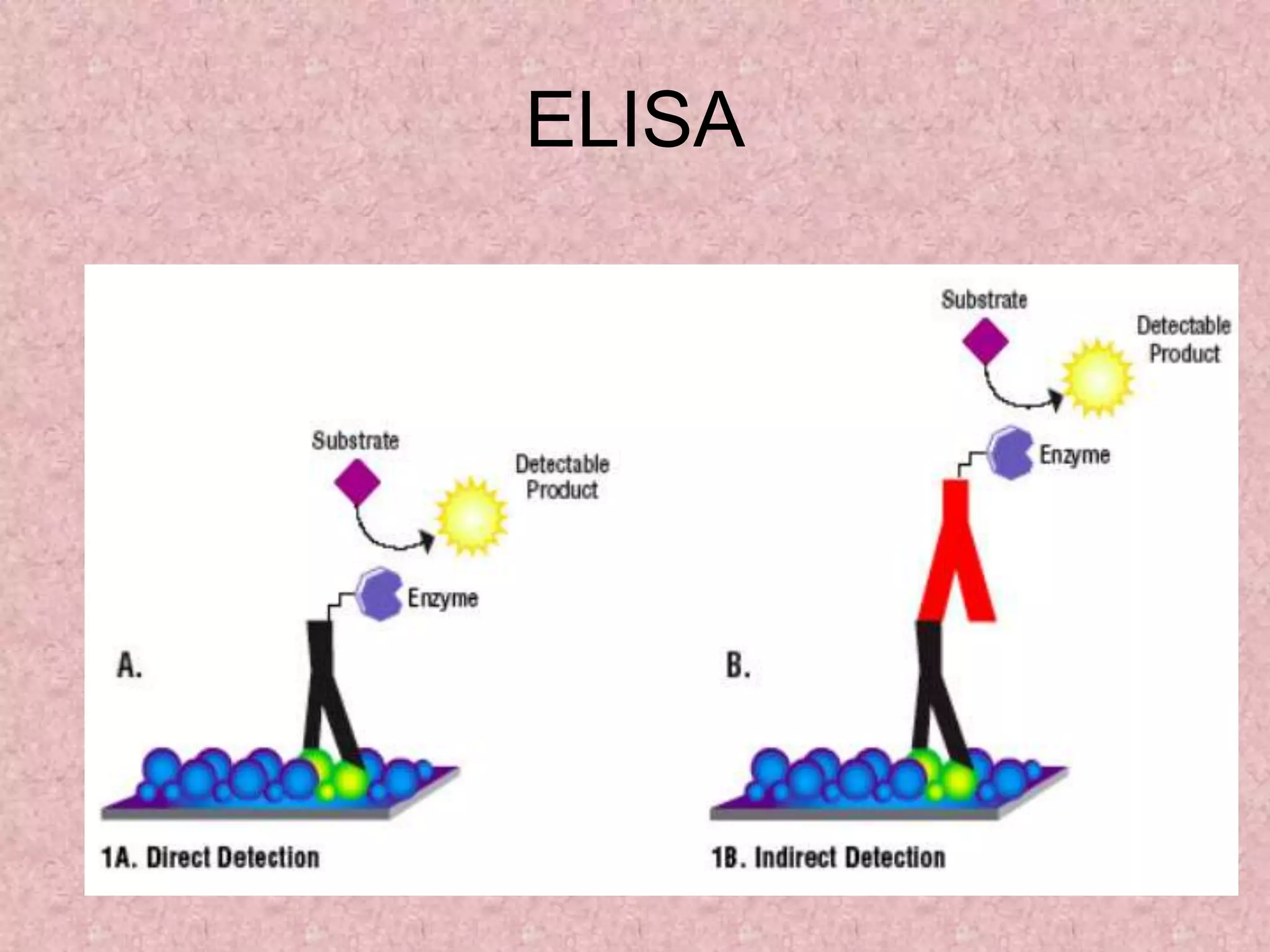
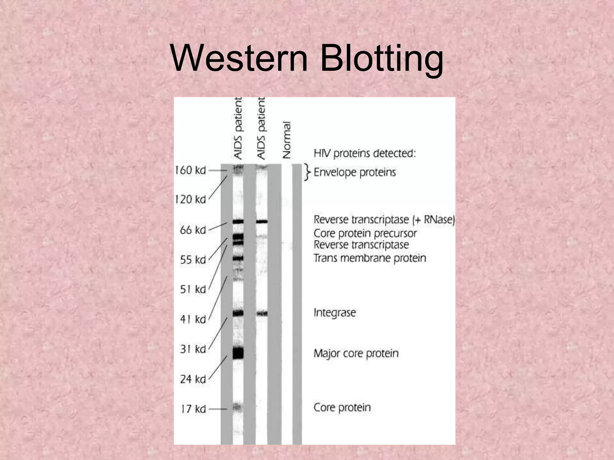
ANTIGEN-ANTIBODY
REACTIONS
• Antigens and antibodies combine with
each other specifically and in an
observable manner.
• In the body, they form the basis of antibody
mediated immunity in infectious diseases,
or hypersensitivity and autoimmune
diseases.
• In laboratory,they help in diagnosis of
infections, & of infectious agents.
• Antigen – antibody reactions in vitro are
known as serological reactions
Stages of Ag – Ab reactions
Primary stage:
• Initial interaction between Ag & Ab –
invisible
Rapid, occurs at low temperatures
Secondary stage:
• Demonstrable events – Precipitation,
agglutination, lysis of cells, killing of live
antigens, neutralization of toxins
Tertiary stage:
• Includes neutralization or destruction of
injurious agents or tissue damage.
• Also includes humoral immunity against
infectious diseases as well as clinical
allergy & other immunological diseases.
GENERAL FEATURES OF Ag – Ab
REACTIONS
1. specific.
2. Entire molecules react and not the fragments
3. The combination occurs at the surface. So
surface antigens are immunologically relevant.
4. There is no denaturation of the antigen or the
antibody during the reaction
MEASUREMENT OF ANTIGEN & ANTIBODY
• Measurement may be in terms of mass or
more commonly as units or titre.
• The Antibody titre of a serum is the
highest dilution of the serum which shows
an observable reactions with the antigen in
a particular test.
Two important parameters in serological tests
– Sensitivity & Specificity
Sensitivity: ability to detect even very
minute quantities of antigen or antibody.
• When the test is highly sensitive, false negative
results may be absent or minimal.
Specificity: Ability to detect reactions
between homologous Ags & Abs only, and
with no other.
• In highly specific test, false positive reactions are
absent or minimal.
Types of Antigen – Antibody Reactions
1. Precipitation reaction
2. Agglutination reaction
3. Neutralization reaction
4. Complement fixation test
5. Immobilization test
6. Immunofluorescence
7. Radioimmuno assay
8. Enzyme immunoassay
9. Complement fixation Test
PRECIPITATION REACTION
PRINCIPLE: When a soluble Ag combines with its
Ab in the presence of electrolytes (NaCl) at a
suitable temperature & pH, the Ag-Ab complex
forms an insoluble precipitate.
• When instead of sedimenting, the precipitate
remains suspended as floccules – Flocculation
reaction
• Precipitation can take place in liquid media or in
gels such as agar, agarose or polyacrylamide.
ZONE PHENOMENON
• The amount of precipitate formed is greatly
influenced by the relative proportions of Ags
& Abs.
• If increasing quantities of Ags are added to
the same amount of antiserum in different
tubes, precipitation is found to occur most
rapidly & abundantly in the middle tubes.
– Preceding tubes – Ab excess (Prozone)
– Middle tubes – Ag & Ab in equivalent
proportions (Zone of equivalence)
– Later tubes – Ag excess (Post zone)
Mechanism of precipitation
• Marrack (1934) proposed the lattice
hypothesis – mechanism of precipitation
• The multivalent antigens combine with
bivalent Abs in varying proportions,
depending on the Ag – Ab ratio on the
reacting mixture.
• Precipitation results when a large lattice is
formed consisting of alternating Ag & Ab.
Types of precipitation rxn
• Precipitation in Fluid
• Precipitation In Gel
1)SLIDE TEST:
• When a drop of Ag & antiserum is placed
on a slide & mixed by shaking, floccules will
appear.
• Eg: VDRL test for syphilis
2) RING TEST:
• Consists of layering Ag solution over a column of
antisera in a narrow tube.
Eg: Grouping of Streptococci by Lancefield
technique
3) TUBE TEST:
• This is employed for the standardization of
toxins & toxoids.
• Serial dilutions of toxin/toxoid are added to
the tubes containing a fixed quantity of
antitoxin.
• The amount of toxin that flocculates
optimally with one unit of the antitoxin – Lf
dose.
• Eg: Kahn test for syphilis.
IMMUNODIFFUSION (precipitation in gel)
Advantages of immunodiffusion:
• Reaction is visible as a distinct band of
precipitation.
• Stable, can be stained for preservation.
• Indicates identity, cross reactions, non
identity between different Ags.
SINGLE DIFFUSION
• In this type of immunodiffusion, only
antigen or antibody diffuses in gel.
1. Single diffusion in one dimension (Oudin
procedure)
• Ab is incorporated in agar gel in a test
tube & Ag solution is layered over it.
• Ag diffuses downward through the agar
gel – forming a line of precipitation.
2. Single diffusion in two dimensions (Radial
immunodiffusion)
• antisera is incorporated in a gel & poured
on a flat surface.
• Wells are cut on the surface to which Ag is
added.
• It diffuses radially from the well & forms
ring shaped bands of precipitation
concentrically around the well.
• Mancini method is routinely used to
quantitate serum levels of by incorporating
class-specific anti-isotype antibody into the
DOUBLE DIFFUSION
• double refers to the diffusion of both
antigens and antibodies
1. Double diffusion in one dimension
(Oakley-Fulthorpe procedure)
• Ab is incorporated in agar gel
• Above which is placed a column of plain
agar.
• The Ag is layered over it.
• The Ag & Ab move towards each other
through the intervening column of plain
agar & form the precipitate.
2. Double diffusion in two dimensions
(Ouchterlony procedure)
• Helps to compare different antisera &
antigens directly.
• Agar gel is poured on a slide & wells are
cut .
• Antiserum – central well
• Different Ags in the surrounding wells.
Reaction of identity
Partial identity
Lack of relatedness
3. Immunoelectrophoresis
• involves the electrophoretic separation of
composite Ag into its constituent proteins,
followed by immunodiffusion of antiserum –
and formation of separate precipitin lines.
• It is performed on an agarose gel with an Ag well
& Ab trough cut on it.
• The test serum is placed in the antigen well &
electrophoresed for about 1 hour.
• Ab against human serum is placed in the trough
& diffusion allowed for 18 – 24 hrs.
• whether a patient overproduces some serum
protein, such as albumin, immunoglobulin, or
transferrin.
Immunoelectrophoresis
3)ELECTROIMMUNODIFFUSION
• The development of precipitin lines can
be speeded up by electrically driving the
Ag & Ab.
• Two types
1. Counterimmunoelectrophoresis (One
dimensional double electroimmunodiffusion)
2. Rocket electrophoresis (One dimensional
single electroimmunodiffusion)
1. Countercurrent immunoelectrophoresis
(CIE)
• This involves simultaneous electrophoresis of Ag
& Ab in gel in opposite directions resulting in
precipitation at a point between them.
• Produce precipitation lines within 30 mins.
• detecting Ags of Cryptococcus & Meningococcus
in the CSF.
2. Rocket electrophoresis
• Used for quantitative estimation of Ags.
• The antiserum to the Ag to be quantitated is
incorporated in agarose gel on a slide.
• Ag in increasing concentrations, is placed in wells
punched in the set gel.
• The Ag is electrophoresed into the Ab containing
agarose.
• The pattern of immunoprecipitation resembles a
ROCKET.
• application of this technique is for quantitative
estimation of antigens
Rocket electrophoresis
Agglutination reaction
• When a particulate Ag combines with its
Ab in the presence of electrolytes (NaCl)
at a suitable temperature & pH, the
antigen are clumped/ aglutinated
Application
SLIDE TEST:
• When a drop of Ag & antiserum is placed
on a slide & mixed by shaking, floccules
will appear.
SLIDE TEST:
• Eg: slide agglutination for identification of
bacterial
Tube agglutination
• Serial dilution with particulate antigen
• eg widal test
Passive Agglutination
• agglutination test done with a solubl
antigen coated onto a particle
• Like carbon particle, polystyrene particle
Haemagglutination Test
• agglutination that involves red blood cells
(RBCs).
• e.g blood typing
Passive haemagglutination test
 red blood cells are used to adsorb
soluble antigen onto their surfaces; the red
blood cells then agglutinate in the
presence of antiserum specific for the
adsorbed antigen
Eg. autoantibody (RA factor) appears in
the serum, which acts as an antibody to
gammaglobulin. The RA factor is able to
agglutinate red cells coated with globulins
Hemagglutination Inhibition
• Definition - measures the ability to inhibit the
agglutination of antigen-coated red blood cells
by antibodies
+
Prior to Test
+ +
Test
Patient’s sample
Hemagglutination inhibition
test
• Quantitative agglutination test
1/2
1/4
1/8
1/16
1/32
1/64
1/128
1/256
1/512
1/1024
Pos.
Neg.
Titer
64
8
512
<2
32
128
32
4
Patient
1
2
3
4
5
6
7
8
Neutralisation test
• Virus neutralisation test: plaque inhibition
test for bacteriophage
• Toxin neutralisation test:
Vitro: Addition of ASO inhibit hemolytic
property of st. pyogenes
Vibo: inject toxin & antitoxin mixture, find
least amt which will prevent dis and death
in animal
Immobilization TEST
• Live treponemes (Nichols strain)
constitute the antigen by means of which
serum is checked for immobilizing
antibody, a mixture of antigen and
antibody is incubation
• the mixture is examined by darkfield
microscopy, and if antibody is present in
serum, the incubation results in an
immobilization of the treponemes
Radioimmunoassay (RIA)
• are assays that are based on the
measurement of radioactivity associated
with immune complexes
• Uses radiolabeled isotopes labeled with a
gamma-emitting isotope such as 125I, but
beta-emitting isotopes such as tritium (3H)
Immunofluorescence
• an antibody labeled with a fluorescent
molecule (fluorescein or rhodamine) is
used to detect the presence of an antigen
in or on a cell or tissue by the fluorescence
emitted by the bound antibody.
Immunofluorescence
• Direct
– Ab to tissue Ag is labeled with fluorochrome
Ag
Fluorochrome
Labeled Ab
Tissue Section
Immunofluorescence
• Indirect
– Ab to tissue Ag is
unlabeled
– Fluorochrome-labeled
anti-Ig is used to detect
binding of the first Ab.
Ag
Fluorochrome
Labeled Anti-Ig
Tissue Section
Unlabeled
Ab
Enzyme immunoassay
• Measure of enzyme
labeled with antigen
hapten or ab
• An enzyme
conjugated with an
antibody reacts with
a colorless
substrate to
generate a colored
reaction product.
+
Prior to Test
Labeled
Ag
.
ELISA
• Use of immunosorbent , an absorbing
material
• Material is labeled with antigen or antibody
• Eg Cellose, polysterene, polyvinyl
ELISA
immunoblotting
• More sensitive
• dentification of a specific protein in a
complex mixture of proteins can be
accomplished by a technique
• EIA,RIA to detect antigen
Western Blotting
polyacrylamide
electrophoresis
blot
React with antibodies
Add substrate
Expose x-ray
film and develop
Western Blotting
Complement fixation test
• Serum is isolated from the patient.
– The serum is heated in such a way that all of the
complement proteins—but none of the antibodies—
within it are destroyed. (
– A known amount of standard complement proteins
are added to the serum.
• The antigen of interest is added to the serum.
• Sheep red blood cells (sRBCs) which have been pre-
bound to anti-sRBC antibodies are added to the serum.
The test is considered positive if RBC are unlysed and
settle to the bottom of the well to form a button
Ag -Ab reactions 1.ppt

Ag -Ab reactions 1.ppt

  • 1.
  • 2.
    • Antigens andantibodies combine with each other specifically and in an observable manner. • In the body, they form the basis of antibody mediated immunity in infectious diseases, or hypersensitivity and autoimmune diseases. • In laboratory,they help in diagnosis of infections, & of infectious agents. • Antigen – antibody reactions in vitro are known as serological reactions
  • 3.
    Stages of Ag– Ab reactions Primary stage: • Initial interaction between Ag & Ab – invisible Rapid, occurs at low temperatures Secondary stage: • Demonstrable events – Precipitation, agglutination, lysis of cells, killing of live antigens, neutralization of toxins
  • 4.
    Tertiary stage: • Includesneutralization or destruction of injurious agents or tissue damage. • Also includes humoral immunity against infectious diseases as well as clinical allergy & other immunological diseases.
  • 5.
    GENERAL FEATURES OFAg – Ab REACTIONS 1. specific. 2. Entire molecules react and not the fragments 3. The combination occurs at the surface. So surface antigens are immunologically relevant. 4. There is no denaturation of the antigen or the antibody during the reaction
  • 6.
    MEASUREMENT OF ANTIGEN& ANTIBODY • Measurement may be in terms of mass or more commonly as units or titre. • The Antibody titre of a serum is the highest dilution of the serum which shows an observable reactions with the antigen in a particular test.
  • 7.
    Two important parametersin serological tests – Sensitivity & Specificity Sensitivity: ability to detect even very minute quantities of antigen or antibody. • When the test is highly sensitive, false negative results may be absent or minimal. Specificity: Ability to detect reactions between homologous Ags & Abs only, and with no other. • In highly specific test, false positive reactions are absent or minimal.
  • 8.
    Types of Antigen– Antibody Reactions 1. Precipitation reaction 2. Agglutination reaction 3. Neutralization reaction 4. Complement fixation test 5. Immobilization test 6. Immunofluorescence 7. Radioimmuno assay 8. Enzyme immunoassay 9. Complement fixation Test
  • 9.
    PRECIPITATION REACTION PRINCIPLE: Whena soluble Ag combines with its Ab in the presence of electrolytes (NaCl) at a suitable temperature & pH, the Ag-Ab complex forms an insoluble precipitate. • When instead of sedimenting, the precipitate remains suspended as floccules – Flocculation reaction • Precipitation can take place in liquid media or in gels such as agar, agarose or polyacrylamide.
  • 10.
    ZONE PHENOMENON • Theamount of precipitate formed is greatly influenced by the relative proportions of Ags & Abs. • If increasing quantities of Ags are added to the same amount of antiserum in different tubes, precipitation is found to occur most rapidly & abundantly in the middle tubes. – Preceding tubes – Ab excess (Prozone) – Middle tubes – Ag & Ab in equivalent proportions (Zone of equivalence) – Later tubes – Ag excess (Post zone)
  • 11.
    Mechanism of precipitation •Marrack (1934) proposed the lattice hypothesis – mechanism of precipitation • The multivalent antigens combine with bivalent Abs in varying proportions, depending on the Ag – Ab ratio on the reacting mixture. • Precipitation results when a large lattice is formed consisting of alternating Ag & Ab.
  • 13.
    Types of precipitationrxn • Precipitation in Fluid • Precipitation In Gel
  • 14.
    1)SLIDE TEST: • Whena drop of Ag & antiserum is placed on a slide & mixed by shaking, floccules will appear. • Eg: VDRL test for syphilis
  • 15.
    2) RING TEST: •Consists of layering Ag solution over a column of antisera in a narrow tube. Eg: Grouping of Streptococci by Lancefield technique
  • 16.
    3) TUBE TEST: •This is employed for the standardization of toxins & toxoids. • Serial dilutions of toxin/toxoid are added to the tubes containing a fixed quantity of antitoxin. • The amount of toxin that flocculates optimally with one unit of the antitoxin – Lf dose. • Eg: Kahn test for syphilis.
  • 18.
    IMMUNODIFFUSION (precipitation ingel) Advantages of immunodiffusion: • Reaction is visible as a distinct band of precipitation. • Stable, can be stained for preservation. • Indicates identity, cross reactions, non identity between different Ags.
  • 19.
    SINGLE DIFFUSION • Inthis type of immunodiffusion, only antigen or antibody diffuses in gel.
  • 20.
    1. Single diffusionin one dimension (Oudin procedure) • Ab is incorporated in agar gel in a test tube & Ag solution is layered over it. • Ag diffuses downward through the agar gel – forming a line of precipitation.
  • 21.
    2. Single diffusionin two dimensions (Radial immunodiffusion) • antisera is incorporated in a gel & poured on a flat surface. • Wells are cut on the surface to which Ag is added. • It diffuses radially from the well & forms ring shaped bands of precipitation concentrically around the well. • Mancini method is routinely used to quantitate serum levels of by incorporating class-specific anti-isotype antibody into the
  • 23.
    DOUBLE DIFFUSION • doublerefers to the diffusion of both antigens and antibodies
  • 24.
    1. Double diffusionin one dimension (Oakley-Fulthorpe procedure) • Ab is incorporated in agar gel • Above which is placed a column of plain agar. • The Ag is layered over it. • The Ag & Ab move towards each other through the intervening column of plain agar & form the precipitate.
  • 26.
    2. Double diffusionin two dimensions (Ouchterlony procedure) • Helps to compare different antisera & antigens directly. • Agar gel is poured on a slide & wells are cut . • Antiserum – central well • Different Ags in the surrounding wells.
  • 27.
    Reaction of identity Partialidentity Lack of relatedness
  • 28.
    3. Immunoelectrophoresis • involvesthe electrophoretic separation of composite Ag into its constituent proteins, followed by immunodiffusion of antiserum – and formation of separate precipitin lines. • It is performed on an agarose gel with an Ag well & Ab trough cut on it. • The test serum is placed in the antigen well & electrophoresed for about 1 hour. • Ab against human serum is placed in the trough & diffusion allowed for 18 – 24 hrs. • whether a patient overproduces some serum protein, such as albumin, immunoglobulin, or transferrin.
  • 30.
  • 31.
    3)ELECTROIMMUNODIFFUSION • The developmentof precipitin lines can be speeded up by electrically driving the Ag & Ab. • Two types 1. Counterimmunoelectrophoresis (One dimensional double electroimmunodiffusion) 2. Rocket electrophoresis (One dimensional single electroimmunodiffusion)
  • 32.
    1. Countercurrent immunoelectrophoresis (CIE) •This involves simultaneous electrophoresis of Ag & Ab in gel in opposite directions resulting in precipitation at a point between them. • Produce precipitation lines within 30 mins. • detecting Ags of Cryptococcus & Meningococcus in the CSF.
  • 33.
    2. Rocket electrophoresis •Used for quantitative estimation of Ags. • The antiserum to the Ag to be quantitated is incorporated in agarose gel on a slide. • Ag in increasing concentrations, is placed in wells punched in the set gel. • The Ag is electrophoresed into the Ab containing agarose. • The pattern of immunoprecipitation resembles a ROCKET. • application of this technique is for quantitative estimation of antigens
  • 34.
  • 35.
    Agglutination reaction • Whena particulate Ag combines with its Ab in the presence of electrolytes (NaCl) at a suitable temperature & pH, the antigen are clumped/ aglutinated Application SLIDE TEST: • When a drop of Ag & antiserum is placed on a slide & mixed by shaking, floccules will appear.
  • 36.
    SLIDE TEST: • Eg:slide agglutination for identification of bacterial
  • 38.
    Tube agglutination • Serialdilution with particulate antigen • eg widal test
  • 40.
    Passive Agglutination • agglutinationtest done with a solubl antigen coated onto a particle • Like carbon particle, polystyrene particle
  • 41.
    Haemagglutination Test • agglutinationthat involves red blood cells (RBCs). • e.g blood typing
  • 43.
    Passive haemagglutination test red blood cells are used to adsorb soluble antigen onto their surfaces; the red blood cells then agglutinate in the presence of antiserum specific for the adsorbed antigen Eg. autoantibody (RA factor) appears in the serum, which acts as an antibody to gammaglobulin. The RA factor is able to agglutinate red cells coated with globulins
  • 44.
    Hemagglutination Inhibition • Definition- measures the ability to inhibit the agglutination of antigen-coated red blood cells by antibodies + Prior to Test + + Test Patient’s sample
  • 45.
    Hemagglutination inhibition test • Quantitativeagglutination test 1/2 1/4 1/8 1/16 1/32 1/64 1/128 1/256 1/512 1/1024 Pos. Neg. Titer 64 8 512 <2 32 128 32 4 Patient 1 2 3 4 5 6 7 8
  • 46.
    Neutralisation test • Virusneutralisation test: plaque inhibition test for bacteriophage • Toxin neutralisation test: Vitro: Addition of ASO inhibit hemolytic property of st. pyogenes Vibo: inject toxin & antitoxin mixture, find least amt which will prevent dis and death in animal
  • 47.
    Immobilization TEST • Livetreponemes (Nichols strain) constitute the antigen by means of which serum is checked for immobilizing antibody, a mixture of antigen and antibody is incubation • the mixture is examined by darkfield microscopy, and if antibody is present in serum, the incubation results in an immobilization of the treponemes
  • 48.
    Radioimmunoassay (RIA) • areassays that are based on the measurement of radioactivity associated with immune complexes • Uses radiolabeled isotopes labeled with a gamma-emitting isotope such as 125I, but beta-emitting isotopes such as tritium (3H)
  • 49.
    Immunofluorescence • an antibodylabeled with a fluorescent molecule (fluorescein or rhodamine) is used to detect the presence of an antigen in or on a cell or tissue by the fluorescence emitted by the bound antibody.
  • 50.
    Immunofluorescence • Direct – Abto tissue Ag is labeled with fluorochrome Ag Fluorochrome Labeled Ab Tissue Section
  • 51.
    Immunofluorescence • Indirect – Abto tissue Ag is unlabeled – Fluorochrome-labeled anti-Ig is used to detect binding of the first Ab. Ag Fluorochrome Labeled Anti-Ig Tissue Section Unlabeled Ab
  • 52.
    Enzyme immunoassay • Measureof enzyme labeled with antigen hapten or ab • An enzyme conjugated with an antibody reacts with a colorless substrate to generate a colored reaction product. + Prior to Test Labeled Ag .
  • 53.
    ELISA • Use ofimmunosorbent , an absorbing material • Material is labeled with antigen or antibody • Eg Cellose, polysterene, polyvinyl
  • 54.
  • 55.
    immunoblotting • More sensitive •dentification of a specific protein in a complex mixture of proteins can be accomplished by a technique • EIA,RIA to detect antigen
  • 56.
    Western Blotting polyacrylamide electrophoresis blot React withantibodies Add substrate Expose x-ray film and develop
  • 57.
  • 58.
    Complement fixation test •Serum is isolated from the patient. – The serum is heated in such a way that all of the complement proteins—but none of the antibodies— within it are destroyed. ( – A known amount of standard complement proteins are added to the serum. • The antigen of interest is added to the serum. • Sheep red blood cells (sRBCs) which have been pre- bound to anti-sRBC antibodies are added to the serum. The test is considered positive if RBC are unlysed and settle to the bottom of the well to form a button

Editor's Notes

  • #28 Fig 2 – lack of relatedness
  • #31 Immunoelectrophoresis