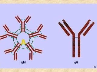
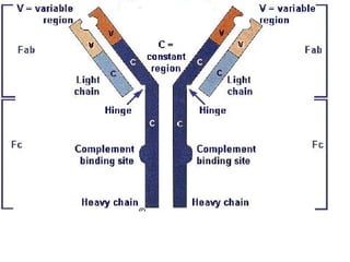
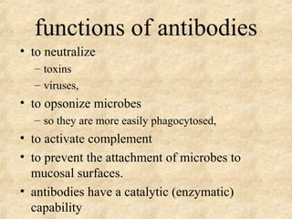
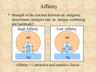
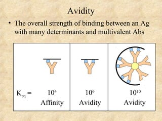
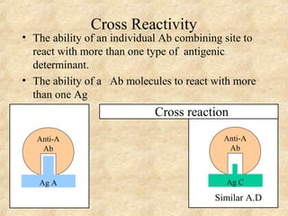
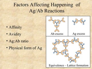
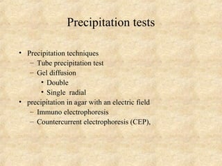
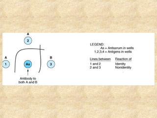
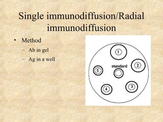
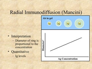
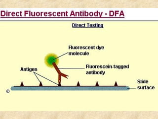
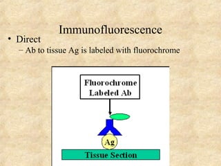
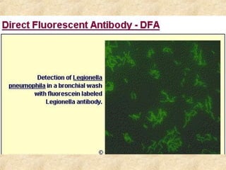
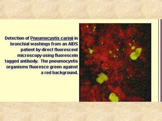
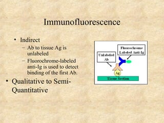
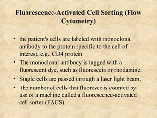
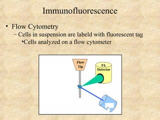
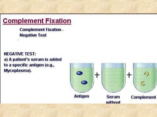
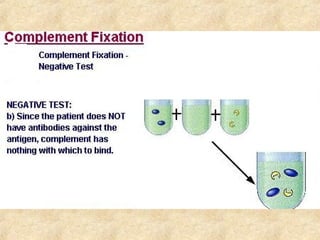
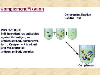
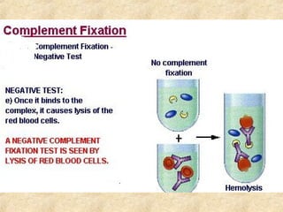
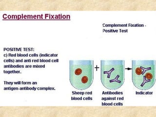
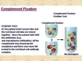
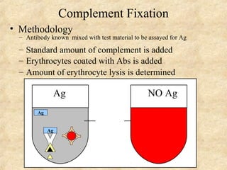
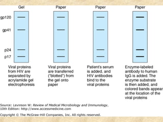
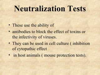
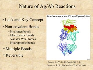
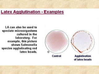
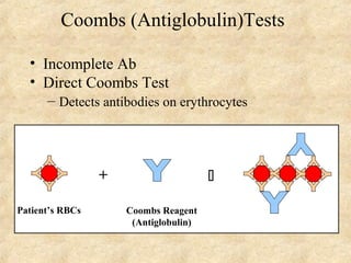
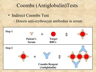
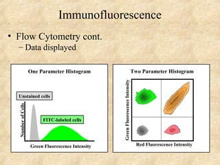
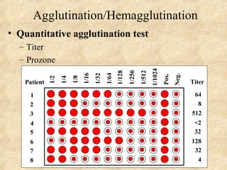
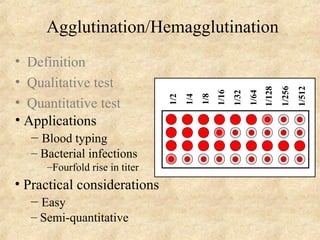
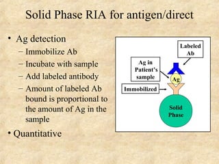
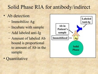
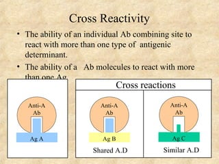
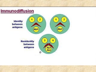
Antigen antibody 
reactions
plasma 
• Albumin 
• Fibrinogen 
• Gobulins 
– Alpha 
– Beta 
– Gammaglobulis
Gammaglobulis 
• Five types 
– IgG 
– IgA 
– IgM 
– IgE 
– IgD
functions of antibodies 
• to neutralize 
– toxins 
– viruses, 
• to opsonize microbes 
– so they are more easily phagocytosed, 
• to activate complement 
• to prevent the attachment of microbes to 
mucosal surfaces. 
• antibodies have a catalytic (enzymatic) 
capability
• Strength of the reaction between an antigenic 
determinant (antigen) and an antigen combining 
site (antibody) 
High Affinity 
Ab 
Ag 
Affinity 
Low Affinity 
Ab 
Ag 
Affinity =  attractive and repulsive forces
Avidity 
• The overall strength of binding between an Ag 
with many determinants and multivalent Abs 
Keq = 104 
Affinity 
106 
Avidity 
1010 
Avidity
Specificity 
• The ability of an antibody combining site to react 
with only an antigenic determinant. 
• The ability of antibody to combine with an antigen
Cross Reactivity 
• The ability of an individual Ab combining site to 
react with more than one type of antigenic 
determinant. 
• The ability of a Ab molecules to react with more 
than one Ag 
Anti-A 
Ab 
Ag A 
Cross reaction 
Anti-A 
Ab 
Ag C 
Similar A.D
Factors Affecting Happening of 
Ag/Ab Reactions 
• Affinity 
• Avidity 
• Ag:Ab ratio 
• Physical form of Ag 
Ab excess Ag excess 
Equivalence – Lattice formation
Tests Based on Ag/Ab Reactions 
• All tests based on Ag/Ab reactions will 
have to depend on lattice formation itself or 
• we will have to utilize ways to detect small 
immune complexes 
• All tests based on Ag/Ab reactions can be 
used to detect either Ag or Ab
Antigen antibody tests 
• Used in both directions 
– Qualitative 
– Quantitative
Uses of antigens/antibody reactions 
• Diagnosis Infectious diseases 
• Diagnosis of autoimmune diseases 
• Determination of blood type and HLA types 
• Determination of chemical levels
Diagnosis of infectious 
diseases 
• When the organism cannot be cultured-or 
difficult to culture hep A B C Syphilis 
• When the organism is too dangerous to 
culture- rickettsial disease 
• When the culture technique is not readily 
available-HIV,EBV 
• When the organism takes too long to grow 
e.g Mycoplasma
Qualitative /quantitative 
• Qualitative 
– determines antigen or antibody is present or 
absent 
• Quantitative 
– determines the quantity of the antibody 
– Titer 
– The highest dilution of the specimen usually 
serum which gives a positive reaction in the 
test
Antigen and antibody reactions in 
the lab 
• Precipitation tests 
• Agglutination 
• Elisa 
• Radioimmunoassay 
• Immunofluorescence 
• Complement Fixation
Precipitation tests 
• The antigen and antibody are in, soluble 
form 
• Combine to form a visible precipitate 
• Presence of electrolytes 
• Positive controls and negative controls
Precipitation tests 
• Precipitation techniques 
– Tube precipitation test 
– Gel diffusion 
• Double 
• Single radial 
• precipitation in agar with an electric field 
– Immuno electrophoresis 
– Countercurrent electrophoresis (CEP),
Precipitation in a capillary tube 
• Streptococcus grouping
Double Diffusion 
• antigen and antibody are placed in different 
wells in agar and 
• allowed to diffuse and form concentration 
gradients. 
• Where optimal proportions occur, lines of 
precipitate form
Double diffusion method (Ouchterlony) 
• indicates whether 
– antigens are identical, 
– Antigens not identical 
– Partially identical
Single immunodiffusion/Radial 
immunodiffusion 
• Method 
– Ab in gel 
– Ag in a well
Radial Immunodiffusion (Mancini) 
• Interpretation 
– Diameter of ring is 
proportional to the 
concentration 
• Quantitative 
– Ig levels 
Ag Concentration 
Ab in gel 
Diameter2 
Ag Ag Ag Ag
Precipitation in agar with an 
electric field
Immunoelectrophoresis 
• Application of electric current 
• Separation of proteins
Interpertation 
• serum proteins are characterized in terms 
of their 
• presence, 
• absence 
• unusual pattern (e.g., human myeloma protein).
Counter- 
Immunoelectrophoresis 
• Method 
– Ag and Ab migrate toward each other by 
electrophoresis 
– Used only when Ag and Ab have opposite charges 
- + 
• Qualitative 
–Rapid 
Ag Ab
uses 
• The meeting of the antigen and antibody is 
greatly accelerated 
• made visible in 30–60 minutes. 
• detection of bacterial and fungal 
polysaccharide antigens in cerebrospinal 
fluid.
Agglutination 
• Visible clumping together of particulate matter by 
antigen combining with its specific antibody. 
• The clumps will be called agglutinates 
• Performed 
– Slide 
– Tube 
– Tile 
– Micrtitration plates
Agglutination 
• Used in both directions 
• Antigen part of a particulate matter 
Salmonella 
• Particulate matter 
– Latex 
– Carbon particles 
– cells 
– Bacteria 
• Stablised staphylococcal cells
Agglutination 
• Active agglutination 
test 
• The antigen part of a 
particulate matter per 
se 
• examples 
• Salmonella, vibrio,
Agglutination 
• Passive agglutination test 
• Antigen or antibody are not part of 
particulate matter but are attached (rided on 
inert particles like latex, carbon,) 
+  Particulat 
matter
coagglutination 
• stabilized staphylococcal cells-protein 
affinity for FC fragment of antibody – 
protein A
Prozone phenomenon 
• Tube agglutination 
• The lower dilutions do not show agglutination 
• the tubes (prior/before the optimum zone ) 
• The tubes in higher dilutions show agglutination 
• Reasons/factors 
– Antibody excess:High level of antibody 
– Non specific inhibitory factors
Haemagglutination 
• Active Haemagglutination test 
– (blood group) 
• Passive haemagglutination (TPHA) 
– Known antigen coated on to treated RBC,s 
– Treated To remove their own antigens 
– Turkey,s RBC,s are used
Immunofluorescence 
• Fluorescent dyes illuminated by ultraviolet 
light are used to show combination of 
antigen antibody . 
• The end point antigen antibody complexes 
are seen fluorescing against a dark 
background 
• Direct 
• Indirect
Immunofluorescence 
• Direct 
– Ab to tissue Ag is labeled with fluorochrome
Immunofluorescence 
• Indirect 
– Ab to tissue Ag is 
unlabeled 
– Fluorochrome-labeled 
anti-Ig is used to detect 
binding of the first Ab. 
• Qualitative to Semi- 
Quantitative
Fluorescence-Activated Cell Sorting (Flow 
Cytometry) 
• the patient's cells are labeled with monoclonal 
antibody to the protein specific to the cell of 
interest, e.g., CD4 protein 
• The monoclonal antibody is tagged with a 
fluorescent dye, such as fluorescein or rhodamine. 
• Single cells are passed through a laser light beam, 
• the number of cells that fluoresce is counted by 
use of a machine called a fluorescence-activated 
cell sorter (FACS).
Immunofluorescence 
• Flow Cytometry 
– Cells in suspension are labeld with fluorescent tag 
•Cells analyzed on a flow cytometer
Assays Based on Complement 
Lattice formation not required
CFT 
• The CFT is used to detect antibodies or 
antigens 
• inference is made by the ability of the 
antibodies to fix the complement 
• The fixation of the complement is measured 
by adding the indicator system 
• Used in diagnosis of viral parasitic and 
rickettsial diseases
Complement Fixation 
• Methodology 
– Antibody known mixed with test material to be assayed for Ag 
– Standard amount of complement is added 
– Erythrocytes coated with Abs is added 
– Amount of erythrocyte lysis is determined 
Ag NO Ag 
Ag 
Ag
RadioImmunoassay 
• The radioactivity of the specific labeled 
antibody or antigen is used to quantify the 
antigen or antibody in patient ,s serum 
• HbsAg 
• Hav IGM
ELISA 
• Uses an enzyme system to show the specific 
combination of antigen antibody 
• An enzyme labeled or linked to a specific antigen 
• A substrate 
• A color reader 
• Double antibody technique to detect and assay 
antigen 
• Indirect technique to Assay antibody
Direct Elisa 
• Ag detection 
– Add labeled antibody 
– Amount of labeled Ab 
bound is proportional 
to the amount of Ag in 
the sample
Indirect ELISA 
• Ab detection 
– Immobilize Ag 
– Incubate with sample 
– Add labeled anti-Ig 
– Amount of labeled Ab 
bound is proportional 
to amount of Ab in the 
sample 
• Quantitative 
Immobilized Ag 
Solid 
Phase 
Ab in 
Patient’s 
sample 
Labeled 
Anti-Ig
Western blot 
• HIV proteins are separated electrophoretically in a gel, 
• discrete bands of viral protein. 
• These proteins are then transferred from the gel, i.e., blotted, 
onto filter paper, and 
• the person's serum is added. 
• If antibodies are present, they bind to the viral proteins 
(primarily gp41 and p24) and can be detected by adding 
antibody to human IgG labeled an enzyme, 
• produces a visible color change when the enzyme substrate is 
added.
Neutralization Tests 
• These use the ability of 
• antibodies to block the effect of toxins or 
the infectivity of viruses. 
• They can be used in cell culture ( inhibition 
of cytopathic effect 
• in host animals ( mouse protection tests).
Extras
Ag-Ab reactions 
Tests for Ag-Ab reactions
Nature of Ag/Ab Reactions 
• Lock and Key Concept 
• Non-covalent Bonds 
– Hydrogen bonds 
– Electrostatic bonds 
– Van der Waal forces 
– Hydrophobic bonds 
• Multiple Bonds 
• Reversible 
http://www.med.sc.edu:85/chime2/lyso-abfr.htm 
Source: Li, Y., Li, H., Smith-Gill, S. J., 
Mariuzza, R. A., Biochemistry 39, 6296, 2000
Coombs (Antiglobulin)Tests 
• Incomplete Ab 
• Direct Coombs Test 
– Detects antibodies on erythrocytes 
+  
Patient’s RBCs Coombs Reagent 
(Antiglobulin)
Coombs (Antiglobulin)Tests 
• Indirect Coombs Test 
– Detects anti-erythrocyte antibodies in serum 
Patient’s 
Serum 
+  
Target 
RBCs 
Step 1 
+  
Coombs Reagent 
(Antiglobulin) 
Step 2
Immunofluorescence 
• Flow Cytometry cont. 
– Data displayed 
One Parameter Histogram 
Unstained cells 
Green Fluorescence Intensity 
Number of Cells 
FITC-labeled cells 
Red Fluorescence Intensity 
Green Fluorescence Intensity 
Two Parameter Histogram
Agglutination Tests 
Lattice Formation
Active Agglutination 
• Definition - tests that have as their endpoint 
the agglutination of a particulate antigen 
– Agglutinin
Agglutination/Hemagglutination 
• Quantitative agglutination test 
– Titer 
– Prozone 
1/2 
1/4 
1/8 
1/16 
1/32 
1/64 
1/128 
1/256 
1/512 
1/1024 
Pos. 
Neg. 
Titer 
64 
8 
512 
<2 
32 
128 
32 
4 
Patient 
1 
2 
3 
4 
5 
6 
7 
8
Agglutination/Hemagglutination 
• Definition 
• Qualitative test 
• Quantitative test 
• Applications 
– Blood typing 
– Bacterial infections 
–Fourfold rise in titer 
• Practical considerations 
– Easy 
– Semi-quantitative 
1/2 
1/4 
1/8 
1/16 
1/32 
1/64 
1/128 
1/256 
1/512
Passive Agglutination/Hemagglutination 
• Definition - agglutination test done with a 
soluble antigen coated onto a particle 
• Applications 
– Measurement of antibodies to soluble antigens
Coombs (Antiglobulin)Tests 
• Applications 
– Detection of anti-Rh Ab 
– Autoimmune hemolytic anemia
Precipitation Tests 
Lattice Formation
Radioimmuoassays (RIA) 
Enzyme-Linked Immunosorbent 
Assays (ELISA) 
Lattice formation not required
Competitive ELISA for antigen 
• Method 
– Determine amount 
of Ab needed to bind 
to a known amount 
of labeled Ag 
+  
Prior to Test 
Labeled 
Ag 
+  
Test 
+ 
Patient’s 
sample 
Labeled 
Ag 
+ 
– Use predetermined 
amounts of labeled 
Ag and Ab and add a 
sample containing 
unlabeled Ag as a 
competitor
Competitive ELISA for Ag 
• Method cont. 
– Determine amount 
of labeled Ag bound 
to Ab 
  NH4SO4 
  anti-Ig 
• Immobilize the Ab 
• Quantitative 
– Most sensitive test 
+  
Test 
+ 
Patient’s 
sample 
Labeled 
Ag 
+ 
Solid 
Phase 
Solid 
Phase 
– Concentration determined from a standard curve 
using known amounts of unlabeled Ag
• Extras
Solid Phase RIA for antigen/direct 
• Ag detection 
– Immobilize Ab 
– Incubate with sample 
– Add labeled antibody 
– Amount of labeled Ab 
bound is proportional to 
the amount of Ag in the 
sample 
• Quantitative 
Ag 
Solid 
Phase 
Ag in 
Patient’s 
sample 
Immobilized 
Labeled 
Ab
Solid Phase RIA for antibody/indirect 
• Ab detection 
– Immobilize Ag 
– Incubate with sample 
– Add labeled anti-Ig 
– Amount of labeled Ab 
bound is proportional 
to amount of Ab in the 
sample 
• Quantitative 
Immobilized Ag 
Solid 
Phase 
Ab in 
Patient’s 
sample 
Labeled 
Anti-Ig
Factors Affecting Measurement of 
Ag/Ab Reactions 
• Affinity 
• Avidity 
• Ag:Ab ratio 
• Physical form of Ag 
Ab excess Ag excess 
Equivalence – Lattice formation
Cross Reactivity 
• The ability of an individual Ab combining site to 
react with more than one type of antigenic 
determinant. 
• The ability of a Ab molecules to react with more 
than one Ag 
Anti-A 
Ab 
Ag A 
Cross reactions 
Anti-A 
Ab 
Ag B 
Shared A.D 
Anti-A 
Ab 
Ag C 
Similar A.D
Immunoelectrophoresis 
• Method 
– Ags are separated by electrophoresis 
– Ab is placed in trough cut in the agar 
+ - 
Ag 
• Interpretation 
Ag 
Ab 
Ag 
Ab 
– Precipitin arc represent individual antigens
Haemagglutination inhibiotion test 
• Certain viruses measles and influenza 
arboviruses agglutinate RBC,s 
• If specific antibodies are included in the 
system a virus is identified
Tests for Cell Associated 
Antigens 
Lattice formation not required
Immunofluorescence 
• Flow Cytometry 
– Cells in suspension are labeld with fluorescent tag 
•Cells analyzed on a flow cytometer
Double Antibody ELISA 
• Ag detection 
– Immobilize Ab 
– Incubate with sample 
– Add labeled antibody 
– Amount of labeled Ab 
bound is proportional to 
the amount of Ag in the 
sample 
• Quantitative 
Ag 
Solid 
Phase 
Ag in 
Patient’s 
sample 
Immobilized 
Labeled 
Ab

Anigen antiboy reactions

  • 1.
  • 2.
    plasma • Albumin • Fibrinogen • Gobulins – Alpha – Beta – Gammaglobulis
  • 3.
    Gammaglobulis • Fivetypes – IgG – IgA – IgM – IgE – IgD
  • 9.
    functions of antibodies • to neutralize – toxins – viruses, • to opsonize microbes – so they are more easily phagocytosed, • to activate complement • to prevent the attachment of microbes to mucosal surfaces. • antibodies have a catalytic (enzymatic) capability
  • 10.
    • Strength ofthe reaction between an antigenic determinant (antigen) and an antigen combining site (antibody) High Affinity Ab Ag Affinity Low Affinity Ab Ag Affinity =  attractive and repulsive forces
  • 11.
    Avidity • Theoverall strength of binding between an Ag with many determinants and multivalent Abs Keq = 104 Affinity 106 Avidity 1010 Avidity
  • 12.
    Specificity • Theability of an antibody combining site to react with only an antigenic determinant. • The ability of antibody to combine with an antigen
  • 13.
    Cross Reactivity •The ability of an individual Ab combining site to react with more than one type of antigenic determinant. • The ability of a Ab molecules to react with more than one Ag Anti-A Ab Ag A Cross reaction Anti-A Ab Ag C Similar A.D
  • 14.
    Factors Affecting Happeningof Ag/Ab Reactions • Affinity • Avidity • Ag:Ab ratio • Physical form of Ag Ab excess Ag excess Equivalence – Lattice formation
  • 15.
    Tests Based onAg/Ab Reactions • All tests based on Ag/Ab reactions will have to depend on lattice formation itself or • we will have to utilize ways to detect small immune complexes • All tests based on Ag/Ab reactions can be used to detect either Ag or Ab
  • 16.
    Antigen antibody tests • Used in both directions – Qualitative – Quantitative
  • 17.
    Uses of antigens/antibodyreactions • Diagnosis Infectious diseases • Diagnosis of autoimmune diseases • Determination of blood type and HLA types • Determination of chemical levels
  • 18.
    Diagnosis of infectious diseases • When the organism cannot be cultured-or difficult to culture hep A B C Syphilis • When the organism is too dangerous to culture- rickettsial disease • When the culture technique is not readily available-HIV,EBV • When the organism takes too long to grow e.g Mycoplasma
  • 19.
    Qualitative /quantitative •Qualitative – determines antigen or antibody is present or absent • Quantitative – determines the quantity of the antibody – Titer – The highest dilution of the specimen usually serum which gives a positive reaction in the test
  • 21.
    Antigen and antibodyreactions in the lab • Precipitation tests • Agglutination • Elisa • Radioimmunoassay • Immunofluorescence • Complement Fixation
  • 22.
    Precipitation tests •The antigen and antibody are in, soluble form • Combine to form a visible precipitate • Presence of electrolytes • Positive controls and negative controls
  • 23.
    Precipitation tests •Precipitation techniques – Tube precipitation test – Gel diffusion • Double • Single radial • precipitation in agar with an electric field – Immuno electrophoresis – Countercurrent electrophoresis (CEP),
  • 24.
    Precipitation in acapillary tube • Streptococcus grouping
  • 25.
    Double Diffusion •antigen and antibody are placed in different wells in agar and • allowed to diffuse and form concentration gradients. • Where optimal proportions occur, lines of precipitate form
  • 28.
    Double diffusion method(Ouchterlony) • indicates whether – antigens are identical, – Antigens not identical – Partially identical
  • 29.
    Single immunodiffusion/Radial immunodiffusion • Method – Ab in gel – Ag in a well
  • 30.
    Radial Immunodiffusion (Mancini) • Interpretation – Diameter of ring is proportional to the concentration • Quantitative – Ig levels Ag Concentration Ab in gel Diameter2 Ag Ag Ag Ag
  • 31.
    Precipitation in agarwith an electric field
  • 32.
    Immunoelectrophoresis • Applicationof electric current • Separation of proteins
  • 34.
    Interpertation • serumproteins are characterized in terms of their • presence, • absence • unusual pattern (e.g., human myeloma protein).
  • 35.
    Counter- Immunoelectrophoresis •Method – Ag and Ab migrate toward each other by electrophoresis – Used only when Ag and Ab have opposite charges - + • Qualitative –Rapid Ag Ab
  • 36.
    uses • Themeeting of the antigen and antibody is greatly accelerated • made visible in 30–60 minutes. • detection of bacterial and fungal polysaccharide antigens in cerebrospinal fluid.
  • 37.
    Agglutination • Visibleclumping together of particulate matter by antigen combining with its specific antibody. • The clumps will be called agglutinates • Performed – Slide – Tube – Tile – Micrtitration plates
  • 39.
    Agglutination • Usedin both directions • Antigen part of a particulate matter Salmonella • Particulate matter – Latex – Carbon particles – cells – Bacteria • Stablised staphylococcal cells
  • 40.
    Agglutination • Activeagglutination test • The antigen part of a particulate matter per se • examples • Salmonella, vibrio,
  • 41.
    Agglutination • Passiveagglutination test • Antigen or antibody are not part of particulate matter but are attached (rided on inert particles like latex, carbon,) +  Particulat matter
  • 42.
    coagglutination • stabilizedstaphylococcal cells-protein affinity for FC fragment of antibody – protein A
  • 43.
    Prozone phenomenon •Tube agglutination • The lower dilutions do not show agglutination • the tubes (prior/before the optimum zone ) • The tubes in higher dilutions show agglutination • Reasons/factors – Antibody excess:High level of antibody – Non specific inhibitory factors
  • 44.
    Haemagglutination • ActiveHaemagglutination test – (blood group) • Passive haemagglutination (TPHA) – Known antigen coated on to treated RBC,s – Treated To remove their own antigens – Turkey,s RBC,s are used
  • 45.
    Immunofluorescence • Fluorescentdyes illuminated by ultraviolet light are used to show combination of antigen antibody . • The end point antigen antibody complexes are seen fluorescing against a dark background • Direct • Indirect
  • 47.
    Immunofluorescence • Direct – Ab to tissue Ag is labeled with fluorochrome
  • 50.
    Immunofluorescence • Indirect – Ab to tissue Ag is unlabeled – Fluorochrome-labeled anti-Ig is used to detect binding of the first Ab. • Qualitative to Semi- Quantitative
  • 51.
    Fluorescence-Activated Cell Sorting(Flow Cytometry) • the patient's cells are labeled with monoclonal antibody to the protein specific to the cell of interest, e.g., CD4 protein • The monoclonal antibody is tagged with a fluorescent dye, such as fluorescein or rhodamine. • Single cells are passed through a laser light beam, • the number of cells that fluoresce is counted by use of a machine called a fluorescence-activated cell sorter (FACS).
  • 52.
    Immunofluorescence • FlowCytometry – Cells in suspension are labeld with fluorescent tag •Cells analyzed on a flow cytometer
  • 53.
    Assays Based onComplement Lattice formation not required
  • 54.
    CFT • TheCFT is used to detect antibodies or antigens • inference is made by the ability of the antibodies to fix the complement • The fixation of the complement is measured by adding the indicator system • Used in diagnosis of viral parasitic and rickettsial diseases
  • 61.
    Complement Fixation •Methodology – Antibody known mixed with test material to be assayed for Ag – Standard amount of complement is added – Erythrocytes coated with Abs is added – Amount of erythrocyte lysis is determined Ag NO Ag Ag Ag
  • 62.
    RadioImmunoassay • Theradioactivity of the specific labeled antibody or antigen is used to quantify the antigen or antibody in patient ,s serum • HbsAg • Hav IGM
  • 64.
    ELISA • Usesan enzyme system to show the specific combination of antigen antibody • An enzyme labeled or linked to a specific antigen • A substrate • A color reader • Double antibody technique to detect and assay antigen • Indirect technique to Assay antibody
  • 65.
    Direct Elisa •Ag detection – Add labeled antibody – Amount of labeled Ab bound is proportional to the amount of Ag in the sample
  • 66.
    Indirect ELISA •Ab detection – Immobilize Ag – Incubate with sample – Add labeled anti-Ig – Amount of labeled Ab bound is proportional to amount of Ab in the sample • Quantitative Immobilized Ag Solid Phase Ab in Patient’s sample Labeled Anti-Ig
  • 67.
    Western blot •HIV proteins are separated electrophoretically in a gel, • discrete bands of viral protein. • These proteins are then transferred from the gel, i.e., blotted, onto filter paper, and • the person's serum is added. • If antibodies are present, they bind to the viral proteins (primarily gp41 and p24) and can be detected by adding antibody to human IgG labeled an enzyme, • produces a visible color change when the enzyme substrate is added.
  • 69.
    Neutralization Tests •These use the ability of • antibodies to block the effect of toxins or the infectivity of viruses. • They can be used in cell culture ( inhibition of cytopathic effect • in host animals ( mouse protection tests).
  • 70.
  • 71.
    Ag-Ab reactions Testsfor Ag-Ab reactions
  • 72.
    Nature of Ag/AbReactions • Lock and Key Concept • Non-covalent Bonds – Hydrogen bonds – Electrostatic bonds – Van der Waal forces – Hydrophobic bonds • Multiple Bonds • Reversible http://www.med.sc.edu:85/chime2/lyso-abfr.htm Source: Li, Y., Li, H., Smith-Gill, S. J., Mariuzza, R. A., Biochemistry 39, 6296, 2000
  • 74.
    Coombs (Antiglobulin)Tests •Incomplete Ab • Direct Coombs Test – Detects antibodies on erythrocytes +  Patient’s RBCs Coombs Reagent (Antiglobulin)
  • 75.
    Coombs (Antiglobulin)Tests •Indirect Coombs Test – Detects anti-erythrocyte antibodies in serum Patient’s Serum +  Target RBCs Step 1 +  Coombs Reagent (Antiglobulin) Step 2
  • 76.
    Immunofluorescence • FlowCytometry cont. – Data displayed One Parameter Histogram Unstained cells Green Fluorescence Intensity Number of Cells FITC-labeled cells Red Fluorescence Intensity Green Fluorescence Intensity Two Parameter Histogram
  • 77.
  • 78.
    Active Agglutination •Definition - tests that have as their endpoint the agglutination of a particulate antigen – Agglutinin
  • 79.
    Agglutination/Hemagglutination • Quantitativeagglutination test – Titer – Prozone 1/2 1/4 1/8 1/16 1/32 1/64 1/128 1/256 1/512 1/1024 Pos. Neg. Titer 64 8 512 <2 32 128 32 4 Patient 1 2 3 4 5 6 7 8
  • 80.
    Agglutination/Hemagglutination • Definition • Qualitative test • Quantitative test • Applications – Blood typing – Bacterial infections –Fourfold rise in titer • Practical considerations – Easy – Semi-quantitative 1/2 1/4 1/8 1/16 1/32 1/64 1/128 1/256 1/512
  • 81.
    Passive Agglutination/Hemagglutination •Definition - agglutination test done with a soluble antigen coated onto a particle • Applications – Measurement of antibodies to soluble antigens
  • 82.
    Coombs (Antiglobulin)Tests •Applications – Detection of anti-Rh Ab – Autoimmune hemolytic anemia
  • 83.
  • 84.
    Radioimmuoassays (RIA) Enzyme-LinkedImmunosorbent Assays (ELISA) Lattice formation not required
  • 85.
    Competitive ELISA forantigen • Method – Determine amount of Ab needed to bind to a known amount of labeled Ag +  Prior to Test Labeled Ag +  Test + Patient’s sample Labeled Ag + – Use predetermined amounts of labeled Ag and Ab and add a sample containing unlabeled Ag as a competitor
  • 86.
    Competitive ELISA forAg • Method cont. – Determine amount of labeled Ag bound to Ab   NH4SO4   anti-Ig • Immobilize the Ab • Quantitative – Most sensitive test +  Test + Patient’s sample Labeled Ag + Solid Phase Solid Phase – Concentration determined from a standard curve using known amounts of unlabeled Ag
  • 87.
  • 88.
    Solid Phase RIAfor antigen/direct • Ag detection – Immobilize Ab – Incubate with sample – Add labeled antibody – Amount of labeled Ab bound is proportional to the amount of Ag in the sample • Quantitative Ag Solid Phase Ag in Patient’s sample Immobilized Labeled Ab
  • 89.
    Solid Phase RIAfor antibody/indirect • Ab detection – Immobilize Ag – Incubate with sample – Add labeled anti-Ig – Amount of labeled Ab bound is proportional to amount of Ab in the sample • Quantitative Immobilized Ag Solid Phase Ab in Patient’s sample Labeled Anti-Ig
  • 91.
    Factors Affecting Measurementof Ag/Ab Reactions • Affinity • Avidity • Ag:Ab ratio • Physical form of Ag Ab excess Ag excess Equivalence – Lattice formation
  • 92.
    Cross Reactivity •The ability of an individual Ab combining site to react with more than one type of antigenic determinant. • The ability of a Ab molecules to react with more than one Ag Anti-A Ab Ag A Cross reactions Anti-A Ab Ag B Shared A.D Anti-A Ab Ag C Similar A.D
  • 94.
    Immunoelectrophoresis • Method – Ags are separated by electrophoresis – Ab is placed in trough cut in the agar + - Ag • Interpretation Ag Ab Ag Ab – Precipitin arc represent individual antigens
  • 97.
    Haemagglutination inhibiotion test • Certain viruses measles and influenza arboviruses agglutinate RBC,s • If specific antibodies are included in the system a virus is identified
  • 98.
    Tests for CellAssociated Antigens Lattice formation not required
  • 99.
    Immunofluorescence • FlowCytometry – Cells in suspension are labeld with fluorescent tag •Cells analyzed on a flow cytometer
  • 101.
    Double Antibody ELISA • Ag detection – Immobilize Ab – Incubate with sample – Add labeled antibody – Amount of labeled Ab bound is proportional to the amount of Ag in the sample • Quantitative Ag Solid Phase Ag in Patient’s sample Immobilized Labeled Ab

Editor's Notes

  • #24 Counter immuno electrophoresis immuno electro osmo phoresis (IEOP)
  • #45 Reverse passive Haemagglutination test(RPHA) Antibody is attached to RBC,s identification of viruses which will not cause haemagglutination per se.