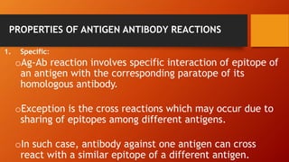
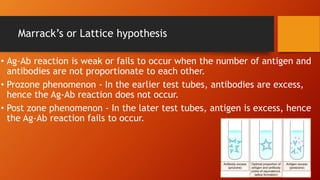
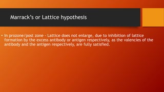
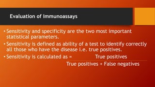
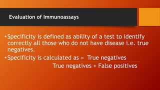
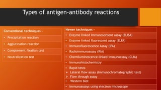
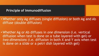
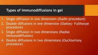
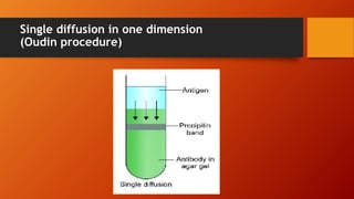
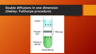
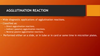
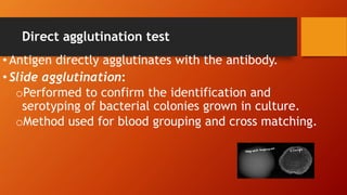
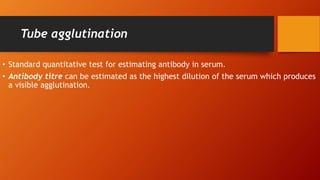
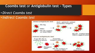
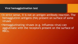
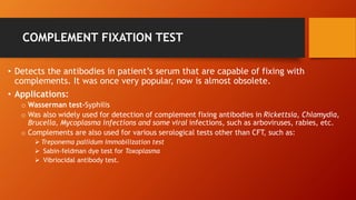
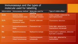
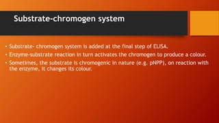
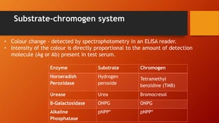
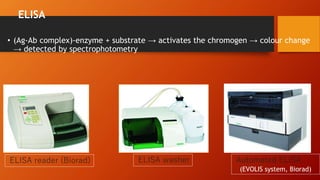
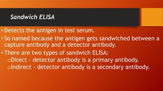
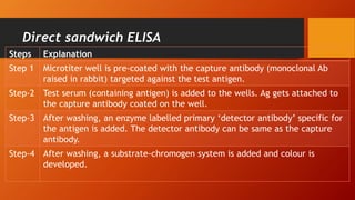
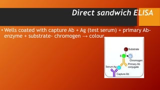
This document provides an overview of antigen-antibody reactions including their general properties, types of reactions, and evaluation of immunoassays. It describes various conventional techniques such as precipitation reactions, agglutination reactions, and complement fixation tests. It also summarizes newer techniques like enzyme-linked immunosorbent assay (ELISA), enzyme-linked fluorescent assay (ELFA), radioimmunoassay (RIA), and chemiluminescence-linked immunoassay (CLIA). The document aims to help students understand antigen-antibody reactions and their applications in diagnostic testing.
serology presentation
Serology is the scientific study of blood serum and other bodily fluids such as semen and saliva.
In practical immunological terms, serology is the diagnostic identification of antibodies in the serum.
Antibodies are typically formed in response to;
An infection, (against a given microorganism),
Other foreign proteins (blood transfusion)
Or to one’s own proteins (autoimmune disease).
The lecture was presented to the students of Saudi board of Community Medicine to help them know about the various serological methods applicable in the diagnosis of infectious diseases in general with attention upon the specificity and sensitivity of various diagnostic modalities. The lecture covers the basic principles of each test and the clinical applications with the advantages and disadvantages of each.
Immunodiffusion -Different Types,Principle,procedureand application. it is a diagnostic technique for the detection or measurements of antibodies and antigens by their precipitation which involves diffusion through a substances such as agar or gel agarose .common types -oudin procedure,oakley fulthorpe procedure ,mancini technique ,ouchterlony double immuno diffusion
Since antigen and antibody reactions are specific, they can be used to identify each other.
These diagnostic tests are particularly useful in diagnosing for examples: infectious diseases, autoimmune diseases, and in typing of blood and tissues prior to transplantation.
This topic describes about antigen-antibody reaction in detail including their classification, mechanism of action, various examples of each reaction with labelled diagrams.
HKRISHNA HOSPITAL, 18TH CROSS R.P ROAD, NANJANGUD
HOSPITAL INFECTION CONTROL & PREVENTION-2023
PRE TEST ON TRIAGING THE PATIENT AND MANAGING EMERGENCY PATIENT
Title: Sense of Smell
Presenter: Dr. Faiza, Assistant Professor of Physiology
Qualifications:
MBBS (Best Graduate, AIMC Lahore)
FCPS Physiology
ICMT, CHPE, DHPE (STMU)
MPH (GC University, Faisalabad)
MBA (Virtual University of Pakistan)
Learning Objectives:
Describe the primary categories of smells and the concept of odor blindness.
Explain the structure and location of the olfactory membrane and mucosa, including the types and roles of cells involved in olfaction.
Describe the pathway and mechanisms of olfactory signal transmission from the olfactory receptors to the brain.
Illustrate the biochemical cascade triggered by odorant binding to olfactory receptors, including the role of G-proteins and second messengers in generating an action potential.
Identify different types of olfactory disorders such as anosmia, hyposmia, hyperosmia, and dysosmia, including their potential causes.
Key Topics:
Olfactory Genes:
3% of the human genome accounts for olfactory genes.
400 genes for odorant receptors.
Olfactory Membrane:
Located in the superior part of the nasal cavity.
Medially: Folds downward along the superior septum.
Laterally: Folds over the superior turbinate and upper surface of the middle turbinate.
Total surface area: 5-10 square centimeters.
Olfactory Mucosa:
Olfactory Cells: Bipolar nerve cells derived from the CNS (100 million), with 4-25 olfactory cilia per cell.
Sustentacular Cells: Produce mucus and maintain ionic and molecular environment.
Basal Cells: Replace worn-out olfactory cells with an average lifespan of 1-2 months.
Bowman’s Gland: Secretes mucus.
Stimulation of Olfactory Cells:
Odorant dissolves in mucus and attaches to receptors on olfactory cilia.
Involves a cascade effect through G-proteins and second messengers, leading to depolarization and action potential generation in the olfactory nerve.
Quality of a Good Odorant:
Small (3-20 Carbon atoms), volatile, water-soluble, and lipid-soluble.
Facilitated by odorant-binding proteins in mucus.
Membrane Potential and Action Potential:
Resting membrane potential: -55mV.
Action potential frequency in the olfactory nerve increases with odorant strength.
Adaptation Towards the Sense of Smell:
Rapid adaptation within the first second, with further slow adaptation.
Psychological adaptation greater than receptor adaptation, involving feedback inhibition from the central nervous system.
Primary Sensations of Smell:
Camphoraceous, Musky, Floral, Pepperminty, Ethereal, Pungent, Putrid.
Odor Detection Threshold:
Examples: Hydrogen sulfide (0.0005 ppm), Methyl-mercaptan (0.002 ppm).
Some toxic substances are odorless at lethal concentrations.
Characteristics of Smell:
Odor blindness for single substances due to lack of appropriate receptor protein.
Behavioral and emotional influences of smell.
Transmission of Olfactory Signals:
From olfactory cells to glomeruli in the olfactory bulb, involving lateral inhibition.
Primitive, less old, and new olfactory systems with different path
These lecture slides, by Dr Sidra Arshad, offer a quick overview of the physiological basis of a normal electrocardiogram.
Learning objectives:
1. Define an electrocardiogram (ECG) and electrocardiography
2. Describe how dipoles generated by the heart produce the waveforms of the ECG
3. Describe the components of a normal electrocardiogram of a typical bipolar lead (limb II)
4. Differentiate between intervals and segments
5. Enlist some common indications for obtaining an ECG
6. Describe the flow of current around the heart during the cardiac cycle
7. Discuss the placement and polarity of the leads of electrocardiograph
8. Describe the normal electrocardiograms recorded from the limb leads and explain the physiological basis of the different records that are obtained
9. Define mean electrical vector (axis) of the heart and give the normal range
10. Define the mean QRS vector
11. Describe the axes of leads (hexagonal reference system)
12. Comprehend the vectorial analysis of the normal ECG
13. Determine the mean electrical axis of the ventricular QRS and appreciate the mean axis deviation
14. Explain the concepts of current of injury, J point, and their significance
Study Resources:
1. Chapter 11, Guyton and Hall Textbook of Medical Physiology, 14th edition
2. Chapter 9, Human Physiology - From Cells to Systems, Lauralee Sherwood, 9th edition
3. Chapter 29, Ganong’s Review of Medical Physiology, 26th edition
4. Electrocardiogram, StatPearls - https://www.ncbi.nlm.nih.gov/books/NBK549803/
5. ECG in Medical Practice by ABM Abdullah, 4th edition
6. Chapter 3, Cardiology Explained, https://www.ncbi.nlm.nih.gov/books/NBK2214/
7. ECG Basics, http://www.nataliescasebook.com/tag/e-c-g-basics
share - Lions, tigers, AI and health misinformation, oh my!.pptxTina Purnat
• Pitfalls and pivots needed to use AI effectively in public health
• Evidence-based strategies to address health misinformation effectively
• Building trust with communities online and offline
• Equipping health professionals to address questions, concerns and health misinformation
• Assessing risk and mitigating harm from adverse health narratives in communities, health workforce and health system
Knee anatomy and clinical tests 2024.pdfvimalpl1234
This includes all relevant anatomy and clinical tests compiled from standard textbooks, Campbell,netter etc..It is comprehensive and best suited for orthopaedicians and orthopaedic residents.
Title: Sense of Taste
Presenter: Dr. Faiza, Assistant Professor of Physiology
Qualifications:
MBBS (Best Graduate, AIMC Lahore)
FCPS Physiology
ICMT, CHPE, DHPE (STMU)
MPH (GC University, Faisalabad)
MBA (Virtual University of Pakistan)
Learning Objectives:
Describe the structure and function of taste buds.
Describe the relationship between the taste threshold and taste index of common substances.
Explain the chemical basis and signal transduction of taste perception for each type of primary taste sensation.
Recognize different abnormalities of taste perception and their causes.
Key Topics:
Significance of Taste Sensation:
Differentiation between pleasant and harmful food
Influence on behavior
Selection of food based on metabolic needs
Receptors of Taste:
Taste buds on the tongue
Influence of sense of smell, texture of food, and pain stimulation (e.g., by pepper)
Primary and Secondary Taste Sensations:
Primary taste sensations: Sweet, Sour, Salty, Bitter, Umami
Chemical basis and signal transduction mechanisms for each taste
Taste Threshold and Index:
Taste threshold values for Sweet (sucrose), Salty (NaCl), Sour (HCl), and Bitter (Quinine)
Taste index relationship: Inversely proportional to taste threshold
Taste Blindness:
Inability to taste certain substances, particularly thiourea compounds
Example: Phenylthiocarbamide
Structure and Function of Taste Buds:
Composition: Epithelial cells, Sustentacular/Supporting cells, Taste cells, Basal cells
Features: Taste pores, Taste hairs/microvilli, and Taste nerve fibers
Location of Taste Buds:
Found in papillae of the tongue (Fungiform, Circumvallate, Foliate)
Also present on the palate, tonsillar pillars, epiglottis, and proximal esophagus
Mechanism of Taste Stimulation:
Interaction of taste substances with receptors on microvilli
Signal transduction pathways for Umami, Sweet, Bitter, Sour, and Salty tastes
Taste Sensitivity and Adaptation:
Decrease in sensitivity with age
Rapid adaptation of taste sensation
Role of Saliva in Taste:
Dissolution of tastants to reach receptors
Washing away the stimulus
Taste Preferences and Aversions:
Mechanisms behind taste preference and aversion
Influence of receptors and neural pathways
Impact of Sensory Nerve Damage:
Degeneration of taste buds if the sensory nerve fiber is cut
Abnormalities of Taste Detection:
Conditions: Ageusia, Hypogeusia, Dysgeusia (parageusia)
Causes: Nerve damage, neurological disorders, infections, poor oral hygiene, adverse drug effects, deficiencies, aging, tobacco use, altered neurotransmitter levels
Neurotransmitters and Taste Threshold:
Effects of serotonin (5-HT) and norepinephrine (NE) on taste sensitivity
Supertasters:
25% of the population with heightened sensitivity to taste, especially bitterness
Increased number of fungiform papillae
263778731218 Abortion Clinic /Pills In Harare ,sisternakatoto
263778731218 Abortion Clinic /Pills In Harare ,ABORTION WOMEN’S CLINIC +27730423979 IN women clinic we believe that every woman should be able to make choices in her pregnancy. Our job is to provide compassionate care, safety,affordable and confidential services. That’s why we have won the trust from all generations of women all over the world. we use non surgical method(Abortion pills) to terminate…Dr.LISA +27730423979women Clinic is committed to providing the highest quality of obstetrical and gynecological care to women of all ages. Our dedicated staff aim to treat each patient and her health concerns with compassion and respect.Our dedicated group ABORTION WOMEN’S CLINIC +27730423979 IN women clinic we believe that every woman should be able to make choices in her pregnancy. Our job is to provide compassionate care, safety,affordable and confidential services. That’s why we have won the trust from all generations of women all over the world. we use non surgical method(Abortion pills) to terminate…Dr.LISA +27730423979women Clinic is committed to providing the highest quality of obstetrical and gynecological care to women of all ages. Our dedicated staff aim to treat each patient and her health concerns with compassion and respect.Our dedicated group of receptionists, nurses, and physicians have worked together as a teamof receptionists, nurses, and physicians have worked together as a team wwww.lisywomensclinic.co.za/
NVBDCP.pptx Nation vector borne disease control programSapna Thakur
NVBDCP was launched in 2003-2004 . Vector-Borne Disease: Disease that results from an infection transmitted to humans and other animals by blood-feeding arthropods, such as mosquitoes, ticks, and fleas. Examples of vector-borne diseases include Dengue fever, West Nile Virus, Lyme disease, and malaria.
Antimicrobial stewardship to prevent antimicrobial resistanceGovindRankawat1
India is among the nations with the highest burden of bacterial infections.
India is one of the largest consumers of antibiotics worldwide.
India carries one of the largest burdens of drug‑resistant pathogens worldwide.
Highest burden of multidrug‑resistant tuberculosis,
Alarmingly high resistance among Gram‑negative and Gram‑positive bacteria even to newer antimicrobials such as carbapenems.
NDM‑1 ( New Delhi Metallo Beta lactamase 1, an enzyme which inactivates majority of Beta lactam antibiotics including carbapenems) was reported in 2008
2. Learning objectives
By the end of this session students should be able to know:
• General properties of antigen antibody reactions
• Types of antigen antibody reactions
• Conventional immunoassays
• Overview of newer techniques
3. Antigen-antibody reaction
• Bimolecular association where the antigen and antibody
combine with each other specifically and in an observable
manner.
oSimilar to an enzyme-substrate interaction.
oOnly difference is, it does not lead to an irreversible
alteration in either antibody or in antigen.
4. PROPERTIES OF ANTIGEN ANTIBODY REACTIONS
1. Specific:
oAg-Ab reaction involves specific interaction of epitope of
an antigen with the corresponding paratope of its
homologous antibody.
oException is the cross reactions which may occur due to
sharing of epitopes among different antigens.
oIn such case, antibody against one antigen can cross
react with a similar epitope of a different antigen.
5. PROPERTIES OF ANTIGEN ANTIBODY REACTIONS
2. Non-covalent interactions:
o Union of antigen and antibody requires formation of large number of non-covalent interactions
between them such as:
Hydrogen bonds
Electrostatic interactions
Hydrophobic interactions
van der Waals forces
6. PROPERTIES OF ANTIGEN ANTIBODY REACTIONS
• 3. Strength- The strength or the firmness of the association is influenced by the
affinity and avidity of the antigen-antibody interaction.
o Affinity
Refers to sum total of non covalent interactions between a single epitope of an antigen with its
corresponding paratope present on antibody.
Affinity can be measured by two methods- i) by equilibrium dialysis and ii) by surface plasmon
resonance method
7. PROPERTIES OF ANTIGEN ANTIBODY REACTIONS
• Avidity: Term used to describe the affinities of all the binding sites when multivalent
antibody reacts with a complex antigen carrying multiple epitopes.
o When a complex antigen carrying multiple epitopes reacts with a multivalent antibody; the total
strength (i.e. avidity) would be much higher than the individual affinity at each binding site.
o Avidity is not exactly equal but lower than the sum of all affinities.
o This difference is primarily due to geometry of antigen antibody binding.
8. PROPERTIES OF ANTIGEN ANTIBODY REACTIONS
oAvidity is a better indicator of strength of an antigen
antibody reaction.
oAvidity of an antibody can compensate for its low affinity.
For example, IgM has a low affinity than IgG but it is
multivalent (10 valencies), therefore has a much higher avidity.
It can bind to an antigen more effectively than IgG.
9. Diagnostic Use of Ag-Ab reactions
•Specific and observable
•The diagnostic tests based on antigen-antibody reactions
are called as immunoassays
•Immunoassays can be broadly categorized into two
types-
oAntigen detection assays
oAntibody detection assays
10. Qualitative assays
• Result is read as ‘positive’ or ‘negative’ based on presence or
absence of antigen or antibody in the clinical specimen.
11. Disadvantages
• Number of antigen or antibody molecules in the reaction are disproportionate to
each other.
o If either antigen or antibody are present in higher quantity - false negative.
To rule out a false negative result, it is ideal to test the series of diluted sera (quantitative test)
instead of just testing the one specimen of undiluted serum.
12. Quantitative assays
• When the qualitative test turns positive, the exact amount of antibody in serum
can be known by serial dilution of the patient’s serum and mixing each dilution of
the serum with a known quantity of antigen.
• The measurement of antibody is expressed in terms of units or more commonly
titer.
14. Marrack’s or Lattice hypothesis
• Ag-Ab reaction is weak or fails to occur when the number of antigen and
antibodies are not proportionate to each other.
• Prozone phenomenon - In the earlier test tubes, antibodies are excess,
hence the Ag-Ab reaction does not occur.
• Post zone phenomenon - In the later test tubes, antigen is excess, hence
the Ag-Ab reaction fails to occur.
15. Marrack’s or Lattice hypothesis
• In prozone/post zone - Lattice does not enlarge, due to inhibition of lattice
formation by the excess antibody or antigen respectively, as the valencies of the
antibody and the antigen respectively, are fully satisfied.
16. Evaluation of Immunoassays
• Sensitivity and specificity are the two most important
statistical parameters.
• Sensitivity is defined as ability of a test to identify correctly
all those who have the disease i.e. true positives.
• Sensitivity is calculated as = True positives
True positives + False negatives
17. Evaluation of Immunoassays
• Specificity is defined as ability of a test to identify
correctly all those who do not have disease i.e. true
negatives.
• Specificity is calculated as = True negatives
True negatives + False positives
18. Types of antigen-antibody reactions
Conventional techniques -
• Precipitation reaction
• Agglutination reaction
• Complement fixation test
• Neutralization test
Newer techniques -
• Enzyme linked immunosorbent assay (ELISA)
• Enzyme linked fluorescent assay (ELFA)
• Immunofluorescence Assay (IFA)
• Radioimmunoassay (RIA)
• Chemiluminescence-linked immunoassay (CLIA)
• Immunohistochemistry
• Rapid tests-
Lateral flow assay (Immunochromatographic test)
Flow through assay
• Western blot
• Immunoassays using electron microscope
19. PRECIPITATION REACTION
• Definition-When a soluble antigen reacts with its antibody
in the presence of optimal temperature, pH and electrolytes
(NaCl), it leads to formation of the antigen-antibody
complex in the form of:
oInsoluble precipitate band when gel containing medium is used
or
oInsoluble floccules when liquid medium is used (precipitate remains
suspended as floccules)
20. Precipitation in liquid medium
• Ring test: In a narrow tube (e.g. capillary tube), antigen
solution is layered over an antiserum
oPrecipitate ring appears at the junction of two liquids.
oExample: Streptococcal grouping by Lancefield
technique, and Ascoli’s thermoprecipitin test done for
anthrax.
21. Precipitation in liquid medium
• Flocculation test- When a drop of antigen is mixed with a
drop of patient's serum, then floccules appear.
oExamples of slide flocculation test- VDRL and RPR tests
used for diagnosis of syphilis
oExamples of tube flocculation test- Kahn test used
previously for syphilis
22. Precipitation in gel (Immunodiffusion)
• Using 1% soft agarose gel for precipitation reaction has
many advantages over liquid medium-
oResults in formation of clearly visible bands instead of
floccules that can be preserved for longer time.
oCan differentiate individual antigens from a mixture as
each antigen forms a separate band after reacting with
specific antibody.
23. Principle of Immunodiffusion
• Whether only Ag diffuses (single diffusion) or both Ag and Ab
diffuse (double diffusion)
• Whether Ag or Ab diffuses in one dimension (i.e. vertical
diffusion when test is done on a tube layered with gel) or
two dimensions (i.e. diffusion in both X and Y axis when test
is done on a slide or a petri dish layered with gel)
24. Types of immunodiffusions in gel
1. Single diffusion in one dimension (Oudin procedure)
2. Double diffusions in one dimension (Oakley- Fulthorpe
procedure)
3. Single diffusion in two dimensions (Radial
immunodiffusion)
4. Double diffusions in two dimensions (Ouchterlony
procedure)
29. Double diffusions in two dimensions
(Ouchterlony procedure)
• Examples of double diffusions in two dimensions include:
oElek's test for detecting toxin of Corynebacterium
diphtheria.
oEiken test to detect toxin of Escherichia coli
30. Precipitation in gel in presence of
electric current
1. Electroimmunodiffusion (EID)
2. CIEP (Countercurrent immunoelectrophoresis)
3. Rocket electrophoresis
31. Electroimmunodiffusion
• Electric current is applied to a
slide layered with gel.
• Helps in identification and
approximate quantitation of
various proteins present in the
serum.
32. Counter-current immunoelectrophoresis
• Test is even faster (takes 30 minutes)
and more sensitive than EID.
• Popular in the past - detecting alpha
fetoprotein in serum and capsular
antigens of Cryptococcus and
meningococcus in the cerebrospinal
fluid.
34. AGGLUTINATION REACTION
• Definition-When a particulate or insoluble antigen is mixed with its antibody in the
presence of electrolytes at a suitable temperature and pH, the particles are
clumped or agglutinated.
• Advantage:
o More sensitive than precipitation test.
o Clumps are better visualized and interpreted than bands or floccules.
35. AGGLUTINATION REACTION
• Wide diagnostic applications of agglutination reactions.
• Classified as:
o Direct agglutination reactions.
o Indirect (passive) agglutination reactions.
o Reverse passive agglutination reactions.
• Performed either on a slide, or in tube or in card or some time in microtiter plates.
36. Direct agglutination test
• Antigen directly agglutinates with the antibody.
• Slide agglutination:
oPerformed to confirm the identification and
serotyping of bacterial colonies grown in culture.
oMethod used for blood grouping and cross matching.
37. Tube agglutination
• Standard quantitative test for estimating antibody in serum.
• Antibody titre can be estimated as the highest dilution of the serum which produces
a visible agglutination.
38. Tube agglutination- Applications
• Enteric fever (Widal test)
• Acute brucellosis (Standard agglutination test)
• Coombs Antiglobulin test (see below)
• Heterophile agglutination tests:
o Typhus fever (Weil Felix reaction)
o Infectious mononucleosis (Paul Bunnell test)
o Mycoplasma pneumoniae (Cold agglutination test)
39. Microscopic agglutination test (MAT)
• Agglutination test is performed on a microtiter plate.
• Result is read under a microscope.
• MAT is done for leptospirosis.
40. Indirect or passive agglutination test
• Indirect hemagglutination test (IHA)
• Latex agglutination test (LAT)
41. Indirect hemagglutination test (IHA)
• Passive agglutination test where RBCs are used as carrier
molecules.
• IHA was used widely in the past, but is less popular at
present.
42. Latex agglutination test (LAT) for antibody detection
• Polystyrene latex particles (0.8 – 1 µm in diameter) are used
as carrier molecules which are capable of adsorbing several
types of antigens.
• For better interpretation of result, the test is performed on
a black colour card.
• Drop of patient’s serum (containing antibody) is added to a
drop of latex solution coated with the antigen and the card
is rocked.
43. Latex agglutination test (LAT) for
antibody detection
• Positive result is indicated by formation of visible clumps.
• LAT is one of the most widely used tests at present as it is
very simple and rapid.
• Used for detection of ASO (antistreptolysin O antibody).
44. Reverse passive agglutination test
(for antigen detection)
• Antibody is coated on a carrier molecule which detects
antigen in the patient’s serum.
Test Carrier
molecule
Clinical applications
RPHA (Reverse passive
hemagglutination assay)
RBCs • Detection of hepatitis B surface antigen (HBsAg) –
not used now.
Latex agglutination test Latex
particles
• Detection of CRP (C reactive protein), RA
(rheumatoid arthritis factor), capsular antigen
detection in CSF (for pneumococcus,
meningococcus and Cryptococcus) and
streptococcal grouping.
45. Reverse passive agglutination test
(for antigen detection)
Test Carrier
molecule
Clinical applications
Coagglutination
test
Staphylococcus
aureus
• Used in past for antigen detection (e.g.
Salmonella antigen detection from blood
and urine).
• Obsolete at present.
46. Hemagglutination test
• Refers to the agglutination tests that use red blood cells (RBCs) as source of
antigen.
• Various types of hemagglutination tests include:
o Direct hemagglutination test
o Indirect hemagglutination test
47. Direct hemagglutination test
• Serum antibodies directly agglutinate with surface antigens of RBCs to produce a
matt. Examples include-
o Paul Bunnell test
o Cold agglutination test
o Blood grouping
o Coombs test or Antiglobulin test
48. Coombs test or Antiglobulin test
• Performed to diagnose Rh incompatibility by detecting Rh
antibody from mother’s and baby’s serum.
• Rh incompatibility is a condition when an Rh negative
mother (Rh Ag -ve and Rh Ab -ve) delivers a Rh positive
baby (Rh Ag +ve and Rh Ab -ve).
• During birth, some Rh Ag +ve RBCs may pass from fetus to
the maternal circulation and may induce Rh Ab formation
in mother and affect future Rh positive pregnancies.
49. Coombs test or Antiglobulin test
• Rh antibodies are incomplete or blocking antibodies of IgG type.
• They can cross placenta and bind to Rh Ag on fetal RBCs.
• Does not result in agglutination; instead they block the sites on fetal RBCs.
• Such reaction can be visualized by adding Coombs reagent (antiglobulin or
antibody to human IgG) which can bind to Fc portion of Rh Ab bound on RBCs,
resulting in visible agglutination.
50. Coombs test or Antiglobulin test - Types
• Direct Coombs test
• Indirect Coombs test
51. Viral hemagglutination test
• In strict sense, it is not an antigen antibody reaction. The
hemagglutinin antigens (HA) present on surface of some
viruses
• Hemagglutinating viruses (e.g. influenza virus) can
agglutinate with the receptors present on the surface of
RBCs.
52. Technical issues in agglutination reactions
• Two main problems pertaining to agglutination - can cause false negative
agglutination test.
o Prozone phenomenon
o Blocking antibodies
53. COMPLEMENT FIXATION TEST
• Detects the antibodies in patient’s serum that are capable of fixing with
complements. It was once very popular, now is almost obsolete.
• Applications:
o Wasserman test-Syphilis
o Was also widely used for detection of complement fixing antibodies in Rickettsia, Chlamydia,
Brucella, Mycoplasma infections and some viral infections, such as arboviruses, rabies, etc.
o Complements are also used for various serological tests other than CFT, such as:
Treponema pallidum immobilization test
Sabin-feldman dye test for Toxoplasma
Vibriocidal antibody test.
54. NEWER TECHNIQUES
• Uses a detector molecule to label antibody or antigen which in turn detects the
corresponding antigen or the antibody in the sample by producing a visible effect.
• Most of the newer techniques use the same principle, but they differ from each other
by the type of labelled molecule used and the type of visible effect produced.
55. Immunoassays and the types of
molecule used for labelling
Abbreviation Immunoassay method Molecules used for
labelling
Type of visible effect
ELISA Enzyme linked
immunosorbent assay
Enzyme- substrate-
chromogen complex
Colour change is detected by
spectrophotometer
ELFA Enzyme linked
fluorescent assay
Enzyme- substrate Fluorometric detection
IFA Immunofluorescence
Assay
Fluorescent dye Emits light, detected by
fluorescence microscope
RIA Radioimmunoassay Radioactive isotope Emits β and γ radiations, detected
by β and γ counters
CLIA Chemiluminescence-
linked immunoassay
Chemiluminescent
compounds
Emits light, detected by
luminometer
56. Immunoassays and the types of
molecule used for labelling
Abbreviation Immunoassay method Molecules used for
labeling
Type of visible effect
IHC Immunohistochemistry Enzyme or Fluorescent
dye
Color change (naked eye) or
Fluorescence microscope
WB Western blot Enzyme Colour band (naked eye)
Rapid test Immunochromatographic
test
Colloidal gold or silver Colour band, (naked eye)
Flow through assay Protein A conjugate Colour band, (naked eye)
IEM Immunoferritin
electron microscopy
Electron dense
molecules (e.g ferritin)
Appears as black dot under electron
microscope
57. ENZYME-LINKED IMMUNOSORBENT
ASSAY (ELISA)
• Enzyme immunoassay (EIA) is a term used to describe all the
tests that detect either antigen or antibodies or haptens in
the specimen, by using enzyme–substrate system for
detection.
• They can be classified as below:
oHomogenous EIA
oHeterogeneous EIA
58. ENZYME-LINKED IMMUNOSORBENT
ASSAY (ELISA)
• Homogenous EIA
o All reagents are added at one step.
o Used for detection of haptens such as drugs (e.g. opiates, cocaine), but not for detection of
microbial antigens and antibodies
• Heterogeneous EIA
o Involves multiple steps with different reagents being added at every step.
o Used for detection of antigens and antibodies.
o ELISA is a classical example.
59. ELISA (ENZYME LINKED IMMUNOSORBENT ASSAY)
• So named because of two of its components-
oImmunosorbent- Adsorbing material used (e.g. polystyrene,
polyvinyl) that specifically adsorbs the antigen or antibody
present in serum.
oEnzyme is used to label one of the components of
immunoassay (i.e. antigen or antibody).
60. Substrate-chromogen system
• Substrate- chromogen system is added at the final step of ELISA.
• Enzyme-substrate reaction in turn activates the chromogen to produce a colour.
• Sometimes, the substrate is chromogenic in nature (e.g. pNPP), on reaction with
the enzyme, it changes its colour.
61. Substrate-chromogen system
Enzyme Substrate Chromogen
Horseradish
Peroxidase
Hydrogen
peroxide
Tetramethyl
benzidine (TMB)
Urease Urea Bromocresol
β-Galactosidase ONPG ONPG
Alkaline
Phosphatase
pNPP* pNPP*
• Colour change - detected by spectrophotometry in an ELISA reader.
• Intensity of the colour is directly proportional to the amount of detection
molecule (Ag or Ab) present in test serum.
63. ELISA
• Performed on a microtiter plate containing 96 wells
(micro-ELISA) or less commonly performed in tubes
(macro-ELISA).
• The microtiter plate or the tubes are made up of
polystyrene, polyvinyl or polycarbonate material.
64. ELISA
• ELISA kits are commercially available; contain all the
necessary reagents (such as enzyme conjugate, dilution
buffer, substrate/chromogen etc).
• Procedure involves a series of steps done sequentially; at
each step, a reagent is being added, and then incubated
followed by washing of the wells (manually or by automated
ELISA washer).
65. Types of ELISA
ELISA type Used for detection of Enzyme is labeled with
Direct ELISA Antigen Primary antibody
Indirect ELISA Antibody or antigen Secondary antibody
Sandwich ELISA Antigen Primary antibody in sandwich direct ELISA
Secondary antibody in sandwich indirect
ELISA
Competitive ELISA Antigen or antibody Secondary antibody
ELISPOT Cells producing antibody
or cytokine
Primary antibody
66. Direct ELISA
• Used for detection of antigen in test serum.
• Primary antibody (targeted against the serum antigen) is
labelled with the enzyme.
Steps Explanation
Step 1 Wells of microtiter plate are empty, not pre-coated with Ag or Ab.
Step-
2
Test serum (containing antigen) is added into the wells. Antigen
becomes attached to the solid phase by passive adsorption.
Step-
3
After washing, the enzyme-labelled primary antibodies (raised in
rabbits) are added.
Step-
4
After washing, a substrate- chromogen system is added and colour is
measured.
67. Direct ELISA
• Well + Ag (test serum) + primary Ab-Enzyme + substrate- chromogen → Colour
change.
68. Indirect ELISA – Antibody detection
• It differs from the direct ELISA in that the secondary antibody (Ab
targeted to Fc region of any human Ig) is labelled with enzyme instead of
primary antibody.
Steps Explanation
Step 1 Solid phase of the wells of microtiter plates are pre-coated with the
Ag.
Step-
2
Test serum (containing primary Ab specific to the Ag) is added to the
wells. Ab gets attached to the Ag coated on the well.
Step-
3
After washing, enzyme-labelled secondary Ab (anti-human
immunoglobulin) is added.
Step-
4
After washing, a substrate-chromogen system is added and colour is
developed.
69. Indirect ELISA
• Wells are coated with Ag + primary Ab (test serum) +
secondary Ab-Enzyme + substrate- chromogen →
development of color.
70. Indirect ELISA – Antigen detection
• Wells are empty, not pre-coated with Ag or Ab.
Steps Explanation
Step 1 Test antigen (serum) is added to the well. The Ag gets absorbed onto the
well.
Step-2 Primary antibody raised in rabbits (reagent) is added. The Ag binds to the
primary antibody.
Step-3 After washing, enzyme-labelled secondary Ab (anti-rabbit Ab) is added.
Step-4 After washing, a substrate- chromogen system is added and colour is
developed.
71. Indirect ELISA
• Ag (test serum) added, gets adsorbed to well + primary Ab +
secondary Ab-Enzyme + substrate- chromogen → development
of color.
72. Sandwich ELISA
• Detects the antigen in test serum.
• So named because the antigen gets sandwiched between a
capture antibody and a detector antibody.
• There are two types of sandwich ELISA:
oDirect - detector antibody is a primary antibody.
oIndirect - detector antibody is a secondary antibody.
73. Direct sandwich ELISA
Steps Explanation
Step 1 Microtiter well is pre-coated with the capture antibody (monoclonal Ab
raised in rabbit) targeted against the test antigen.
Step-2 Test serum (containing antigen) is added to the wells. Ag gets attached to
the capture antibody coated on the well.
Step-3 After washing, an enzyme labelled primary ‘detector antibody’ specific for
the antigen is added. The detector antibody can be same as the capture
antibody.
Step-4 After washing, a substrate-chromogen system is added and colour is
developed.
74. • Wells coated with capture Ab + Ag (test serum) + primary Ab-
enzyme + substrate- chromogen → colour
Direct sandwich ELISA
75. Indirect sandwich ELISA
• Primary antibody and the capture antibody belong to different
species. More so, the primary antibody is not labelled with enzyme.
• Another enzyme-labelled secondary antibody targeted against the
primary antibody is added.
• More specific than direct sandwich ELISA.
76. • Wells coated with capture Ab + Ag (test serum) + primary Ab +
secondary Ab- enzyme + substrate- chromogen → color.
Indirect sandwich ELISA
77. Steps Explanation
Step 1 Primary antibody is first incubated in a solution with a serum sample
containing the test antigen.
Step-2 Antigen-antibody mixture is then added to the microtiter well pre-coated
with the same type of antigen.
Step-3 Free antibodies bind to the antigen coated on the well. More the test
antigens present in the sample, lesser free antibodies will be available to
bind to the antigens coated onto well.
Competitive ELISA
• Example: Indirect competitive ELISA format used for antigen
detection.
78. Steps Explanation
Step-4 After washing (to remove free antibodies and antigens), enzyme-conjugated
secondary antibody is added.
Step-
5-
After washing, a substrate- chromogen system is added and colour is
developed. Intensity of the colour is inversely proportional to the amount
of antigen present in the test serum.
Competitive ELISA
79. Competitive ELISA
• Can also be used for the detection of antibody in serum.
• Different formats are available such as direct, indirect and sandwich formats.
80. Advantage of ELISA
• Method of choice for detection of antigens/antibodies in serum -
in big laboratories as large number of samples can be tested
together using the 96 well microtiter plate.
• Economical, takes 2-3 hours for performing the assay.
• Most sensitive immunoassay - commonly used for performing
screening test at blood banks and tertiary care sites.
• Specificity used to be low - But now, with use of more purified
recombinant and synthetic antigens, and monoclonal antibodies,
ELISA has become more specific.
81. Disadvantage of ELISA
• Small laboratories with less sample load - ELISA is less
preferred than rapid tests as the later can be done on
individual samples.
• More time (2-3 hours) compared to rapid tests which take 10-
20 minutes.
• Needs expensive equipments such as ELISA washer and
reader.
82. Applications of ELISA
• ELISA used for antigen detection -hepatitis B [hepatitis B
surface antigen (HBsAg) and pre-core antigen (HBeAg)], NS1
antigen for dengue etc.
• ELISA can also be used for antibody detection against
hepatitis B, hepatitis C, HIV, dengue, EBV, HSV,
toxoplasmosis, leishmaniasis, etc.
83. IMMUNOFLUORESCENCE ASSAY
• Technique similar to ELISA, but differs by some important
features:
oFluorescent dye is used instead of enzyme for labelling of
antibody.
oDetects cell surface antigens or antibodies bound to cell
surface antigens, unlike ELISA which detects free antigen
or antibody.
84. IMMUNOFLUORESCENCE ASSAY - Principle
• Fluorescence refers to absorbing high energy-shorter
wavelength ultraviolet light rays by the fluorescent
compounds and in turn emitting visible light rays with a low
energy-longer wavelength.
• Fluorescent dye (FITC) is used to conjugate the antibody and
such labelled antibody can be used to detect the antigens or
antigen-antibody complex on the cell surface.
• Types of Immunofluorescence Assays : Direct and Indirect.
85. Direct Immunofluorescence Assay
Steps Explanation
Step-1 Sample containing cells carrying surface antigens is smeared on a
slide.
Step-2 Primary antibody specific to the antigen, tagged with fluorescent
dye is added.
Step-3 Slide is washed to remove the unbound antibodies and then viewed
under a fluorescence microscope.
86. Indirect Immunofluorescence Assay
Steps Explanation
Step-1 Test serum containing primary antibody is added to the slide.
Step-2 Slide is washed to remove the unbound antibodies. A secondary antibody
(anti human antibody conjugated with fluorescent dye) is added.
Step-3 Slide is washed and then viewed under a fluorescence microscope
• Detects antibodies in sample. Slides smeared with cells carrying known antigens are
commercially available.
87. Applications
• Detection of autoantibodies (e.g. antinuclear antibody) in
autoimmune diseases.
• Detecting microbial antigens, e.g. Rabies antigen in corneal
smear
• Detection of viral antigens in cell lines inoculated with the
specimens