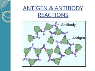
INTRODUCTION
Antigens & antibodiescombine specifically with each other.
This interaction between them is called ‘Antigen-Antibody
reaction’.
- Abbreviated as Ag – Ab reaction.
- They form the basis for humoral/antibody mediated
immunity.
- They are used for detection of disease , Onset of
disease(from concentration of antibodies),causing agents &
some non-specific Ag’s like enzymes,immune reaction,
treatment & prevention.
3.
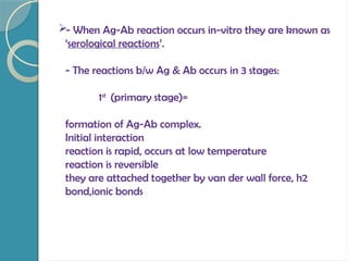
- When Ag-Abreaction occurs in-vitro they are known as
‘serological reactions’.
- The reactions b/w Ag & Ab occurs in 3 stages:
1st
(primary stage)=
formation of Ag-Ab complex.
Initial interaction
reaction is rapid, occurs at low temperature
reaction is reversible
they are attached together by van der wall force, h2
bond,ionic bonds
4.
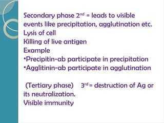
Secondary phase 2nd
=leads to visible
events like precipitation, agglutination etc.
Lysis of cell
Killing of live antigen
Example
•Precipitin-ab participate in precipitation
•Agglitinin-ab participate in agglutination
(Tertiary phase) 3rd
= destruction of Ag or
its neutralization.
Visible immunity
5.
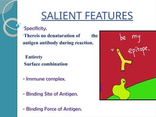
SALIENT FEATURES
-Specificity.
-Thereis nodenaturation of the
antigen antibody during reaction.
Entirety
Surface combination
- Immune complex.
- Binding Site of Antigen.
- Binding Force of Antigen.
6.
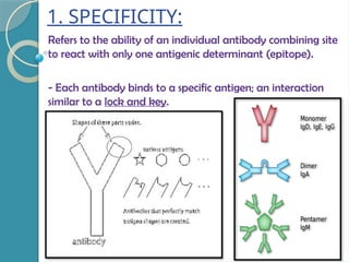
1. SPECIFICITY:
Refers tothe ability of an individual antibody combining site
to react with only one antigenic determinant (epitope).
- Each antibody binds to a specific antigen; an interaction
similar to a lock and key.
7.
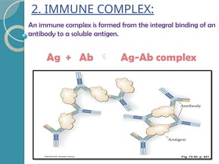
2. IMMUNE COMPLEX:
Animmune complex is formed from the integral binding of an
antibody to a soluble antigen.
Ag + Ab Ag-Ab complex
8.
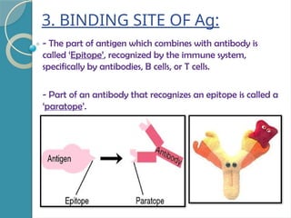
3. BINDING SITEOF Ag:
- The part of antigen which combines with antibody is
called ‘Epitope’, recognized by the immune system,
specifically by antibodies, B cells, or T cells.
- Part of an antibody that recognizes an epitope is called a
‘paratope’.
9.
4. BINDING FORCEOF Ag:
- The binding b/w Ag & Ab in Ag – Ab reaction is due to
three factors namely:
* Closeness b/w Ag & Ab -> more close = good
strength of binding.
* Non – covalent bonds or Intermolecular forces ->
hydrogen bonds, vander walls forces, hydrophobic bonds.
* Affinity of antibody -> strength of reaction b/w a
single epitope & single paratope.
* VALENCY- antibodies are bivalent in nature, both are
multivalent
10.
•ANTIBODY AVIDITY:
it isthe overall strength of ag – ab
complex,dependes upon
affinity
valency
oerall structure
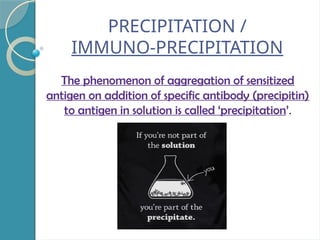
DEFINITION
CHEMICAL INTERACTIONB/W
MULTIVALENT ANTIGEN & BIVALENT
ANTIBODY, LEADSTO FORM THE
INSOLUABLE PRECIPITATES UNDER
FAVORABLE CONDITION INTHE
PRESENCE OF ELECTROLYTES
16.
- Precipitation occursin two media:
* Liquid
* Gel
1. Precipitation in Liquids:
- Place constant amount of Ab in a series of tubes.
- Add increased amount of antigen.
- Antigen – Antibody reacts together resulting in
precipitation.
- Plotting the amount of precipitate against increasing
antigen conc. yields a ‘precipitin curve’.
17.
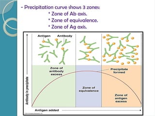
- Precipitation curveshows 3 zones:
* Zone of Ab axis.
* Zone of equivalence.
* Zone of Ag axis.
18.
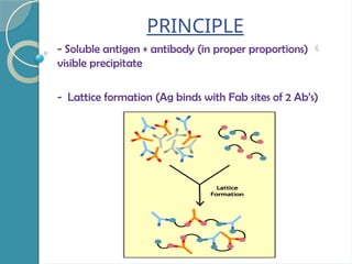
PRINCIPLE
- Soluble antigen+ antibody (in proper proportions)
visible precipitate
- Lattice formation (Ag binds with Fab sites of 2 Ab’s)
19.
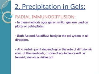
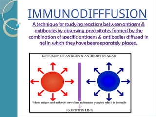
2. Precipitation inGels:
RADIAL IMMUNODIFFUSION:
- In these methods agar gel or similar gels are used on
plates or petri-plates.
- Both Ag and Ab diffuse freely in the gel system in all
directions.
- At a certain point depending on the rate of diffusion &
conc. of the reactants, a zone of equivalence will be
formed, seen as a visible ppt.
20.
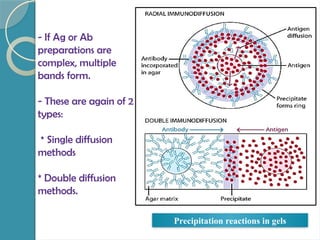
- If Agor Ab
preparations are
complex, multiple
bands form.
- These are again of 2
types:
* Single diffusion
methods
* Double diffusion
methods.
Precipitation reactions in gels
21.
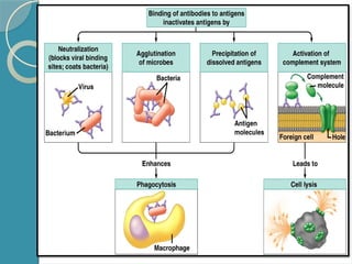
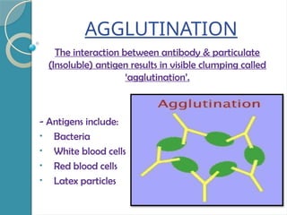
AGGLUTINATION
The interaction betweenantibody & particulate
(Insoluble) antigen results in visible clumping called
‘agglutination’.
- Antigens include:
• Bacteria
• White blood cells
• Red blood cells
• Latex particles
22.
- The Abof the serum causes the cellular Ag’s to
form clumps and these are called ‘Agglutinins’.
- The particulate antigens that are aggregated
are termed ‘Agglutinogens’.
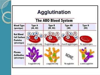
- Agglutination can be performed in a tube or
on a glass slide e.g. ABO blood grouping.
- Ab is divalent and cross links the multivalent
antigen to form clumps.
23.
TUBE AGGLUTINATION:
- Serumcontaining Ab is diluted serially with saline in small
test tubes, a constant volume of Ag suspension is added.
- Control tube is kept which has no antiserum.
- The tubes are incubated until visible agglutination is
observed.
- The tube showing highest agglutination is referred to as
the ‘titre’.
APPLICATION -> Widal test is used for the estimation
of typhoid fever
24.
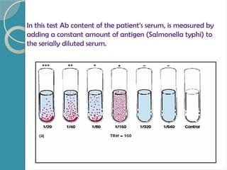
In this testAb content of the patient’s serum, is measured by
adding a constant amount of antigen (Salmonella typhi) to
the serially diluted serum.
25.
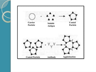
PASSIVE AGGLUTINATION:
- Agis coated on the surface of a carrier particle.
- This helps to convert a precipitation reaction to an
agglutination reaction making the reaction more
sensitive.
- The carrier particles used can be RBC, latex particles or
bentonite.
- When patients serum is mixed with these, it leads to
agglutination.
APPLICATION -> diagnosis of Rheumatoid arthritis.
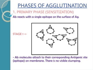
PHASES OF AGGLUTINATION
1.PRIMARY PHASE (SENSITIZATION)
Ab reacts with a single epitope on the surface of Ag.
STAGE 1 ->
- Ab molecules attach to their corresponding Antigenic site
(epitope) on membrane. There is no visible clumping.
28.
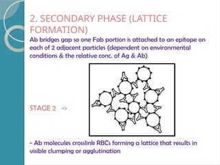
2. SECONDARY PHASE(LATTICE
FORMATION)
Ab bridges gap so one Fab portion is attached to an epitope on
each of 2 adjacent particles (dependent on environmental
conditions & the relative conc. of Ag & Ab)
STAGE 2 ->
- Ab molecules crosslink RBCs forming a lattice that results in
visible clumping or agglutination
29.
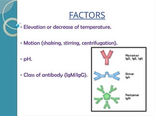
FACTORS
- Elevation ordecrease of temperature.
- Motion (shaking, stirring, centrifugation).
- pH.
- Class of antibody (IgM/IgG).
30.
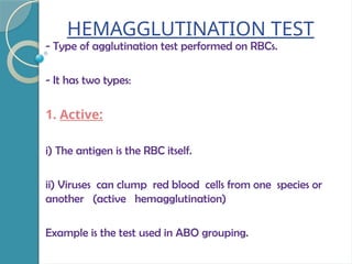
HEMAGGLUTINATION TEST
- Typeof agglutination test performed on RBCs.
- It has two types:
1. Active:
i) The antigen is the RBC itself.
ii) Viruses can clump red blood cells from one species or
another (active hemagglutination)
Example is the test used in ABO grouping.
32.
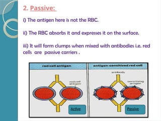
2. Passive:
i) Theantigen here is not the RBC.
ii) The RBC absorbs it and expresses it on the surface.
iii) It will form clumps when mixed with antibodies i.e. red
cells are passive carriers .
Active Passive
33.
APPLICATIONS:
- Blood typing.
-Bacterial infections
LIMITATIONS:
- Time consuming (1 day)
- Cannot distinguish IgG from IgM.
ADVANTAGES:
a) Precipitin bandis visible which can be stained for
preservation.
b) It can be used to detect identity, cross-reaction &
non-identity b/w antigens in a mixture.
TYPES OF IMMUNODIFFUSION
They are classified on the basis of:
a) Number of reactants diffusing
b) Direction of diffusion
36.
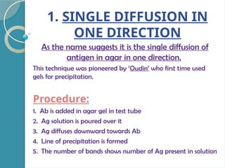
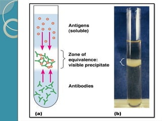
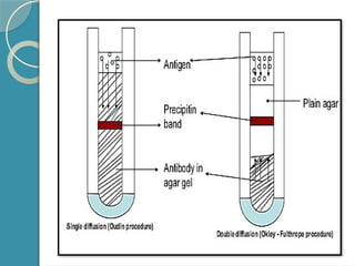
1. SINGLE DIFFUSIONIN
ONE DIRECTION
As the name suggests it is the single diffusion of
antigen in agar in one direction.
This technique was pioneered by ‘Oudin’ who first time used
gels for precipitation.
Procedure:
1. Ab is added in agar gel in test tube
2. Ag solution is poured over it
3. Ag diffuses downward towards Ab
4. Line of precipitation is formed
5. The number of bands shows number of Ag present in solution
38.
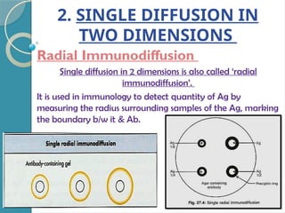
2. SINGLE DIFFUSIONIN
TWO DIMENSIONS
Radial Immunodiffusion
Single diffusion in 2 dimensions is also called ‘radial
immunodiffusion’.
It is used in immunology to detect quantity of Ag by
measuring the radius surrounding samples of the Ag, marking
the boundary b/w it & Ab.
39.
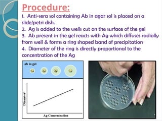
Procedure:
1. Anti-sera solcontaining Ab in agar sol is placed on a
slide/petri dish.
2. Ag is added to the wells cut on the surface of the gel
3. Ab present in the gel reacts with Ag which diffuses radially
from well & forms a ring shaped band of precipitation
4. Diameter of the ring is directly proportional to the
concentration of the Ag
40.
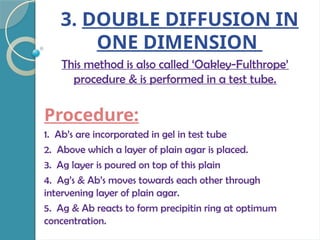
3. DOUBLE DIFFUSIONIN
ONE DIMENSION
This method is also called ‘Oakley-Fulthrope’
procedure & is performed in a test tube.
Procedure:
1. Ab’s are incorporated in gel in test tube
2. Above which a layer of plain agar is placed.
3. Ag layer is poured on top of this plain
4. Ag’s & Ab’s moves towards each other through
intervening layer of plain agar.
5. Ag & Ab reacts to form precipitin ring at optimum
concentration.
42.
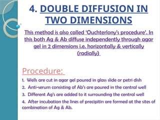
4. DOUBLE DIFFUSIONIN
TWO DIMENSIONS
This method is also called ‘Ouchterlony’s procedure’. In
this both Ag & Ab diffuse independently through agar
gel in 2 dimensions i.e. horizontally & vertically
(radially)
Procedure:
1. Wells are cut in agar gel poured in glass slide or petri dish
2. Anti-serum consisting of Ab’s are poured in the central well
3. Different Ag’s are added to it surrounding the central well
4. After incubation the lines of precipitin are formed at the sites of
combination of Ag & Ab.
43.
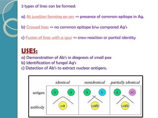
3 types oflines can be formed:
a) At junction forming an arc => presence of common epitope in Ag.
b) Crossed lines => no common epitope b/w compared Ag’s
c) Fusion of lines with a spur => cross-reaction or partial identity
USES:
a) Demonstration of Ab’s in diagnosis of small pox
b) Identification of fungal Ag’s
c) Detection of Ab’s to extract nuclear antigens.
44.
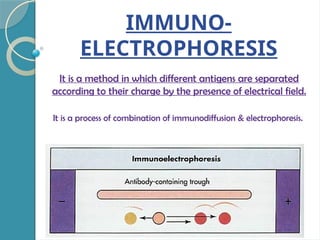
IMMUNO-
ELECTROPHORESIS
It is amethod in which different antigens are separated
according to their charge by the presence of electrical field.
It is a process of combination of immunodiffusion & electrophoresis.
45.
Procedure:
1. A dropof Ag is placed into a well in agar on glass slide.
2. Electric current is passed through agar
3. Ag move in the electric field according to their size & charge.
4. A trough is cut into agar & Ab is poured to it & diffusion is
allowed to occur.
5. As the Ag & Ab diffuse they form series of lines
ADVANTAGE -> Number of Ag’s can be identified in serum.
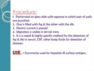
Procedure:
1. Performed onglass slide with agarose in which pair of wells
are punched.
2. One is filled with Ag & the other with the Ab
3. Electric current is passed
4. Migration is visible in 30-60 mins.
5. It is a rapid & highly specific method for the detection of
Ag & Ab in serum, CSF, other body fluids for detection of
diseases.
USE -> Commonly used for hepatitis-B surface antigen.
48.
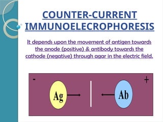
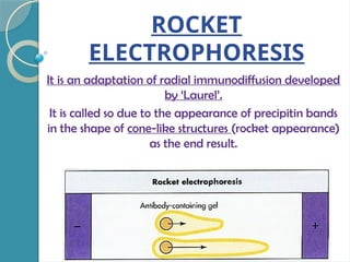
ROCKET
ELECTROPHORESIS
It is anadaptation of radial immunodiffusion developed
by ‘Laurel’.
It is called so due to the appearance of precipitin bands
in the shape of cone-like structures (rocket appearance)
as the end result.
49.
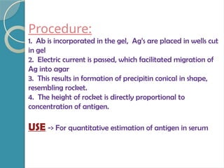
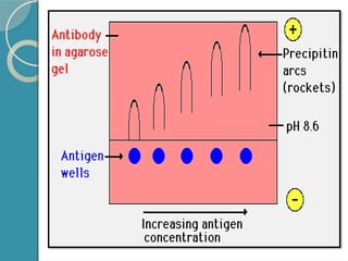
Procedure:
1. Ab isincorporated in the gel, Ag’s are placed in wells cut
in gel
2. Electric current is passed, which facilitated migration of
Ag into agar
3. This results in formation of precipitin conical in shape,
resembling rocket.
4. The height of rocket is directly proportional to
concentration of antigen.
USE -> For quantitative estimation of antigen in serum
51.
CONCLUSION
Thus we herebyconclude with the fact that
antigen-antibody reactions are very important
for serological testing of human beings, as they
give you a complete picture of all the immune
responses occurring the body & helps
determining the immunological disorders by the
antigen (either self or non-self).