Downloaded 210 times
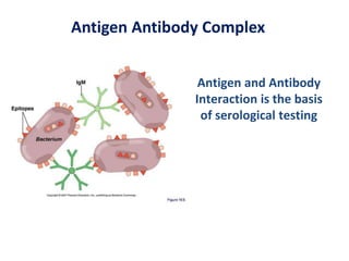
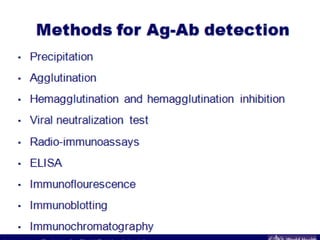
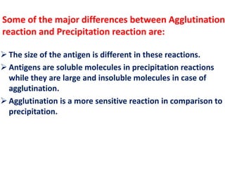

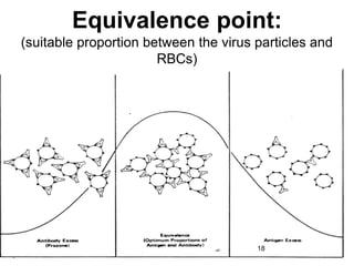
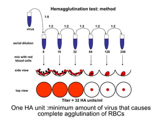
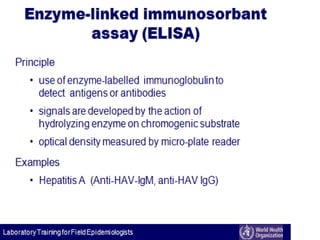
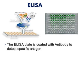
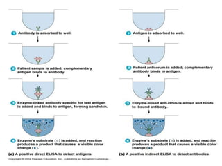
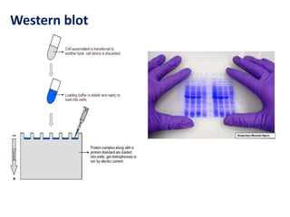
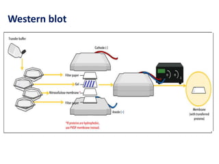
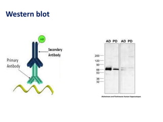
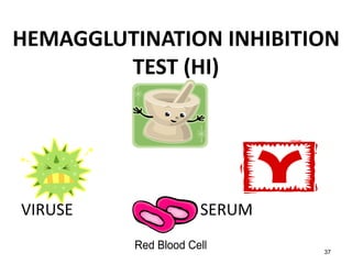
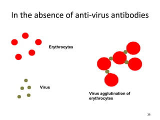
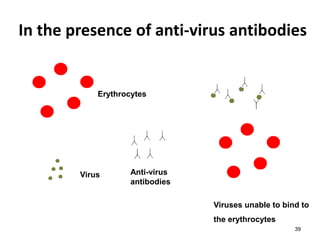
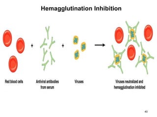
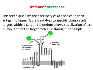
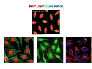


Antigen and antibody interaction is the basis of serological testing. There are several types of serological tests that detect this interaction, including precipitation, agglutination, hemagglutination, enzyme-linked immunosorbent assay (ELISA), Western blot, hemagglutination inhibition, and immunofluorescence. These tests exploit the formation of antigen-antibody complexes to diagnose diseases, identify pathogens, and detect proteins.


![Immunochemical techniques]](https://cdn.slidesharecdn.com/ss_thumbnails/immunochemicaltechniques1-200402171215-thumbnail.jpg?width=640&height=640&fit=bounds)


