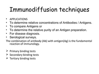
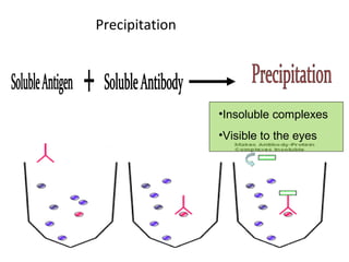
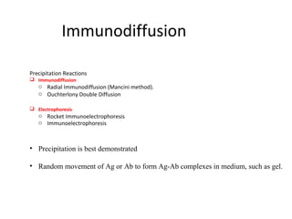
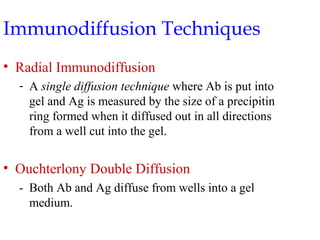
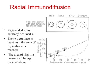
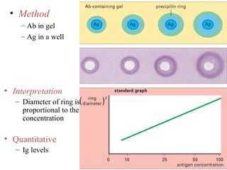
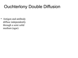
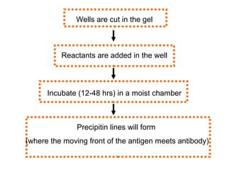
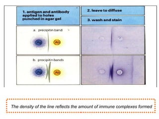
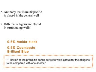
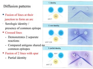
Immunodiffusion techniques
• APPLICATIONS
• To determine relative concentrations of Antibodies / Antigens.
• To compare Antigens or
• To determine the relative purity of an Antigen preparation.
• For disease diagnosis.
• Serological surveys.
The combination of antibody (Ab) with antigen(Ag) is the fundamental
  reaction of immunology.

 Primary binding tests
 Secondary binding tests
 Tertiary binding tests
Principle
• Soluble Ab & soluble Ag interacting in aqueous solution form
  lattice that develops into insoluble visible precipitate.
• Soluble Ag: Toxins, toxoids, proteins, carbohydrates, glycoproteins,
   Liproproteins
• Both qualitatively & quantitatively in solutions & gels.


• Formation of Ag-Ab lattice depends on the valency of both Ag &
  Ab:
  Ab must be bivalent Fab fragments
  -   Ag must be bivalent / polyvalent.
Precipitation


                •Insoluble complexes
                •Visible to the eyes
Precipitation Curve

                • Zone of Equivalence
                  optimum precipitation
                • Prozone
                 excess antibody is present
                • Postzone
                  excess antigen is present

                       Prozone and Postzone
                      phenomena are negative
                             reactions.
Immunodiffusion

Precipitation Reactions
 Immunodiffusion
   o Radial Immunodiffusion (Mancini method).
   o Ouchterlony Double Diffusion

 Electrophoresis
   o Rocket Immunoelectrophoresis
   o Immunoelectrophoresis


• Precipitation is best demonstrated

• Random movement of Ag or Ab to form Ag-Ab complexes in medium, such as gel.
MEDIUM
 Agar - high molecular weight complex polysaccharide
        - from seaweed
 Agarose- purified agar
- Used to help stabilize the diffusion process
  and allow visualization of precipitin bands.


 0.3 – 1.5 % Agar concentration: diffusion of most reactants

 Agarose- more preferred than agar
     Agar has strong negative charge;
     Agarose has almost none (no charge)
     - interactions between gel and reactants are minimized.
Passive Immunodiffusion
•    Passive diffusion method in which a concentration gradient is
     established for an antigen and/or antibody
     - diffusion of reactants to form Ag - Ab reactions without
     electric current to speed up reaction.


Rate of Diffusion
1.   Size of particles
2.   Temperature
3.   Gel viscosity
4.   Amount of hydration
5.   Interactions between matrix and reactants.
Immunodiffusion Techniques
• Radial Immunodiffusion
  - A single diffusion technique where Ab is put into
    gel and Ag is measured by the size of a precipitin
    ring formed when it diffused out in all directions
    from a well cut into the gel.


• Ouchterlony Double Diffusion
  - Both Ab and Ag diffuse from wells into a gel
    medium.
Radial Immunodiffusion


• Ag is added to an
  antibody rich media.
• The two continue to
  react until the zone of
  equivalence is
  reached.
• The area of ring is a
  measure of the Ag
  concentration.
• Method
    – Ab in gel
    – Ag in a well


• Interpretation
   – Diameter of ring is
     proportional to the
     concentration

• Quantitative
   – Ig levels
Ouchterlony Double Diffusion

• Antigen and antibody
  diffuse independently
  through a semi solid
  medium (agar)
Wells are cut in the gel


          Reactants are added in the well


      Incubate (12-48 hrs) in a moist chamber


               Precipitin lines will form
(where the moving front of the antigen meets antibody)
The density of the line reflects the amount of immune complexes formed
Ouchterlony

• Both antigen and
  antibody can diffuse
  independently.
• Antibody that is multispecific
  is placed in the central well

• Different antigens are placed
  in surrounding wells


    0.5% Amido-black
    0.5% Coomassie
    Brilliant Blue

    **Position of the precipitin bands between wells allows for the antigens
    to be compared with one another.
Diffusion patterns

 Fusion  of lines at their
  junction to form an arc
- Serologic identity /

   presence of common epitope
 Crossed lines

   - Demonstrates 2 separate
     reactions
   - Compared antigens shared no
     common epitopes
 Fusion of 2 lines with spur

   - Partial identity
IMMUNOELECTROPHORESIS

• Double-diffusion technique that utilizes electric current to enhance
  results.
• SPEED, Specificity.
• Introduced by Grabar and Williams in 1953.

•   Combine immunodiffusion with electrophoresis
•   Can be used for semiquantitaion of wide range of antigens
•   Qualitative
•   Antigen source: serum.
Electrophoresis Techniques

• Electrophoresis separates molecules according to differences in
  their electrical charge.
   – Rocket Immunoelectrophoresis
   – Countercurrent Immunoelectrophoresis
Principle
2 step process

                                                Trough is cut in
                                                 Trough is cut in
                                                the gel parallel
                                                 the gel parallel
        Antigen from
        Antigen from       electrophoresed
                            electrophoresed      to the line of
                                                  to the line of
           serum
            serum                                 separation
                                                   separation


      ••doublediffusion
       double diffusion                          Antiserum is
                                                  Antiserum is
        occurs at right
         occurs at right
                               Incubate: 18-
                                Incubate: 18-
                                  24 hours       placed in the
                                                  placed in the
         angles to the
          angles to the            24 hours
                                                    trough
                                                     trough
       electrophoresis
        electrophoresis
          separation
           separation
       ••Precipitinlines
        Precipitin lines
     develop where Ag-
      develop where Ag-           Lines or arcs shape, intensity and
                                   Lines or arcs shape, intensity and
       Ab combination
        Ab combination         location: compared with normal serum
                                location: compared with normal serum
         takes place.
          takes place.                          control
                                                 control
Immunoeletrophoresis
• Method
  – Ags are separated by electrophoresis
  – Ab is placed in trough cut in the agar


 • Interpretation
     – Precipitin arc represent individual antigens
Uses
• Immunodeficiencies can be detected by this procedure, if no
  precipitin band is formed for a particular Ig.
• Overproduction of serum proteins.
• Deficiencies in complement can also be detected.
• Identification of monoclonal protein
   – Free kappa and lambda light chains
• May be used to identify urine proteins.
• Testing normal & abnormal proteins in serum/urine.
• Purity of Ag.
Countercurrent electrophoresis
Method
– Ag and Ab migrate toward each other by electrophoresis
– Used only when Ag and Ab have opposite charges

         -                                          +
                  Ag                        Ab


• Qualitative- Rapid.
• Detection of Antinuclear- Ribonuclear protein.
Rocket Immunoelectrophoresis
( LAURELL TECHNIQUE )
• One dimension electroimmunodiffusion
   - Adaptation of RID

• Electrophoresis is used to facilitate migration of antigen in agar
  - Ag diffuses out of the well: precipitation begins
  - Change in Ag concentration: dissolution and reformation of
    precipitate.

  End result: precipitin line = conical-shaped, resembles a racket
• . The height of the racket is measured
  - Height of the racket is directly proportional to the amount of
     antigen present
• Racket electrophoresis is more rapid than RID.
Rocket electrophoresis
Uses of Racket Immunoelectrophoresis

• Quantitate Igs
  - (using a buffer = pH 8.6)

• Assay of proteins
  - When concentration is too
    low to be detected by
    nephelometry and too high
    for RID
  - Ex: alpha-fetoprotein, Igs
    in urine and spinal fluid,
    Complement components
    in body fluid.
Applications

• Screening tool for differentiation of more than 30 serum proteins.
  - Major classes of immunoglobulins
• Used for the detection of:
  - Myelomas
  - Malignant lypmhomas
  - Other lymphoproliferative disorder.

Immunodiffusion principles and application

  • 1.
    Immunodiffusion techniques • APPLICATIONS •To determine relative concentrations of Antibodies / Antigens. • To compare Antigens or • To determine the relative purity of an Antigen preparation. • For disease diagnosis. • Serological surveys. The combination of antibody (Ab) with antigen(Ag) is the fundamental reaction of immunology.  Primary binding tests  Secondary binding tests  Tertiary binding tests
  • 2.
    Principle • Soluble Ab& soluble Ag interacting in aqueous solution form lattice that develops into insoluble visible precipitate. • Soluble Ag: Toxins, toxoids, proteins, carbohydrates, glycoproteins, Liproproteins • Both qualitatively & quantitatively in solutions & gels. • Formation of Ag-Ab lattice depends on the valency of both Ag & Ab: Ab must be bivalent Fab fragments - Ag must be bivalent / polyvalent.
  • 3.
    Precipitation •Insoluble complexes •Visible to the eyes
  • 4.
    Precipitation Curve • Zone of Equivalence optimum precipitation • Prozone excess antibody is present • Postzone excess antigen is present Prozone and Postzone phenomena are negative reactions.
  • 5.
    Immunodiffusion Precipitation Reactions  Immunodiffusion o Radial Immunodiffusion (Mancini method). o Ouchterlony Double Diffusion  Electrophoresis o Rocket Immunoelectrophoresis o Immunoelectrophoresis • Precipitation is best demonstrated • Random movement of Ag or Ab to form Ag-Ab complexes in medium, such as gel.
  • 6.
    MEDIUM  Agar -high molecular weight complex polysaccharide - from seaweed  Agarose- purified agar - Used to help stabilize the diffusion process and allow visualization of precipitin bands.  0.3 – 1.5 % Agar concentration: diffusion of most reactants  Agarose- more preferred than agar Agar has strong negative charge; Agarose has almost none (no charge) - interactions between gel and reactants are minimized.
  • 7.
    Passive Immunodiffusion • Passive diffusion method in which a concentration gradient is established for an antigen and/or antibody - diffusion of reactants to form Ag - Ab reactions without electric current to speed up reaction. Rate of Diffusion 1. Size of particles 2. Temperature 3. Gel viscosity 4. Amount of hydration 5. Interactions between matrix and reactants.
  • 8.
    Immunodiffusion Techniques • RadialImmunodiffusion - A single diffusion technique where Ab is put into gel and Ag is measured by the size of a precipitin ring formed when it diffused out in all directions from a well cut into the gel. • Ouchterlony Double Diffusion - Both Ab and Ag diffuse from wells into a gel medium.
  • 9.
    Radial Immunodiffusion • Agis added to an antibody rich media. • The two continue to react until the zone of equivalence is reached. • The area of ring is a measure of the Ag concentration.
  • 10.
    • Method – Ab in gel – Ag in a well • Interpretation – Diameter of ring is proportional to the concentration • Quantitative – Ig levels
  • 12.
    Ouchterlony Double Diffusion •Antigen and antibody diffuse independently through a semi solid medium (agar)
  • 13.
    Wells are cutin the gel Reactants are added in the well Incubate (12-48 hrs) in a moist chamber Precipitin lines will form (where the moving front of the antigen meets antibody)
  • 14.
    The density ofthe line reflects the amount of immune complexes formed
  • 15.
    Ouchterlony • Both antigenand antibody can diffuse independently.
  • 16.
    • Antibody thatis multispecific is placed in the central well • Different antigens are placed in surrounding wells 0.5% Amido-black 0.5% Coomassie Brilliant Blue **Position of the precipitin bands between wells allows for the antigens to be compared with one another.
  • 17.
    Diffusion patterns  Fusion of lines at their junction to form an arc - Serologic identity / presence of common epitope  Crossed lines - Demonstrates 2 separate reactions - Compared antigens shared no common epitopes  Fusion of 2 lines with spur - Partial identity
  • 18.
    IMMUNOELECTROPHORESIS • Double-diffusion techniquethat utilizes electric current to enhance results. • SPEED, Specificity. • Introduced by Grabar and Williams in 1953. • Combine immunodiffusion with electrophoresis • Can be used for semiquantitaion of wide range of antigens • Qualitative • Antigen source: serum.
  • 19.
    Electrophoresis Techniques • Electrophoresisseparates molecules according to differences in their electrical charge. – Rocket Immunoelectrophoresis – Countercurrent Immunoelectrophoresis
  • 20.
    Principle 2 step process Trough is cut in Trough is cut in the gel parallel the gel parallel Antigen from Antigen from electrophoresed electrophoresed to the line of to the line of serum serum separation separation ••doublediffusion double diffusion Antiserum is Antiserum is occurs at right occurs at right Incubate: 18- Incubate: 18- 24 hours placed in the placed in the angles to the angles to the 24 hours trough trough electrophoresis electrophoresis separation separation ••Precipitinlines Precipitin lines develop where Ag- develop where Ag- Lines or arcs shape, intensity and Lines or arcs shape, intensity and Ab combination Ab combination location: compared with normal serum location: compared with normal serum takes place. takes place. control control
  • 21.
  • 23.
    • Method – Ags are separated by electrophoresis – Ab is placed in trough cut in the agar • Interpretation – Precipitin arc represent individual antigens
  • 24.
    Uses • Immunodeficiencies canbe detected by this procedure, if no precipitin band is formed for a particular Ig. • Overproduction of serum proteins. • Deficiencies in complement can also be detected. • Identification of monoclonal protein – Free kappa and lambda light chains • May be used to identify urine proteins. • Testing normal & abnormal proteins in serum/urine. • Purity of Ag.
  • 25.
    Countercurrent electrophoresis Method – Agand Ab migrate toward each other by electrophoresis – Used only when Ag and Ab have opposite charges - + Ag Ab • Qualitative- Rapid. • Detection of Antinuclear- Ribonuclear protein.
  • 26.
    Rocket Immunoelectrophoresis ( LAURELLTECHNIQUE ) • One dimension electroimmunodiffusion - Adaptation of RID • Electrophoresis is used to facilitate migration of antigen in agar - Ag diffuses out of the well: precipitation begins - Change in Ag concentration: dissolution and reformation of precipitate. End result: precipitin line = conical-shaped, resembles a racket • . The height of the racket is measured - Height of the racket is directly proportional to the amount of antigen present • Racket electrophoresis is more rapid than RID.
  • 27.
  • 28.
    Uses of RacketImmunoelectrophoresis • Quantitate Igs - (using a buffer = pH 8.6) • Assay of proteins - When concentration is too low to be detected by nephelometry and too high for RID - Ex: alpha-fetoprotein, Igs in urine and spinal fluid, Complement components in body fluid.
  • 29.
    Applications • Screening toolfor differentiation of more than 30 serum proteins. - Major classes of immunoglobulins • Used for the detection of: - Myelomas - Malignant lypmhomas - Other lymphoproliferative disorder.

Editor's Notes

  • #6 Performed in an agarose gel supported by a glass slide or polyester film
  • #10 Area of ring: measure of Ag concentration: compared to a standard curve using Ags of known concentration
  • #19 Why would we want to combine immunodiffusion with electrophoresis? SPEED, Specificity Two step process
  • #27 Modification of IEP technique - Antigen ( proteins ) undergo electrophoresis in a supporting agarose gel with specific antibody previously mixed into the gel. As antigen moves thru the gel , antigen-antibody complexes form creating visible precipitation lines in the shape of long arches or “rockets”. The length of these “rockets” is proportional to the concentration of antigen.