SlideShare a Scribd company logo
1
◘ Some Clinical LaboratoryMeasurement of Immune Functions.
 Many clinical routine test
procedures are antibody-based.
 These tests rely upon the
ability of antibodies to
aggregate (agglutination)
particulate antigens (e.g., blood
typing) or to precipitate soluble
antigens, e.g.,
1. Radial immunodiffusion,
Ouchterlony or
2. Double diffusion, immunoelectrophoresis).
 Other assays rely upon chemically modified
antibodies to quantitate antigens (e.g.,
1.radioimmunoassay and 2.immunosorbent
assays) with exquisite specificity and
sensitivity.
 Additional assays (e.g., 1.immunofluorescence and 2.flow cytometry)
utilize fluorochrome-labeled antibodies to assess antigen expression
both within and on the surface of cells.
 Immune function may be assessed in the laboratory (e.g., 1.complement
fixation, 2.proliferation, and 3.cytotoxic T-lymphocyte assay) or in a
clinical setting (assessment of hypersensitivity).
I. Qualitative Detection of Antigens and Antibodies
 Many clinical tests are based upon the specificity of antibodies for
antigen and their ability to recognize epitopes (very small portions of an
antigen).
 Antibody-based assays are epitope-detecting tools, and most are based
upon the quantitative precipitin curve (Fig. 1and Fig. 2).
2
A. Particulate Antigens.
 Particulate antigens such as erythrocytes, bacteria, or even antigen-
coated latex beads are normally evenly dispersed in suspension.
 Cross-linking of antigen-bearing particles by antibodies causes
clumping of the particles, also known as agglutination (Fig.2).
 If the particulate antigen is an erythrocyte (hemagglutination), whether
IgM antibodies efficiently cross-link the particles (direct
agglutination), or whether an anti-immunoglobulin (indirect or passive
agglutination) is used to cross-link antigen-bound antibodies.
1-Direct agglutination:
 This reaction usually involves IgM antibodies that cross-link epitopes
on cells or particles.
 As IgM is the largest immunoglobulin, it has 10 epitope-binding sites
(valence).
 It very efficient at cross-linking epitopes on adjacent particles (Figs.1&
2).
 Other Igs, because of their smaller size (lesser valence) are less efficient
in direct agglutination.
 Too much antibody inhibits agglutination (equivalent to the zone of
antibody excess; prozone).
 To avoid the prozone effect, twofold or serial dilutions of antibody are
prepared; each dilution is half as concentrated as the preceding one
(Fig.1).
 Titers are relative measures of antibody activity and are often expressed
as the reciprocal of the dilution (e.g., 1:16, 1:32, 1:64).
Fig.2 Direct agglutination reaction (ABO blood typing) occurs in 15-30 sec.
3
Precipitin curve Preparation of serum serial dilution
Figure (1) Quantitative precipitin curve.
- Antibody preparation: serially diluted antibody-containing serum is prepared
(expressed as 1:2. 1:4, 1:8, 1:16, .. etc.).
- Antigen-antibody reaction: antigen (containing multiple epitopes) in equal
concentrations is added to the antibody dilutions resulting in differing degrees
of antigen- antibody complex formation.
- In the equivalence zone, both antigen and antibody are at concentrations that
result in maximal lattice formation, causing precipitation of antigen-antibody
complexes.
- In the zone of antibody excess (prozone)
antibody molecules are more than the
available epitopes, and precipitating
complexes are not formed.
- In the zone of antigen excess, available
epitopes are more than the antibody-binding
sites, and precipitating complexes are not
formed.
- The point at which cross-linking of the
particulate antigen is no longer observed is
called the titer.
4
2-Indirect or passive agglutination:
 This technique is often used to detect non-IgM antibodies or
antibodies in concentrations too low to be detected by direct
agglutination.
 Human antibodies may not directly agglutinate antigen-bearing particles
(e.g., bacteria, erythrocytes, latex particles) or show agglutination of
very low titer.
 The sensitivity of the agglutination test may be enhanced by the
addition of an anti-immunoglobulin reagent (e.g., rabbit anti-human
immunoglobulin) in the so-called indirect or passive agglutination
technique.
 Addition of these second-step antibodies is used to increase binding
over a greater span and to increase valence by virtue of their ability to
bind to the primary antibody (Fig. 3).
Fig.3 Indirect agglutination
5
3-Coombs' test
 Antibodies against self blood group antigens occur in some
autoimmune diseases (hemolytic anemia).
 Afflicted individuals produce antibodies to their own erythrocytes but in
isotypes or quantities that do not directly
agglutinate their erythrocytes.
a- In the direct Coombs' test, is used to test for
autoimmune hemolytic anemia autoantibodies are
detected by the addition of antihuman
immunoglobulin (secondary antibody or Coombs
reagent).
 If this produces agglutination of RBCs, the direct Coombs test is
positive, a visual indication that antibodies (and/or complement
proteins) are bound to the surface of red blood cells.
b- In the indirect Coombs' assay, is used in
prenatal testing of pregnant women, and in testing
blood prior to a blood transfusion.
 It detects antibodies against RBCs that are
present unbound in the patient's serum.
 Serum sample taken from the patient.
 Then, the serum is incubated with RBCs of known antigenicity from other
patient blood samples.
 Antihuman immunoglobulin is then added, if agglutination occurs, the indirect
Coombs test is positive
Figure (4) the indirect Coombs' assay
6
B. Soluble Antigens
 Often, epitopes present on soluble molecules will precipitate from
solution upon reaction with the "right" amount of antibody.
 Several simple modifications to the quan-titative precipitin reaction
(Fig.1; Fig. 2) allow visualization of immune precipitates in agar, a
semisolid growth medium.
♣ Radial immune-diffusion:
 Also called the Mancini technique, this test is
based upon the diffusion of soluble antigen
within an agar gel that contains a uniform
concentration of antibody.
 Antibody-containing molten agar is poured onto a
glass slide or plastic dish.
 When the agar cools and solidifies, wells are cut
into the gel matrix, and soluble antigen is placed into the well (Fig. 5).
 Antigen diffuses radially from the well, forming a precipitin ring at
equivalence.
 The diameter of the ring is directly proportional to the amount of
antigen loaded into the well.
 The concentration of antigen in a test sample can be accurately
determined by comparing its diameter with a standard calibration curve.
 This technique allows for the rapid and precise determination of the
quantity of antigen loaded into the well.
7
2-Double-diffusion (Ouchterlony technique):
 This test is based upon the diffusion of both antigen (loaded in one
well) and antibody (loaded in another well) through an agar gel.
 A precipitin line forms at equivalence (Fig. 6).
 Solubility, molecular size of the antibody, and detection of epitopes on
antigens of different molecular size all influence precipitin formation
such that multiple precipitin lines often develop.
♣ An advantage of this technique:
• Is that several antigens or antibodies can be compared to determine
identity, partial identity, and nonidentity of antigens and/or antibodies.
• In contrast to radial immunodiffusion, this is a qualitative technique.
• Wells are cut into a solidified agar gel.
• Soluble antigen(s) are loaded into one or more wells, and antibodies are
loaded into another well(s), from which they diffuse through the gel.
• A precipitin band is formed at the equivalence zone.
Figure (6): double –diffusion (Ouchterlony) technique.
8
♦ Immunoelectrophoresis (IEP):
 This technique is a modification of double
diffusion.
 Antigens are loaded into a well within the agar,
an electrical current is applied, and antigens
migrate according to both their size and their electrical charge (Fig. 7).
♣ The electrical current
 Is removed, a trough is cut into the agar, and antiserum is placed in the
trough.
 IEP is qualitative.
II. Quantitative Detection by Antibodies.
 The specificity of antibody molecules
makes them ideal probes for detection of a
wide variety of epitopes.
 Antibodies or the antigens they detect (sometimes referred to as ligands)
may be labeled with radioactive molecules, fluorescent molecules,
enzymes, or heavy metals.
 Antibody or antigen binding is then readily detectable and quantifiable.
a. Radioimmunoassay [RIA]
 RIA has been widely used in clinical diagnostic laboratories.
 Antigens of primary antibodies may be directly labeled with a
radionuclide and form the basis for direct RIA.
 Alternatively, anti-immunoglobulin antibody (secondary antibody) is
radio-labeled and used in the indirect RIA.
 RIA is sensitive but presents problems owing to the potential exposure
of laboratory personnel to radioactivity and radioactive waste disposal.
9
1. Direct RIA:
 This technique utilizes radiolabeled antibody or its ligand (antigen).
 Antibody is incubated with ligand, and unbound reactants are removed
(phase separation) from the (quantitative precipitin reaction), system
(Fig.7).
 Phase separation may utilize precipitation of bound reactants particulate
antigens (such as bacteria that may be separated by centrifugation), the
immobilization of the nonradioactive reactant onto a solid matrix (such
as plastic), and so on.
2. Indirect RIA:
 This technique uses radiolabeled secondary antibody (anti-
immunoglobulin) to detect the binding of a primary antibody.
 As with direct RIA, a phase separation method must be employed to
remove unbound radiolabeled secondary antibody.
10
b. Enzyme-linked immunosorbent assay [ELISA]
 Enzyme-linked immunosorbent assay [ELISA, also called enzyme
immunoassay (EIA)] has replaced RIA in a number of tests.
 ELISA offers the advantage of safety and speed.
 Because there is no radioactive decay, the reagents that are used are
relatively stable.
ELISAs are both specific and quantitative.
 Its sensitivity is often equal to or greater than that of RIA or fluorescent
immunosorbent (FIA) assay, because an enzyme-labeled reactant is
used to turn a chromogenic substrate from colorless to a color (Fig. 8).
 Color change of the substrate indicates that an enzyme-labeled reactant
has bound.
 Increasing substrate incubation time allows low-concentration enzyme
to convert more substrate to enhance test sensitivity (within limits).
Figure (8): Enzyme-linked immunosorbent assay (ELISA)
11
C. Fluorescent immunosorbent assay
 Fluorescent immunosorbent assay (FIA) relies upon antibodies or their
ligands labeled with a variety of fluorescent dyes such as fluorescein
isothiocyanate (FITC) or phycoerythrin (PE).
 The FIA design is similar to the ELISA.
1. The assay is performed in, 96-well polystyrene plates.
2. Soluble antigen is added and noncovalently binds to the plastic.
3. Unbound antigen is washed from the well.
4. Unlabeled primary antibodies (often sera to be tested) are added to the
well and allowed to bind.
5. Unbound primary antibodies are washed from the well.
6. FITC-labeled anti-immunoglobulin antibodies are added to the well and
allowed to bind.
7. Unbound labeled antibodies are
washed from the well.
8. Fluorescence indicates the
presence of epitopes.
12
III. EPITOPE detection IN and ON cells.
 Epitopes expressed both within and on the surface of cells may be
detected by using radio-, enzyme-, or fluorochrome-labeled antibodies
(direct or indirect).
 The outline of two techniques that have extensive application in a
clinical setting: immunofluorescence and flow cytometry.
A. Immunofluorescence (IF)
 (IF) utilizes fluorescent dyes (e.g., fluorescein isothiocyanate FITC)
that are covalently coupled to antibody.
 A thin, frozen section of tissue is prepared and mounted on a glass slide.
The frozen section is then bathed in a solution containing FITC-labeled
antibody (A direct IF) or a solution containing a primary antibody and is
then washed.
 An FITC-labeled anti-immunoglobulin is added (B indirect IF).
 The presence of epitopes is visualized with a fluorescent microscope.
13
B. Monoclonal antibodies (mAb)
 Antibody responses normally derive from multiple B cells or plasma
cells; their antibodies often differ in epitopes that are recognized,
affinity, and isotype.
♣ Antibody responses that arise from multiple cells are termed
“polyclonal antibody” responses.
 This antibody diversity is very important in combating microbial
infection.
 Although polyclonal antibodies can be used in the clinical laboratory, in
1975, scientists fused antibody-secreting plasma cells with myeloma
(myeloid-origin tumor cells).
 The resulting immortalized cells, or hybridomas, secreted antibodies
of single specificity and isotype and were termed monoclonal
antibodies because of their origin from a single antibody-producing
cell.
 Monoclonal antibodies (mAb) are antibodies that are identical because
they were produced by one type of immune cell, all clones of a single
parent cell.
 Vast quantities of monoclonal antibodies can be produced.
 Because monoclonal antibodies produced by any given hybridoma are
unique, they can be used together with fluorescent dyes or other
markers to distinguish individual epitopes on an antigen or cell.
14
IV. ASSESSMENT OF IMMUNE FUNCTION
 The functional capacity of phagocytic cells can be assessed by their
ability in ingest antibody- or opsonin-coated particles.
 Stimulating lymphocytes to increase in number or proliferate in
response to a specific antigen or to a substance that causes polyclonal
mitogenesis (a mitogen) is often used to assess immune function.
 Phagocyte function can be assessed by incubating phagocytic cells
with coated particles (e.g., latex beads or antibody-bound cells) or with
bacteria for 30 to 120 minutes.
 Particle inclusion within the cell is assessed by microscopy.
 Enzymatic activity of phagocytes can be assessed by measuring the
levels of individual degradative or oxidative enzymes (e.g., NADPH
oxidase) produced by these cells.
Fig: phagocytic function
15
B. Proliferation
 Peripheral blood mononuclear cells (lymphocytes, monocytes, and
dendritic cells) are isolated and placed in tissue culture for 48 to 72
hours.
 A specific stimulator (antigen) to which the individual may have been
previously exposed is added to the culture.
 Alternatively, a nonspecific stimulant (mitogen) is added to assess the
ability of a particular subpopulation of leukocytes to respond.
 A radionuclide (such as 3H-thymidine) is added for the final 18 to 24
hours of cultures
 Incorporation of 3H-thymidine into nascent DNA is taken as a measure
of proliferative ability.
V. ASSESSMENT OF HYPERSENSITIVITY
 Hypersensitivity which is an immune-mediated damage to host tissues.
There are four categories of hypersensitivity reactions.
 Type I reactions are called immediate hypersensitivity reactions because
they occur within minutes to hours of antigen exposure.
 Type II reactions involve complement activation in response to
immunoglobulin binding to membranes or the intracellular matrix.
 Type III reactions involve complement activation in response to
"soluble" antigen-antibody complexes.
♠ Both type II and type III reactions occur within hours to days.
 Type IV reactions are "delayed," occurring two to four days after
antigen exposure.
16
A. Allergy skin testing (type I hypersensitivity)
 Sensitivities to allergens (antigens) [e.g., pet dander, mold and pollens
("hay fever"), or certain foods] are common allergic disorders.
 Sensitivity arises from the development of allergen-specific IgE
antibodies that decorate the surfaces of tissue mast cells.
 Intradermal injection of a small amount of diluted allergen tests an
individual's reaction to an allergen.
 In some cases, a scratch test can be used where the diluted allergen is
administered by scratching the skin surface (percutaneous) rather than
being injected into the dermis.
 Sensitive (atopic) individuals develop a wheal-and-flare (redness and
swelling) reaction within 20 minutes after re-exposure to a specific
allergen.
 The test relies upon inflammation caused by allergen-lgE induced
degranulation of mast cells in the dermis.
 Because there is a possibility of the occurrence of a severe allergic
reaction, antihistamine or epinephrine should be available during
testing.
Figure 15. Allergy testing.
17
• These tests assess Type I hypersensitivities to a variety of potential
allergens.
1. Testing is often performed on the ventral side of the arm.
2. A grid is marked and small quantities of substances to be tested are
injected into the dermis.
3. Positive reactions are indicated as redness and swelling within 20 to 30
minutes after re-exposure to the allergen.
C. Contact dermatitis and delayed hypersensitivity (type IV).
 Application of antigen to the surface of the skin (contact dermatitis) or
injected intradermally [delayed-type hypersensitivity, DTH] is used to
measure type IV hypersensitivity.
 In this test, antigen is applied to the surface of the skin under a
nonabrasive dermal patch.
 These tests evaluate whether an individual has had prior exposure to a
specific antigen.
 In contrast to immediate hypersensitivity reactions, type IV
hypersensitivity reactions are delayed; wheal-and-flare reactions are
evident only 24 to 72 hours after antigen challenge.

More Related Content

What's hot

Immune response
Immune responseImmune response
Immune response
raghunathp
 
Cell mediated and antibody mediated immunity by waqar ahmed baber
Cell mediated and antibody mediated immunity by waqar ahmed baberCell mediated and antibody mediated immunity by waqar ahmed baber
Cell mediated and antibody mediated immunity by waqar ahmed baber
Babar Rajput
 
2. antigen antibody reaction
2. antigen antibody reaction2. antigen antibody reaction
2. antigen antibody reaction
Saugat Chapagain
 
Antigen
AntigenAntigen
Antigen
Natasha Sayal
 
HLA and antigen presentation
HLA and antigen presentationHLA and antigen presentation
HLA and antigen presentation
Pooja Dhaka
 
Immunity & infection
Immunity & infectionImmunity & infection
Immunity & infection
nidhi maurya
 
Excellnet serological tests in identification of infectious agents
Excellnet serological tests in identification of infectious agentsExcellnet serological tests in identification of infectious agents
Excellnet serological tests in identification of infectious agentsBruno Mmassy
 
Monoclonal and polyclonal antibody
Monoclonal and polyclonal antibodyMonoclonal and polyclonal antibody
Monoclonal and polyclonal antibody
Amjad Afridi
 
Antigen
AntigenAntigen
Cancer immunology
Cancer immunologyCancer immunology
Cancer immunology
naveenagirish
 
Autoimmunity
AutoimmunityAutoimmunity
Autoimmunity
PrashantSharma807
 
Antigen
AntigenAntigen
Antigen
kamilKhan63
 
Autoimmunity
AutoimmunityAutoimmunity
Autoimmunity
Yahya Noori, Ph.D
 
Introduction and Types of immunology
Introduction and Types of immunologyIntroduction and Types of immunology
Introduction and Types of immunology
Atifa Ambreen
 
Antigen & Antibody Interactions
Antigen & Antibody InteractionsAntigen & Antibody Interactions
Antigen & Antibody Interactions
Guddeti Prashanth Kumar
 
Hiv lab diagnosis
Hiv lab diagnosis Hiv lab diagnosis
Hiv lab diagnosis
Mostafa Mahmoud
 
Major Histocompatibility Complex (MHC)
Major Histocompatibility Complex (MHC)Major Histocompatibility Complex (MHC)
Major Histocompatibility Complex (MHC)
Atifa Ambreen
 
Adjuvants and thier applications
Adjuvants and thier applicationsAdjuvants and thier applications
Adjuvants and thier applications
Ankita Gurao
 

What's hot (20)

Immune response
Immune responseImmune response
Immune response
 
Cell mediated and antibody mediated immunity by waqar ahmed baber
Cell mediated and antibody mediated immunity by waqar ahmed baberCell mediated and antibody mediated immunity by waqar ahmed baber
Cell mediated and antibody mediated immunity by waqar ahmed baber
 
2. antigen antibody reaction
2. antigen antibody reaction2. antigen antibody reaction
2. antigen antibody reaction
 
Antigen
AntigenAntigen
Antigen
 
HLA and antigen presentation
HLA and antigen presentationHLA and antigen presentation
HLA and antigen presentation
 
Immunity & infection
Immunity & infectionImmunity & infection
Immunity & infection
 
Hla typing
Hla typingHla typing
Hla typing
 
Basic immunology
Basic immunologyBasic immunology
Basic immunology
 
Excellnet serological tests in identification of infectious agents
Excellnet serological tests in identification of infectious agentsExcellnet serological tests in identification of infectious agents
Excellnet serological tests in identification of infectious agents
 
Monoclonal and polyclonal antibody
Monoclonal and polyclonal antibodyMonoclonal and polyclonal antibody
Monoclonal and polyclonal antibody
 
Antigen
AntigenAntigen
Antigen
 
Cancer immunology
Cancer immunologyCancer immunology
Cancer immunology
 
Autoimmunity
AutoimmunityAutoimmunity
Autoimmunity
 
Antigen
AntigenAntigen
Antigen
 
Autoimmunity
AutoimmunityAutoimmunity
Autoimmunity
 
Introduction and Types of immunology
Introduction and Types of immunologyIntroduction and Types of immunology
Introduction and Types of immunology
 
Antigen & Antibody Interactions
Antigen & Antibody InteractionsAntigen & Antibody Interactions
Antigen & Antibody Interactions
 
Hiv lab diagnosis
Hiv lab diagnosis Hiv lab diagnosis
Hiv lab diagnosis
 
Major Histocompatibility Complex (MHC)
Major Histocompatibility Complex (MHC)Major Histocompatibility Complex (MHC)
Major Histocompatibility Complex (MHC)
 
Adjuvants and thier applications
Adjuvants and thier applicationsAdjuvants and thier applications
Adjuvants and thier applications
 

Similar to Some Clinical Laboratory Measurement of Immune Functions

Serological tests
Serological testsSerological tests
Serological testsjessicaeo
 
Biochemistry Immunochemical techniques part 1.pdf
Biochemistry Immunochemical techniques part 1.pdfBiochemistry Immunochemical techniques part 1.pdf
Biochemistry Immunochemical techniques part 1.pdf
RinaDas9
 
Antigen ab reactions
Antigen ab reactionsAntigen ab reactions
Antigen ab reactionsBruno Mmassy
 
Serological tests
Serological testsSerological tests
Serological testsdream10f
 
Ag-Ab interaction.pptx
Ag-Ab interaction.pptxAg-Ab interaction.pptx
Ag-Ab interaction.pptx
NaniGopalSaha
 
Immunodiffusion
Immunodiffusion Immunodiffusion
Immunodiffusion
HannaLawrencePereira
 
Antibody validation
Antibody validationAntibody validation
Antibody validation
Md. Nazrul Islam
 
Immunoassay( theoretical basis and optimization of immunoassay)
Immunoassay( theoretical basis and optimization of immunoassay)Immunoassay( theoretical basis and optimization of immunoassay)
Immunoassay( theoretical basis and optimization of immunoassay)
Rashmi116
 
Radio immunoassay (ria)
Radio immunoassay (ria)Radio immunoassay (ria)
Radio immunoassay (ria)
Vipin Shukla
 
Serological assays
Serological assaysSerological assays
Serological assays
Tariq Mustafa Mohamed Ali
 
Immunochemical techniques]
Immunochemical techniques]Immunochemical techniques]
Immunochemical techniques]
Komal Kp
 
Immunodiagnostic methods for bacterial infection By Prabhat Dhakal
Immunodiagnostic methods for bacterial infection  By Prabhat DhakalImmunodiagnostic methods for bacterial infection  By Prabhat Dhakal
Immunodiagnostic methods for bacterial infection By Prabhat Dhakal
Tribhuwan university
 
6_Immunoassays.pdfImmunoassays unlabeled techniques
6_Immunoassays.pdfImmunoassays unlabeled techniques6_Immunoassays.pdfImmunoassays unlabeled techniques
6_Immunoassays.pdfImmunoassays unlabeled techniques
Amany Elshamy
 
serological techniques for detection of plant virus.pptx
serological techniques for detection of plant virus.pptxserological techniques for detection of plant virus.pptx
serological techniques for detection of plant virus.pptx
ReddykumarAv
 
principle instrumentation and application of capillary electrophoresis
principle instrumentation and application of capillary electrophoresisprinciple instrumentation and application of capillary electrophoresis
principle instrumentation and application of capillary electrophoresis
Animikh Ray
 
12. Immunological techniques_MWM.pptx
12. Immunological techniques_MWM.pptx12. Immunological techniques_MWM.pptx
12. Immunological techniques_MWM.pptx
NayeemulIslam13
 
Antigen antibody reaction (part: 2)
Antigen antibody reaction (part: 2)Antigen antibody reaction (part: 2)
Antigen antibody reaction (part: 2)
Dr. Nimra Khan
 
Some immunological techniques
Some immunological techniquesSome immunological techniques
Some immunological techniques
Abhishek M
 

Similar to Some Clinical Laboratory Measurement of Immune Functions (20)

Serological tests
Serological testsSerological tests
Serological tests
 
Biochemistry Immunochemical techniques part 1.pdf
Biochemistry Immunochemical techniques part 1.pdfBiochemistry Immunochemical techniques part 1.pdf
Biochemistry Immunochemical techniques part 1.pdf
 
Antigen ab reactions
Antigen ab reactionsAntigen ab reactions
Antigen ab reactions
 
Serological tests
Serological testsSerological tests
Serological tests
 
Ag-Ab interaction.pptx
Ag-Ab interaction.pptxAg-Ab interaction.pptx
Ag-Ab interaction.pptx
 
Immunodiffusion
Immunodiffusion Immunodiffusion
Immunodiffusion
 
Antibody validation
Antibody validationAntibody validation
Antibody validation
 
Antigen Antibody techniques 6 lecture
Antigen Antibody techniques 6 lectureAntigen Antibody techniques 6 lecture
Antigen Antibody techniques 6 lecture
 
Immunoassay( theoretical basis and optimization of immunoassay)
Immunoassay( theoretical basis and optimization of immunoassay)Immunoassay( theoretical basis and optimization of immunoassay)
Immunoassay( theoretical basis and optimization of immunoassay)
 
Radio immunoassay (ria)
Radio immunoassay (ria)Radio immunoassay (ria)
Radio immunoassay (ria)
 
Serological assays
Serological assaysSerological assays
Serological assays
 
Immunochemical techniques]
Immunochemical techniques]Immunochemical techniques]
Immunochemical techniques]
 
Immunodiagnostic methods for bacterial infection By Prabhat Dhakal
Immunodiagnostic methods for bacterial infection  By Prabhat DhakalImmunodiagnostic methods for bacterial infection  By Prabhat Dhakal
Immunodiagnostic methods for bacterial infection By Prabhat Dhakal
 
6_Immunoassays.pdfImmunoassays unlabeled techniques
6_Immunoassays.pdfImmunoassays unlabeled techniques6_Immunoassays.pdfImmunoassays unlabeled techniques
6_Immunoassays.pdfImmunoassays unlabeled techniques
 
serological techniques for detection of plant virus.pptx
serological techniques for detection of plant virus.pptxserological techniques for detection of plant virus.pptx
serological techniques for detection of plant virus.pptx
 
principle instrumentation and application of capillary electrophoresis
principle instrumentation and application of capillary electrophoresisprinciple instrumentation and application of capillary electrophoresis
principle instrumentation and application of capillary electrophoresis
 
12. Immunological techniques_MWM.pptx
12. Immunological techniques_MWM.pptx12. Immunological techniques_MWM.pptx
12. Immunological techniques_MWM.pptx
 
Antigen antibody reaction (part: 2)
Antigen antibody reaction (part: 2)Antigen antibody reaction (part: 2)
Antigen antibody reaction (part: 2)
 
Some immunological techniques
Some immunological techniquesSome immunological techniques
Some immunological techniques
 
ODD for human blood-M. Asif
ODD for human blood-M. AsifODD for human blood-M. Asif
ODD for human blood-M. Asif
 

More from Amany Elsayed

Urine analysis & Hematology Slides
Urine analysis & Hematology Slides Urine analysis & Hematology Slides
Urine analysis & Hematology Slides
Amany Elsayed
 
Nucleic acids
Nucleic acidsNucleic acids
Nucleic acids
Amany Elsayed
 
PhenylKetonuria & Alkaptomuria & Albinism & Cystinuria & Maple Syrup Urine ...
 PhenylKetonuria &  Alkaptomuria & Albinism & Cystinuria & Maple Syrup Urine ... PhenylKetonuria &  Alkaptomuria & Albinism & Cystinuria & Maple Syrup Urine ...
PhenylKetonuria & Alkaptomuria & Albinism & Cystinuria & Maple Syrup Urine ...
Amany Elsayed
 
Protein Electrophoresis & Gas Liquid Chromatography & HPLC Applications
 Protein Electrophoresis  & Gas Liquid Chromatography &  HPLC Applications  Protein Electrophoresis  & Gas Liquid Chromatography &  HPLC Applications
Protein Electrophoresis & Gas Liquid Chromatography & HPLC Applications
Amany Elsayed
 
Biosynthetic reactions of amino acids and Gel Electrophoresis
Biosynthetic reactions of amino acids and Gel Electrophoresis Biosynthetic reactions of amino acids and Gel Electrophoresis
Biosynthetic reactions of amino acids and Gel Electrophoresis
Amany Elsayed
 
Macromolecules of life (Nucleic acids & Proteins)
 Macromolecules of life (Nucleic acids & Proteins) Macromolecules of life (Nucleic acids & Proteins)
Macromolecules of life (Nucleic acids & Proteins)
Amany Elsayed
 
Chemistry of protein
Chemistry of protein  Chemistry of protein
Chemistry of protein
Amany Elsayed
 
Cancer treatment
Cancer treatment Cancer treatment
Cancer treatment
Amany Elsayed
 
PROGRAMMED CELL DEATH (APOPTOSIS )
PROGRAMMED CELL DEATH (APOPTOSIS ) PROGRAMMED CELL DEATH (APOPTOSIS )
PROGRAMMED CELL DEATH (APOPTOSIS )
Amany Elsayed
 
ONCOGENES
ONCOGENESONCOGENES
ONCOGENES
Amany Elsayed
 
Gene mutation & Chromosomal Mutations
Gene mutation & Chromosomal Mutations Gene mutation & Chromosomal Mutations
Gene mutation & Chromosomal Mutations
Amany Elsayed
 
Cancer Biology
Cancer Biology Cancer Biology
Cancer Biology
Amany Elsayed
 
Molecular Theory and techniques and polymerase chain reaction
Molecular Theory and techniques and polymerase chain reactionMolecular Theory and techniques and polymerase chain reaction
Molecular Theory and techniques and polymerase chain reaction
Amany Elsayed
 
Chemiluminescence immunoassay and Immunofluorescence Assay
Chemiluminescence immunoassay and Immunofluorescence AssayChemiluminescence immunoassay and Immunofluorescence Assay
Chemiluminescence immunoassay and Immunofluorescence Assay
Amany Elsayed
 
Labeled antibody techniques , ELISA
Labeled antibody techniques , ELISA Labeled antibody techniques , ELISA
Labeled antibody techniques , ELISA
Amany Elsayed
 
Spectrophotometric Instruments (Detector) and Application of UV – VIS spectro...
Spectrophotometric Instruments (Detector) and Application of UV – VIS spectro...Spectrophotometric Instruments (Detector) and Application of UV – VIS spectro...
Spectrophotometric Instruments (Detector) and Application of UV – VIS spectro...
Amany Elsayed
 
Cytokines and HYPERSENSITIVITY and Disorder of Human Immunity
Cytokines and HYPERSENSITIVITY and Disorder of Human ImmunityCytokines and HYPERSENSITIVITY and Disorder of Human Immunity
Cytokines and HYPERSENSITIVITY and Disorder of Human Immunity
Amany Elsayed
 
Lymphatic System and ANTIBODIES (Abs) and complement system
Lymphatic System and ANTIBODIES (Abs) and complement systemLymphatic System and ANTIBODIES (Abs) and complement system
Lymphatic System and ANTIBODIES (Abs) and complement system
Amany Elsayed
 
Immunology (Innate and adaptive immune systems) (ANTIGENS (Ag))
Immunology (Innate and adaptive immune systems) (ANTIGENS (Ag)) Immunology (Innate and adaptive immune systems) (ANTIGENS (Ag))
Immunology (Innate and adaptive immune systems) (ANTIGENS (Ag))
Amany Elsayed
 
Endocrine glands and Adrenal (suprarenal) gland
Endocrine glands and Adrenal (suprarenal) glandEndocrine glands and Adrenal (suprarenal) gland
Endocrine glands and Adrenal (suprarenal) gland
Amany Elsayed
 

More from Amany Elsayed (20)

Urine analysis & Hematology Slides
Urine analysis & Hematology Slides Urine analysis & Hematology Slides
Urine analysis & Hematology Slides
 
Nucleic acids
Nucleic acidsNucleic acids
Nucleic acids
 
PhenylKetonuria & Alkaptomuria & Albinism & Cystinuria & Maple Syrup Urine ...
 PhenylKetonuria &  Alkaptomuria & Albinism & Cystinuria & Maple Syrup Urine ... PhenylKetonuria &  Alkaptomuria & Albinism & Cystinuria & Maple Syrup Urine ...
PhenylKetonuria & Alkaptomuria & Albinism & Cystinuria & Maple Syrup Urine ...
 
Protein Electrophoresis & Gas Liquid Chromatography & HPLC Applications
 Protein Electrophoresis  & Gas Liquid Chromatography &  HPLC Applications  Protein Electrophoresis  & Gas Liquid Chromatography &  HPLC Applications
Protein Electrophoresis & Gas Liquid Chromatography & HPLC Applications
 
Biosynthetic reactions of amino acids and Gel Electrophoresis
Biosynthetic reactions of amino acids and Gel Electrophoresis Biosynthetic reactions of amino acids and Gel Electrophoresis
Biosynthetic reactions of amino acids and Gel Electrophoresis
 
Macromolecules of life (Nucleic acids & Proteins)
 Macromolecules of life (Nucleic acids & Proteins) Macromolecules of life (Nucleic acids & Proteins)
Macromolecules of life (Nucleic acids & Proteins)
 
Chemistry of protein
Chemistry of protein  Chemistry of protein
Chemistry of protein
 
Cancer treatment
Cancer treatment Cancer treatment
Cancer treatment
 
PROGRAMMED CELL DEATH (APOPTOSIS )
PROGRAMMED CELL DEATH (APOPTOSIS ) PROGRAMMED CELL DEATH (APOPTOSIS )
PROGRAMMED CELL DEATH (APOPTOSIS )
 
ONCOGENES
ONCOGENESONCOGENES
ONCOGENES
 
Gene mutation & Chromosomal Mutations
Gene mutation & Chromosomal Mutations Gene mutation & Chromosomal Mutations
Gene mutation & Chromosomal Mutations
 
Cancer Biology
Cancer Biology Cancer Biology
Cancer Biology
 
Molecular Theory and techniques and polymerase chain reaction
Molecular Theory and techniques and polymerase chain reactionMolecular Theory and techniques and polymerase chain reaction
Molecular Theory and techniques and polymerase chain reaction
 
Chemiluminescence immunoassay and Immunofluorescence Assay
Chemiluminescence immunoassay and Immunofluorescence AssayChemiluminescence immunoassay and Immunofluorescence Assay
Chemiluminescence immunoassay and Immunofluorescence Assay
 
Labeled antibody techniques , ELISA
Labeled antibody techniques , ELISA Labeled antibody techniques , ELISA
Labeled antibody techniques , ELISA
 
Spectrophotometric Instruments (Detector) and Application of UV – VIS spectro...
Spectrophotometric Instruments (Detector) and Application of UV – VIS spectro...Spectrophotometric Instruments (Detector) and Application of UV – VIS spectro...
Spectrophotometric Instruments (Detector) and Application of UV – VIS spectro...
 
Cytokines and HYPERSENSITIVITY and Disorder of Human Immunity
Cytokines and HYPERSENSITIVITY and Disorder of Human ImmunityCytokines and HYPERSENSITIVITY and Disorder of Human Immunity
Cytokines and HYPERSENSITIVITY and Disorder of Human Immunity
 
Lymphatic System and ANTIBODIES (Abs) and complement system
Lymphatic System and ANTIBODIES (Abs) and complement systemLymphatic System and ANTIBODIES (Abs) and complement system
Lymphatic System and ANTIBODIES (Abs) and complement system
 
Immunology (Innate and adaptive immune systems) (ANTIGENS (Ag))
Immunology (Innate and adaptive immune systems) (ANTIGENS (Ag)) Immunology (Innate and adaptive immune systems) (ANTIGENS (Ag))
Immunology (Innate and adaptive immune systems) (ANTIGENS (Ag))
 
Endocrine glands and Adrenal (suprarenal) gland
Endocrine glands and Adrenal (suprarenal) glandEndocrine glands and Adrenal (suprarenal) gland
Endocrine glands and Adrenal (suprarenal) gland
 

Recently uploaded

Earliest Galaxies in the JADES Origins Field: Luminosity Function and Cosmic ...
Earliest Galaxies in the JADES Origins Field: Luminosity Function and Cosmic ...Earliest Galaxies in the JADES Origins Field: Luminosity Function and Cosmic ...
Earliest Galaxies in the JADES Origins Field: Luminosity Function and Cosmic ...
Sérgio Sacani
 
Richard's aventures in two entangled wonderlands
Richard's aventures in two entangled wonderlandsRichard's aventures in two entangled wonderlands
Richard's aventures in two entangled wonderlands
Richard Gill
 
Viksit bharat till 2047 India@2047.pptx
Viksit bharat till 2047  India@2047.pptxViksit bharat till 2047  India@2047.pptx
Viksit bharat till 2047 India@2047.pptx
rakeshsharma20142015
 
Seminar of U.V. Spectroscopy by SAMIR PANDA
 Seminar of U.V. Spectroscopy by SAMIR PANDA Seminar of U.V. Spectroscopy by SAMIR PANDA
Seminar of U.V. Spectroscopy by SAMIR PANDA
SAMIR PANDA
 
Unveiling the Energy Potential of Marshmallow Deposits.pdf
Unveiling the Energy Potential of Marshmallow Deposits.pdfUnveiling the Energy Potential of Marshmallow Deposits.pdf
Unveiling the Energy Potential of Marshmallow Deposits.pdf
Erdal Coalmaker
 
Mammalian Pineal Body Structure and Also Functions
Mammalian Pineal Body Structure and Also FunctionsMammalian Pineal Body Structure and Also Functions
Mammalian Pineal Body Structure and Also Functions
YOGESH DOGRA
 
Lateral Ventricles.pdf very easy good diagrams comprehensive
Lateral Ventricles.pdf very easy good diagrams comprehensiveLateral Ventricles.pdf very easy good diagrams comprehensive
Lateral Ventricles.pdf very easy good diagrams comprehensive
silvermistyshot
 
Multi-source connectivity as the driver of solar wind variability in the heli...
Multi-source connectivity as the driver of solar wind variability in the heli...Multi-source connectivity as the driver of solar wind variability in the heli...
Multi-source connectivity as the driver of solar wind variability in the heli...
Sérgio Sacani
 
Anemia_ different types_causes_ conditions
Anemia_ different types_causes_ conditionsAnemia_ different types_causes_ conditions
Anemia_ different types_causes_ conditions
muralinath2
 
ESR_factors_affect-clinic significance-Pathysiology.pptx
ESR_factors_affect-clinic significance-Pathysiology.pptxESR_factors_affect-clinic significance-Pathysiology.pptx
ESR_factors_affect-clinic significance-Pathysiology.pptx
muralinath2
 
justice-and-fairness-ethics with example
justice-and-fairness-ethics with examplejustice-and-fairness-ethics with example
justice-and-fairness-ethics with example
azzyixes
 
FAIR & AI Ready KGs for Explainable Predictions
FAIR & AI Ready KGs for Explainable PredictionsFAIR & AI Ready KGs for Explainable Predictions
FAIR & AI Ready KGs for Explainable Predictions
Michel Dumontier
 
The ASGCT Annual Meeting was packed with exciting progress in the field advan...
The ASGCT Annual Meeting was packed with exciting progress in the field advan...The ASGCT Annual Meeting was packed with exciting progress in the field advan...
The ASGCT Annual Meeting was packed with exciting progress in the field advan...
Health Advances
 
filosofia boliviana introducción jsjdjd.pptx
filosofia boliviana introducción jsjdjd.pptxfilosofia boliviana introducción jsjdjd.pptx
filosofia boliviana introducción jsjdjd.pptx
IvanMallco1
 
Hemoglobin metabolism_pathophysiology.pptx
Hemoglobin metabolism_pathophysiology.pptxHemoglobin metabolism_pathophysiology.pptx
Hemoglobin metabolism_pathophysiology.pptx
muralinath2
 
Structural Classification Of Protein (SCOP)
Structural Classification Of Protein  (SCOP)Structural Classification Of Protein  (SCOP)
Structural Classification Of Protein (SCOP)
aishnasrivastava
 
Orion Air Quality Monitoring Systems - CWS
Orion Air Quality Monitoring Systems - CWSOrion Air Quality Monitoring Systems - CWS
Orion Air Quality Monitoring Systems - CWS
Columbia Weather Systems
 
extra-chromosomal-inheritance[1].pptx.pdfpdf
extra-chromosomal-inheritance[1].pptx.pdfpdfextra-chromosomal-inheritance[1].pptx.pdfpdf
extra-chromosomal-inheritance[1].pptx.pdfpdf
DiyaBiswas10
 
Cancer cell metabolism: special Reference to Lactate Pathway
Cancer cell metabolism: special Reference to Lactate PathwayCancer cell metabolism: special Reference to Lactate Pathway
Cancer cell metabolism: special Reference to Lactate Pathway
AADYARAJPANDEY1
 
platelets- lifespan -Clot retraction-disorders.pptx
platelets- lifespan -Clot retraction-disorders.pptxplatelets- lifespan -Clot retraction-disorders.pptx
platelets- lifespan -Clot retraction-disorders.pptx
muralinath2
 

Recently uploaded (20)

Earliest Galaxies in the JADES Origins Field: Luminosity Function and Cosmic ...
Earliest Galaxies in the JADES Origins Field: Luminosity Function and Cosmic ...Earliest Galaxies in the JADES Origins Field: Luminosity Function and Cosmic ...
Earliest Galaxies in the JADES Origins Field: Luminosity Function and Cosmic ...
 
Richard's aventures in two entangled wonderlands
Richard's aventures in two entangled wonderlandsRichard's aventures in two entangled wonderlands
Richard's aventures in two entangled wonderlands
 
Viksit bharat till 2047 India@2047.pptx
Viksit bharat till 2047  India@2047.pptxViksit bharat till 2047  India@2047.pptx
Viksit bharat till 2047 India@2047.pptx
 
Seminar of U.V. Spectroscopy by SAMIR PANDA
 Seminar of U.V. Spectroscopy by SAMIR PANDA Seminar of U.V. Spectroscopy by SAMIR PANDA
Seminar of U.V. Spectroscopy by SAMIR PANDA
 
Unveiling the Energy Potential of Marshmallow Deposits.pdf
Unveiling the Energy Potential of Marshmallow Deposits.pdfUnveiling the Energy Potential of Marshmallow Deposits.pdf
Unveiling the Energy Potential of Marshmallow Deposits.pdf
 
Mammalian Pineal Body Structure and Also Functions
Mammalian Pineal Body Structure and Also FunctionsMammalian Pineal Body Structure and Also Functions
Mammalian Pineal Body Structure and Also Functions
 
Lateral Ventricles.pdf very easy good diagrams comprehensive
Lateral Ventricles.pdf very easy good diagrams comprehensiveLateral Ventricles.pdf very easy good diagrams comprehensive
Lateral Ventricles.pdf very easy good diagrams comprehensive
 
Multi-source connectivity as the driver of solar wind variability in the heli...
Multi-source connectivity as the driver of solar wind variability in the heli...Multi-source connectivity as the driver of solar wind variability in the heli...
Multi-source connectivity as the driver of solar wind variability in the heli...
 
Anemia_ different types_causes_ conditions
Anemia_ different types_causes_ conditionsAnemia_ different types_causes_ conditions
Anemia_ different types_causes_ conditions
 
ESR_factors_affect-clinic significance-Pathysiology.pptx
ESR_factors_affect-clinic significance-Pathysiology.pptxESR_factors_affect-clinic significance-Pathysiology.pptx
ESR_factors_affect-clinic significance-Pathysiology.pptx
 
justice-and-fairness-ethics with example
justice-and-fairness-ethics with examplejustice-and-fairness-ethics with example
justice-and-fairness-ethics with example
 
FAIR & AI Ready KGs for Explainable Predictions
FAIR & AI Ready KGs for Explainable PredictionsFAIR & AI Ready KGs for Explainable Predictions
FAIR & AI Ready KGs for Explainable Predictions
 
The ASGCT Annual Meeting was packed with exciting progress in the field advan...
The ASGCT Annual Meeting was packed with exciting progress in the field advan...The ASGCT Annual Meeting was packed with exciting progress in the field advan...
The ASGCT Annual Meeting was packed with exciting progress in the field advan...
 
filosofia boliviana introducción jsjdjd.pptx
filosofia boliviana introducción jsjdjd.pptxfilosofia boliviana introducción jsjdjd.pptx
filosofia boliviana introducción jsjdjd.pptx
 
Hemoglobin metabolism_pathophysiology.pptx
Hemoglobin metabolism_pathophysiology.pptxHemoglobin metabolism_pathophysiology.pptx
Hemoglobin metabolism_pathophysiology.pptx
 
Structural Classification Of Protein (SCOP)
Structural Classification Of Protein  (SCOP)Structural Classification Of Protein  (SCOP)
Structural Classification Of Protein (SCOP)
 
Orion Air Quality Monitoring Systems - CWS
Orion Air Quality Monitoring Systems - CWSOrion Air Quality Monitoring Systems - CWS
Orion Air Quality Monitoring Systems - CWS
 
extra-chromosomal-inheritance[1].pptx.pdfpdf
extra-chromosomal-inheritance[1].pptx.pdfpdfextra-chromosomal-inheritance[1].pptx.pdfpdf
extra-chromosomal-inheritance[1].pptx.pdfpdf
 
Cancer cell metabolism: special Reference to Lactate Pathway
Cancer cell metabolism: special Reference to Lactate PathwayCancer cell metabolism: special Reference to Lactate Pathway
Cancer cell metabolism: special Reference to Lactate Pathway
 
platelets- lifespan -Clot retraction-disorders.pptx
platelets- lifespan -Clot retraction-disorders.pptxplatelets- lifespan -Clot retraction-disorders.pptx
platelets- lifespan -Clot retraction-disorders.pptx
 

Some Clinical Laboratory Measurement of Immune Functions

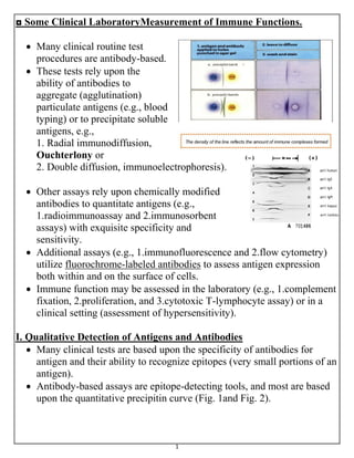
  • 1. 1 ◘ Some Clinical LaboratoryMeasurement of Immune Functions.  Many clinical routine test procedures are antibody-based.  These tests rely upon the ability of antibodies to aggregate (agglutination) particulate antigens (e.g., blood typing) or to precipitate soluble antigens, e.g., 1. Radial immunodiffusion, Ouchterlony or 2. Double diffusion, immunoelectrophoresis).  Other assays rely upon chemically modified antibodies to quantitate antigens (e.g., 1.radioimmunoassay and 2.immunosorbent assays) with exquisite specificity and sensitivity.  Additional assays (e.g., 1.immunofluorescence and 2.flow cytometry) utilize fluorochrome-labeled antibodies to assess antigen expression both within and on the surface of cells.  Immune function may be assessed in the laboratory (e.g., 1.complement fixation, 2.proliferation, and 3.cytotoxic T-lymphocyte assay) or in a clinical setting (assessment of hypersensitivity). I. Qualitative Detection of Antigens and Antibodies  Many clinical tests are based upon the specificity of antibodies for antigen and their ability to recognize epitopes (very small portions of an antigen).  Antibody-based assays are epitope-detecting tools, and most are based upon the quantitative precipitin curve (Fig. 1and Fig. 2).
  • 2. 2 A. Particulate Antigens.  Particulate antigens such as erythrocytes, bacteria, or even antigen- coated latex beads are normally evenly dispersed in suspension.  Cross-linking of antigen-bearing particles by antibodies causes clumping of the particles, also known as agglutination (Fig.2).  If the particulate antigen is an erythrocyte (hemagglutination), whether IgM antibodies efficiently cross-link the particles (direct agglutination), or whether an anti-immunoglobulin (indirect or passive agglutination) is used to cross-link antigen-bound antibodies. 1-Direct agglutination:  This reaction usually involves IgM antibodies that cross-link epitopes on cells or particles.  As IgM is the largest immunoglobulin, it has 10 epitope-binding sites (valence).  It very efficient at cross-linking epitopes on adjacent particles (Figs.1& 2).  Other Igs, because of their smaller size (lesser valence) are less efficient in direct agglutination.  Too much antibody inhibits agglutination (equivalent to the zone of antibody excess; prozone).  To avoid the prozone effect, twofold or serial dilutions of antibody are prepared; each dilution is half as concentrated as the preceding one (Fig.1).  Titers are relative measures of antibody activity and are often expressed as the reciprocal of the dilution (e.g., 1:16, 1:32, 1:64). Fig.2 Direct agglutination reaction (ABO blood typing) occurs in 15-30 sec.
  • 3. 3 Precipitin curve Preparation of serum serial dilution Figure (1) Quantitative precipitin curve. - Antibody preparation: serially diluted antibody-containing serum is prepared (expressed as 1:2. 1:4, 1:8, 1:16, .. etc.). - Antigen-antibody reaction: antigen (containing multiple epitopes) in equal concentrations is added to the antibody dilutions resulting in differing degrees of antigen- antibody complex formation. - In the equivalence zone, both antigen and antibody are at concentrations that result in maximal lattice formation, causing precipitation of antigen-antibody complexes. - In the zone of antibody excess (prozone) antibody molecules are more than the available epitopes, and precipitating complexes are not formed. - In the zone of antigen excess, available epitopes are more than the antibody-binding sites, and precipitating complexes are not formed. - The point at which cross-linking of the particulate antigen is no longer observed is called the titer.
  • 4. 4 2-Indirect or passive agglutination:  This technique is often used to detect non-IgM antibodies or antibodies in concentrations too low to be detected by direct agglutination.  Human antibodies may not directly agglutinate antigen-bearing particles (e.g., bacteria, erythrocytes, latex particles) or show agglutination of very low titer.  The sensitivity of the agglutination test may be enhanced by the addition of an anti-immunoglobulin reagent (e.g., rabbit anti-human immunoglobulin) in the so-called indirect or passive agglutination technique.  Addition of these second-step antibodies is used to increase binding over a greater span and to increase valence by virtue of their ability to bind to the primary antibody (Fig. 3). Fig.3 Indirect agglutination
  • 5. 5 3-Coombs' test  Antibodies against self blood group antigens occur in some autoimmune diseases (hemolytic anemia).  Afflicted individuals produce antibodies to their own erythrocytes but in isotypes or quantities that do not directly agglutinate their erythrocytes. a- In the direct Coombs' test, is used to test for autoimmune hemolytic anemia autoantibodies are detected by the addition of antihuman immunoglobulin (secondary antibody or Coombs reagent).  If this produces agglutination of RBCs, the direct Coombs test is positive, a visual indication that antibodies (and/or complement proteins) are bound to the surface of red blood cells. b- In the indirect Coombs' assay, is used in prenatal testing of pregnant women, and in testing blood prior to a blood transfusion.  It detects antibodies against RBCs that are present unbound in the patient's serum.  Serum sample taken from the patient.  Then, the serum is incubated with RBCs of known antigenicity from other patient blood samples.  Antihuman immunoglobulin is then added, if agglutination occurs, the indirect Coombs test is positive Figure (4) the indirect Coombs' assay
  • 6. 6 B. Soluble Antigens  Often, epitopes present on soluble molecules will precipitate from solution upon reaction with the "right" amount of antibody.  Several simple modifications to the quan-titative precipitin reaction (Fig.1; Fig. 2) allow visualization of immune precipitates in agar, a semisolid growth medium. ♣ Radial immune-diffusion:  Also called the Mancini technique, this test is based upon the diffusion of soluble antigen within an agar gel that contains a uniform concentration of antibody.  Antibody-containing molten agar is poured onto a glass slide or plastic dish.  When the agar cools and solidifies, wells are cut into the gel matrix, and soluble antigen is placed into the well (Fig. 5).  Antigen diffuses radially from the well, forming a precipitin ring at equivalence.  The diameter of the ring is directly proportional to the amount of antigen loaded into the well.  The concentration of antigen in a test sample can be accurately determined by comparing its diameter with a standard calibration curve.  This technique allows for the rapid and precise determination of the quantity of antigen loaded into the well.
  • 7. 7 2-Double-diffusion (Ouchterlony technique):  This test is based upon the diffusion of both antigen (loaded in one well) and antibody (loaded in another well) through an agar gel.  A precipitin line forms at equivalence (Fig. 6).  Solubility, molecular size of the antibody, and detection of epitopes on antigens of different molecular size all influence precipitin formation such that multiple precipitin lines often develop. ♣ An advantage of this technique: • Is that several antigens or antibodies can be compared to determine identity, partial identity, and nonidentity of antigens and/or antibodies. • In contrast to radial immunodiffusion, this is a qualitative technique. • Wells are cut into a solidified agar gel. • Soluble antigen(s) are loaded into one or more wells, and antibodies are loaded into another well(s), from which they diffuse through the gel. • A precipitin band is formed at the equivalence zone. Figure (6): double –diffusion (Ouchterlony) technique.
  • 8. 8 ♦ Immunoelectrophoresis (IEP):  This technique is a modification of double diffusion.  Antigens are loaded into a well within the agar, an electrical current is applied, and antigens migrate according to both their size and their electrical charge (Fig. 7). ♣ The electrical current  Is removed, a trough is cut into the agar, and antiserum is placed in the trough.  IEP is qualitative. II. Quantitative Detection by Antibodies.  The specificity of antibody molecules makes them ideal probes for detection of a wide variety of epitopes.  Antibodies or the antigens they detect (sometimes referred to as ligands) may be labeled with radioactive molecules, fluorescent molecules, enzymes, or heavy metals.  Antibody or antigen binding is then readily detectable and quantifiable. a. Radioimmunoassay [RIA]  RIA has been widely used in clinical diagnostic laboratories.  Antigens of primary antibodies may be directly labeled with a radionuclide and form the basis for direct RIA.  Alternatively, anti-immunoglobulin antibody (secondary antibody) is radio-labeled and used in the indirect RIA.  RIA is sensitive but presents problems owing to the potential exposure of laboratory personnel to radioactivity and radioactive waste disposal.
  • 9. 9 1. Direct RIA:  This technique utilizes radiolabeled antibody or its ligand (antigen).  Antibody is incubated with ligand, and unbound reactants are removed (phase separation) from the (quantitative precipitin reaction), system (Fig.7).  Phase separation may utilize precipitation of bound reactants particulate antigens (such as bacteria that may be separated by centrifugation), the immobilization of the nonradioactive reactant onto a solid matrix (such as plastic), and so on. 2. Indirect RIA:  This technique uses radiolabeled secondary antibody (anti- immunoglobulin) to detect the binding of a primary antibody.  As with direct RIA, a phase separation method must be employed to remove unbound radiolabeled secondary antibody.
  • 10. 10 b. Enzyme-linked immunosorbent assay [ELISA]  Enzyme-linked immunosorbent assay [ELISA, also called enzyme immunoassay (EIA)] has replaced RIA in a number of tests.  ELISA offers the advantage of safety and speed.  Because there is no radioactive decay, the reagents that are used are relatively stable. ELISAs are both specific and quantitative.  Its sensitivity is often equal to or greater than that of RIA or fluorescent immunosorbent (FIA) assay, because an enzyme-labeled reactant is used to turn a chromogenic substrate from colorless to a color (Fig. 8).  Color change of the substrate indicates that an enzyme-labeled reactant has bound.  Increasing substrate incubation time allows low-concentration enzyme to convert more substrate to enhance test sensitivity (within limits). Figure (8): Enzyme-linked immunosorbent assay (ELISA)
  • 11. 11 C. Fluorescent immunosorbent assay  Fluorescent immunosorbent assay (FIA) relies upon antibodies or their ligands labeled with a variety of fluorescent dyes such as fluorescein isothiocyanate (FITC) or phycoerythrin (PE).  The FIA design is similar to the ELISA. 1. The assay is performed in, 96-well polystyrene plates. 2. Soluble antigen is added and noncovalently binds to the plastic. 3. Unbound antigen is washed from the well. 4. Unlabeled primary antibodies (often sera to be tested) are added to the well and allowed to bind. 5. Unbound primary antibodies are washed from the well. 6. FITC-labeled anti-immunoglobulin antibodies are added to the well and allowed to bind. 7. Unbound labeled antibodies are washed from the well. 8. Fluorescence indicates the presence of epitopes.
  • 12. 12 III. EPITOPE detection IN and ON cells.  Epitopes expressed both within and on the surface of cells may be detected by using radio-, enzyme-, or fluorochrome-labeled antibodies (direct or indirect).  The outline of two techniques that have extensive application in a clinical setting: immunofluorescence and flow cytometry. A. Immunofluorescence (IF)  (IF) utilizes fluorescent dyes (e.g., fluorescein isothiocyanate FITC) that are covalently coupled to antibody.  A thin, frozen section of tissue is prepared and mounted on a glass slide. The frozen section is then bathed in a solution containing FITC-labeled antibody (A direct IF) or a solution containing a primary antibody and is then washed.  An FITC-labeled anti-immunoglobulin is added (B indirect IF).  The presence of epitopes is visualized with a fluorescent microscope.
  • 13. 13 B. Monoclonal antibodies (mAb)  Antibody responses normally derive from multiple B cells or plasma cells; their antibodies often differ in epitopes that are recognized, affinity, and isotype. ♣ Antibody responses that arise from multiple cells are termed “polyclonal antibody” responses.  This antibody diversity is very important in combating microbial infection.  Although polyclonal antibodies can be used in the clinical laboratory, in 1975, scientists fused antibody-secreting plasma cells with myeloma (myeloid-origin tumor cells).  The resulting immortalized cells, or hybridomas, secreted antibodies of single specificity and isotype and were termed monoclonal antibodies because of their origin from a single antibody-producing cell.  Monoclonal antibodies (mAb) are antibodies that are identical because they were produced by one type of immune cell, all clones of a single parent cell.  Vast quantities of monoclonal antibodies can be produced.  Because monoclonal antibodies produced by any given hybridoma are unique, they can be used together with fluorescent dyes or other markers to distinguish individual epitopes on an antigen or cell.
  • 14. 14 IV. ASSESSMENT OF IMMUNE FUNCTION  The functional capacity of phagocytic cells can be assessed by their ability in ingest antibody- or opsonin-coated particles.  Stimulating lymphocytes to increase in number or proliferate in response to a specific antigen or to a substance that causes polyclonal mitogenesis (a mitogen) is often used to assess immune function.  Phagocyte function can be assessed by incubating phagocytic cells with coated particles (e.g., latex beads or antibody-bound cells) or with bacteria for 30 to 120 minutes.  Particle inclusion within the cell is assessed by microscopy.  Enzymatic activity of phagocytes can be assessed by measuring the levels of individual degradative or oxidative enzymes (e.g., NADPH oxidase) produced by these cells. Fig: phagocytic function
  • 15. 15 B. Proliferation  Peripheral blood mononuclear cells (lymphocytes, monocytes, and dendritic cells) are isolated and placed in tissue culture for 48 to 72 hours.  A specific stimulator (antigen) to which the individual may have been previously exposed is added to the culture.  Alternatively, a nonspecific stimulant (mitogen) is added to assess the ability of a particular subpopulation of leukocytes to respond.  A radionuclide (such as 3H-thymidine) is added for the final 18 to 24 hours of cultures  Incorporation of 3H-thymidine into nascent DNA is taken as a measure of proliferative ability. V. ASSESSMENT OF HYPERSENSITIVITY  Hypersensitivity which is an immune-mediated damage to host tissues. There are four categories of hypersensitivity reactions.  Type I reactions are called immediate hypersensitivity reactions because they occur within minutes to hours of antigen exposure.  Type II reactions involve complement activation in response to immunoglobulin binding to membranes or the intracellular matrix.  Type III reactions involve complement activation in response to "soluble" antigen-antibody complexes. ♠ Both type II and type III reactions occur within hours to days.  Type IV reactions are "delayed," occurring two to four days after antigen exposure.
  • 16. 16 A. Allergy skin testing (type I hypersensitivity)  Sensitivities to allergens (antigens) [e.g., pet dander, mold and pollens ("hay fever"), or certain foods] are common allergic disorders.  Sensitivity arises from the development of allergen-specific IgE antibodies that decorate the surfaces of tissue mast cells.  Intradermal injection of a small amount of diluted allergen tests an individual's reaction to an allergen.  In some cases, a scratch test can be used where the diluted allergen is administered by scratching the skin surface (percutaneous) rather than being injected into the dermis.  Sensitive (atopic) individuals develop a wheal-and-flare (redness and swelling) reaction within 20 minutes after re-exposure to a specific allergen.  The test relies upon inflammation caused by allergen-lgE induced degranulation of mast cells in the dermis.  Because there is a possibility of the occurrence of a severe allergic reaction, antihistamine or epinephrine should be available during testing. Figure 15. Allergy testing.
  • 17. 17 • These tests assess Type I hypersensitivities to a variety of potential allergens. 1. Testing is often performed on the ventral side of the arm. 2. A grid is marked and small quantities of substances to be tested are injected into the dermis. 3. Positive reactions are indicated as redness and swelling within 20 to 30 minutes after re-exposure to the allergen. C. Contact dermatitis and delayed hypersensitivity (type IV).  Application of antigen to the surface of the skin (contact dermatitis) or injected intradermally [delayed-type hypersensitivity, DTH] is used to measure type IV hypersensitivity.  In this test, antigen is applied to the surface of the skin under a nonabrasive dermal patch.  These tests evaluate whether an individual has had prior exposure to a specific antigen.  In contrast to immediate hypersensitivity reactions, type IV hypersensitivity reactions are delayed; wheal-and-flare reactions are evident only 24 to 72 hours after antigen challenge.