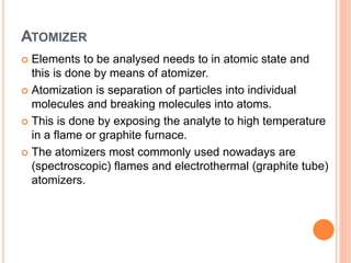
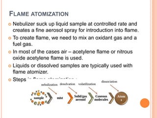
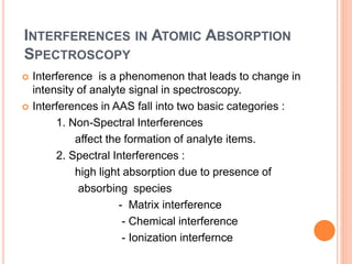
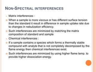
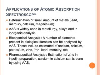
ATOMIC ABSORPTION SPECTROSCOPY
GUIDED BY
Dr. S. S. PAWAR
PREPARED BY
VINAYAK R BODHANKAR
M PHARM (SEM-I)
P’CEUTICAL QUALITYASSURANCE
ROLL NO. : 01
Sanjivani College of Pharmaceutical Education &
Research, Kopargaon
CONTENTS
 Introduction
 History
 Principle
 Instrumentation
 Interferences
 Applications
INTRODUCTION
 Atomic Absorption Spectroscopy is a very common
technique for detecting metals and metalloids in
samples.
 It is very reliable and simple to use.
 It also measures the concentration of metals in the
sample.
 Atomic Absorption Spectroscopy is an analytical
technique that measures the concentration of an
element by measuring the amount of light that is
absorbed at a characteristic wavelength when it
passes through cloud of atoms
 As the number of atoms in the light path increases, the
amount of light absorbed increases.
HISTORY OF ATOMIC ABSORPTION
SPECTROSCOPY
 The Atomic Absorption Spectroscopy was first used
as analytical technique in the second half of 19th
century by Robert Bunsen and Robert Kirchhoff.
 The modern form of Atomic Absorption
Spectroscopy was largely developed during the
1950s by a team of Australian chemists.
 They were led by Sir Alan Walsh at the Common
wealth Scientific and Industrial Research
Organization (CSIRO)
PRINCIPLE OF ATOMIC ABSORPTION
SPECTROSCOPY
 The technique uses basically the principle that free
atoms generated in an atomizer can absorb radiation
at specific frequency.
 Atomic Absorption Spectroscopy quantifies the
absorption of ground state atoms in the gaseous
state.
 The atoms absorb UV or visible light & make
transition to higher electronic energy level. The
analyte concentration is determined from the amount
of absorption.
 Concentration measurements are usually determined
from a working curve after the instrument with
standards of known concentration.
INSTRUMENTATION
 Parts of Atomic Absorption Spectrophotometer :
 Light source
 Nebulizer
 Atomizer
 Monochromator
 Detector and amplifier
 Read out system
Schematic diagram of Atomic Absorption
Spectrophotometer
LIGHT SOURCE
 Hollow cathode lamp are the most common radiation
source in AAS.
 It contains a tungsten anode and a hollow cylindrical
cathode .
 These are sealed in a glass tube filled with an inert gas.
(mainly neon or argon)
 Each elements has its own unique lamp which must be
used for that analysis
NEBULIZER
 Nebulizer suck up liquid samples at controlled rate.
 Create a fine aerosol spray for introduction into the flame.
 Mix the aerosol and fuel and oxidant thoroughly for
introduction into flame.
ATOMIZER
 Elements to be analysed needs to in atomic state and
this is done by means of atomizer.
 Atomization is separation of particles into individual
molecules and breaking molecules into atoms.
 This is done by exposing the analyte to high temperature
in a flame or graphite furnace.
 The atomizers most commonly used nowadays are
(spectroscopic) flames and electrothermal (graphite tube)
atomizers.
FLAME ATOMIZATION
 Nebulizer suck up liquid sample at controlled rate and
creates a fine aerosol spray for introduction into flame.
 To create flame, we need to mix an oxidant gas and a
fuel gas.
 In most of the cases air – acetylene flame or nitrous
oxide acetylene flame is used.
 Liquids or dissolved samples are typically used with
flame atomizer.
 Steps in flame atomization :
ELECTRO THERMAL ATOMIZATION
 It uses a graphite coated furnace to vaporize the sample.
 Samples are deposited in a small graphite coated tube
which then heated to vaporize and atomize the analyte.
 The graphite tubes are heated using a high current power
supply.
 Steps in electro thermal atomization : Drying
Pyrolysis
Atomization
Cleaning
MONOCHROMATOR
 This is very important part in an AAS.
 It is used to separate out all of the thousand of
lines.
 A monochromator is used to select the specific
wavelength of light which is absorbed by the
sample and to remove other wavelengths.
 The selection of the specific light allows the
determination of the selected element in the
presence of others.
DETECTOR AND AMPLIFIER
 The light selected by the monochromator is directed onto
a detector whose function is convert the light signal into
an electrical signal.
 Photomultiplier tube detector is mainly used.
 The processing of electrical signal is fulfilled by a signal
amplifier.
 The amplified signal is then displayed on read out system
or fed into a data station for printout by the requested
format.
CALIBRATION CURVE
 A calibration curve is used to determine the unknown
concentration of an element in a sample.
 The instrument is calibrated using several solutions of
known concentrations.
 The absorbance of each known solution is measured &
then a calibration curve of concentrations vs absorbance
is plotted.
 The sample solution is fed into instrument & the
absorbance of the element in the solution is measured.
 The unknown concentration of element is then calculated
from the calibration curve.
INTERFERENCES IN ATOMIC ABSORPTION
SPECTROSCOPY
 Interference is a phenomenon that leads to change in
intensity of analyte signal in spectroscopy.
 Interferences in AAS fall into two basic categories :
1. Non-Spectral Interferences
affect the formation of analyte items.
2. Spectral Interferences :
high light absorption due to presence of
absorbing species
- Matrix interference
- Chemical interference
- Ionization interfernce
NON-SPECTRAL INTERFERENCES
 Matrix interferences :
 When a sample is more viscous or has different surface tension
than the standard it result in difference in sample uptake rate due
to changes in nebulization efficiency.
 Such interferences are minimized by matching the matrix
composition of standard and sample
 Chemical interferences :
 If a sample contains a species which forms a thermally stable
compound with analyte that is not completely decomposed by the
flame energy then chemical interferences exist.
 Such interferences are minimized by using higher flame temp. to
provide higher dissociation energy.
IONIZATION INTERFERENCE
 It is more common in hot flames.
 The dissociation process doesn’t stop at formation of
ground state atoms.
 Excess energy of the flame lead to excitation of ground
state atoms to ionic state by loss of electrons thereby
resulting in depletion of ground state atoms.
 Ionization interference is eliminated by an excess of an
element which is easily ionized thereby creating a large
number of electrons in the flame & suppressing the
ionization of the analyte.
SPECTRAL INTERFERENCES
 Spectral interferences are caused by presence of another
atomic absorption line or a molecular absorbance band
close to the spectral line of element of interest.
 Most of these interferences are due to molecular
emission from oxides of other element is a sample.
APPLICATIONS OF ATOMIC ABSORPTION
SPECTROSCOPY
 Determination of small amount of metals (lead,
mercury, calcium, magnesium)
 AAS is widely used in metallurgy, alloys and in
inorganic analysis.
 Biochemical Analysis : A number of elements
present in biological samples can be analysed by
AAS. These include estimated of sodium, calcium,
potassium, zinc, iron, lead, mercury, etc.
 Pharmaceutical Analysis : Estimation of zinc in
insulin preparation, calcium in calcium salt is done
by using AAS.
• Sodium, potassium, calcium in saline and ringer
solution are estimated by AAS.
• Analysis of ash for determining the content of sodium,
potassium, calcium, magnesium and iron is done by
AAS.
• Atomic absorption spectroscopy is used in assay of
a) Intraperitoneal dialysis of fluid for calcium &
magnesium.
b) Activated charcoal for zinc.
c) Cisplastin for liver.
References :
• Pharmaceutical analysis Instrumental methods
Volume II by Dr. A.V. Kasture, Dr. S.G. Wadodkar,
Nirali prakashan page no. 23.9 - 23.12
• Instrumental methods of Chemical analysis by
G.R. Chatwal, S.K. Anand, Himalaya publishing
house, fifth edition page no. 2.340 - 2.360
THANK YOU

Atomic absorption spectroscopy

  • 1.
    ATOMIC ABSORPTION SPECTROSCOPY GUIDEDBY Dr. S. S. PAWAR PREPARED BY VINAYAK R BODHANKAR M PHARM (SEM-I) P’CEUTICAL QUALITYASSURANCE ROLL NO. : 01 Sanjivani College of Pharmaceutical Education & Research, Kopargaon
  • 2.
    CONTENTS  Introduction  History Principle  Instrumentation  Interferences  Applications
  • 3.
    INTRODUCTION  Atomic AbsorptionSpectroscopy is a very common technique for detecting metals and metalloids in samples.  It is very reliable and simple to use.  It also measures the concentration of metals in the sample.  Atomic Absorption Spectroscopy is an analytical technique that measures the concentration of an element by measuring the amount of light that is absorbed at a characteristic wavelength when it passes through cloud of atoms  As the number of atoms in the light path increases, the amount of light absorbed increases.
  • 4.
    HISTORY OF ATOMICABSORPTION SPECTROSCOPY  The Atomic Absorption Spectroscopy was first used as analytical technique in the second half of 19th century by Robert Bunsen and Robert Kirchhoff.  The modern form of Atomic Absorption Spectroscopy was largely developed during the 1950s by a team of Australian chemists.  They were led by Sir Alan Walsh at the Common wealth Scientific and Industrial Research Organization (CSIRO)
  • 5.
    PRINCIPLE OF ATOMICABSORPTION SPECTROSCOPY  The technique uses basically the principle that free atoms generated in an atomizer can absorb radiation at specific frequency.  Atomic Absorption Spectroscopy quantifies the absorption of ground state atoms in the gaseous state.  The atoms absorb UV or visible light & make transition to higher electronic energy level. The analyte concentration is determined from the amount of absorption.  Concentration measurements are usually determined from a working curve after the instrument with standards of known concentration.
  • 6.
    INSTRUMENTATION  Parts ofAtomic Absorption Spectrophotometer :  Light source  Nebulizer  Atomizer  Monochromator  Detector and amplifier  Read out system
  • 7.
    Schematic diagram ofAtomic Absorption Spectrophotometer
  • 8.
    LIGHT SOURCE  Hollowcathode lamp are the most common radiation source in AAS.  It contains a tungsten anode and a hollow cylindrical cathode .  These are sealed in a glass tube filled with an inert gas. (mainly neon or argon)  Each elements has its own unique lamp which must be used for that analysis
  • 9.
    NEBULIZER  Nebulizer suckup liquid samples at controlled rate.  Create a fine aerosol spray for introduction into the flame.  Mix the aerosol and fuel and oxidant thoroughly for introduction into flame.
  • 10.
    ATOMIZER  Elements tobe analysed needs to in atomic state and this is done by means of atomizer.  Atomization is separation of particles into individual molecules and breaking molecules into atoms.  This is done by exposing the analyte to high temperature in a flame or graphite furnace.  The atomizers most commonly used nowadays are (spectroscopic) flames and electrothermal (graphite tube) atomizers.
  • 11.
    FLAME ATOMIZATION  Nebulizersuck up liquid sample at controlled rate and creates a fine aerosol spray for introduction into flame.  To create flame, we need to mix an oxidant gas and a fuel gas.  In most of the cases air – acetylene flame or nitrous oxide acetylene flame is used.  Liquids or dissolved samples are typically used with flame atomizer.  Steps in flame atomization :
  • 12.
    ELECTRO THERMAL ATOMIZATION It uses a graphite coated furnace to vaporize the sample.  Samples are deposited in a small graphite coated tube which then heated to vaporize and atomize the analyte.  The graphite tubes are heated using a high current power supply.  Steps in electro thermal atomization : Drying Pyrolysis Atomization Cleaning
  • 13.
    MONOCHROMATOR  This isvery important part in an AAS.  It is used to separate out all of the thousand of lines.  A monochromator is used to select the specific wavelength of light which is absorbed by the sample and to remove other wavelengths.  The selection of the specific light allows the determination of the selected element in the presence of others.
  • 14.
    DETECTOR AND AMPLIFIER The light selected by the monochromator is directed onto a detector whose function is convert the light signal into an electrical signal.  Photomultiplier tube detector is mainly used.  The processing of electrical signal is fulfilled by a signal amplifier.  The amplified signal is then displayed on read out system or fed into a data station for printout by the requested format.
  • 15.
    CALIBRATION CURVE  Acalibration curve is used to determine the unknown concentration of an element in a sample.  The instrument is calibrated using several solutions of known concentrations.  The absorbance of each known solution is measured & then a calibration curve of concentrations vs absorbance is plotted.  The sample solution is fed into instrument & the absorbance of the element in the solution is measured.  The unknown concentration of element is then calculated from the calibration curve.
  • 16.
    INTERFERENCES IN ATOMICABSORPTION SPECTROSCOPY  Interference is a phenomenon that leads to change in intensity of analyte signal in spectroscopy.  Interferences in AAS fall into two basic categories : 1. Non-Spectral Interferences affect the formation of analyte items. 2. Spectral Interferences : high light absorption due to presence of absorbing species - Matrix interference - Chemical interference - Ionization interfernce
  • 17.
    NON-SPECTRAL INTERFERENCES  Matrixinterferences :  When a sample is more viscous or has different surface tension than the standard it result in difference in sample uptake rate due to changes in nebulization efficiency.  Such interferences are minimized by matching the matrix composition of standard and sample  Chemical interferences :  If a sample contains a species which forms a thermally stable compound with analyte that is not completely decomposed by the flame energy then chemical interferences exist.  Such interferences are minimized by using higher flame temp. to provide higher dissociation energy.
  • 18.
    IONIZATION INTERFERENCE  Itis more common in hot flames.  The dissociation process doesn’t stop at formation of ground state atoms.  Excess energy of the flame lead to excitation of ground state atoms to ionic state by loss of electrons thereby resulting in depletion of ground state atoms.  Ionization interference is eliminated by an excess of an element which is easily ionized thereby creating a large number of electrons in the flame & suppressing the ionization of the analyte.
  • 19.
    SPECTRAL INTERFERENCES  Spectralinterferences are caused by presence of another atomic absorption line or a molecular absorbance band close to the spectral line of element of interest.  Most of these interferences are due to molecular emission from oxides of other element is a sample.
  • 20.
    APPLICATIONS OF ATOMICABSORPTION SPECTROSCOPY  Determination of small amount of metals (lead, mercury, calcium, magnesium)  AAS is widely used in metallurgy, alloys and in inorganic analysis.  Biochemical Analysis : A number of elements present in biological samples can be analysed by AAS. These include estimated of sodium, calcium, potassium, zinc, iron, lead, mercury, etc.  Pharmaceutical Analysis : Estimation of zinc in insulin preparation, calcium in calcium salt is done by using AAS.
  • 21.
    • Sodium, potassium,calcium in saline and ringer solution are estimated by AAS. • Analysis of ash for determining the content of sodium, potassium, calcium, magnesium and iron is done by AAS. • Atomic absorption spectroscopy is used in assay of a) Intraperitoneal dialysis of fluid for calcium & magnesium. b) Activated charcoal for zinc. c) Cisplastin for liver.
  • 22.
    References : • Pharmaceuticalanalysis Instrumental methods Volume II by Dr. A.V. Kasture, Dr. S.G. Wadodkar, Nirali prakashan page no. 23.9 - 23.12 • Instrumental methods of Chemical analysis by G.R. Chatwal, S.K. Anand, Himalaya publishing house, fifth edition page no. 2.340 - 2.360
  • 23.