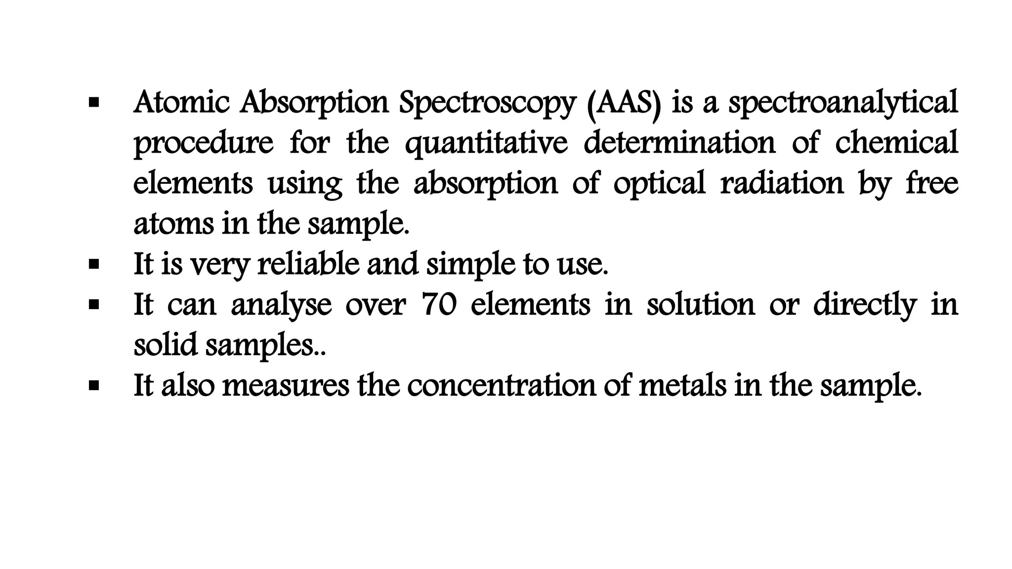
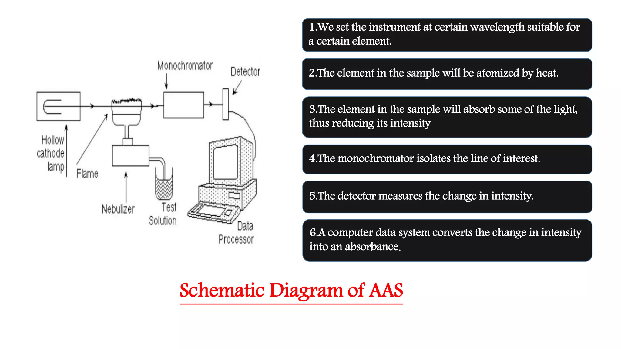
ATOMIC ABSORPTION
SPECTROSCOPY
 Atomic Absorption Spectroscopy (AAS) is a spectroanalytical
procedure for the quantitative determination of chemical
elements using the absorption of optical radiation by free
atoms in the sample.
 It is very reliable and simple to use.
 It can analyse over 70 elements in solution or directly in
solid samples..
 It also measures the concentration of metals in the sample.
History
• Atomic absorption spectroscopy was first established in the
second half of the 19th century by Robert Wilhelm Bunsen and
Gustav Robert Kirchhoff, both professors at the University of
Heidelberg, Germany.
• The modern form of AAS was largely developed during the
1950s by a team of Australian chemists.They were led by Sir
Alan Walsh at the Commonwealth Scientific and Industrial
Research Organisation (CSIRO),Division of Chemical Physics,in
Melbourne,Australia.
Principle
• The technique uses basically the principle that free
atoms generated in an atomizer can absorb radiation at
specific frequency.
• Atomic absorption spectroscopy quantifies the
absorption of ground state atoms in the gaseous state.
• The atoms absorb UV or visible light and make
transitions to higher electronic energy levels.The
analyte concentration is determined from the amount
of absorption.
• Concentration measurements are usually determined
from a working curve after calibrating the instrument
with standards of known concentration.
Atomic
Absorption
Spectroscopy
Hollow Cathode
Lamp
NebulizerDetector
AtomizerMonochromator
Components of AAS
Light source
 Hollow Cathode Lamp are the most common radiation
source in AAS.
 It contains a tungsten anode and a hollow cylindrical
cathode made of the element to be determined.
 These are sealed in a glass tube filled with an inert gas
(neon or argon).
 Each element has its own unique lamp which must be used
for that analysis.
Nebulizer
• Nebulizer suck up liquid samples at controlled rate.
• It create a fine aerosol spray for introduction into flame.
• Mix the aerosol,fuel and oxidant thoroughly for
introduction into flame.
Atomizer
• Elements to be analysed needs to be in atomic state.
• Atomisation is separation of particles into individual
molecules and breaking molecules into atoms.This is
done by exposing the analyte to high temperatures in a
flame or graphite furnace.
Atomizers
Flame Atomizer
• To create flame,we need to mix an
oxidant gas and a fuel gas.
• In most of the cases air – acetylene flame
or nitrous oxide – acetylene flame is
used.
• Liquid or dissolved samples are typically
used with flame atomizer.
Graphite Tube Atomizer
• It uses a graphite coated furnace to
vaporize the sample.
• In GFAAS sample,samples are deposited in
a small graphite coated tube which can
then be heated to vaporize and atomize
the analyte.
• The graphite tubes are heated using a high
current power supply.
Monochromator
• It is used in AAS to separate out all of the thousands of
lines.
• A Monochromator is used to select the specific
wavelength of light which is absorbed by the sample,and
to exclude other wavelengths.
• The selection of the specific light allows the
determination of the selected element in the presence of
others.
Detector
• The light selected by the monochromator is directed that is
typically a photomultiplier tube,whose function is to convert
the light signal into an electrical signal proportional to the
light intensity.
• The processing of electrical signal is fulfilled by a signal
amplifier.The signal could be displayed for readout ,or further
fed into a data station for printout by the requested formal.
Calibration curve
• A calibration curve is used to determine the unknown
concentration of an element in a solution. The instrument is
calibrated using several solutions of known concentrations.The
absorbance of each known solution is measured and then a
calibration curve of concentration v/s absorbance is plotted.
• The sample solution is fed into the instrument,and the
absorbance of the element in this solution is measured.The
unknown concentration of the element is then calculated from
the calibration curve.
Schematic Diagram of AAS
1.We set the instrument at certain wavelength suitable for
a certain element.
2.The element in the sample will be atomized by heat.
3.The element in the sample will absorb some of the light,
thus reducing its intensity
4.The monochromator isolates the line of interest.
5.The detector measures the change in intensity.
6.A computer data system converts the change in intensity
into an absorbance.
Applications
• Determination of even small amounts of metals (lead, mercury,
calcium,magnesium,etc.)as follows:
Environmental studies: drinking water, ocean water, soil.
Food industry.
Pharmaceutical industry.
• Atomic absorption spectroscopy has many uses in different areas of
metals in biological fluids and tissues such as whole blood,plasma,urine,
saliva, brain tissue, liver,musle tissue,semen,in some pharmaceutical
manufacturing processes,minute quantities of a catalyst that remain in
the final drug product,and analyzing water for its metal content.
Atomic absorption spectroscopy

Atomic absorption spectroscopy

  • 1.
  • 2.
     Atomic AbsorptionSpectroscopy (AAS) is a spectroanalytical procedure for the quantitative determination of chemical elements using the absorption of optical radiation by free atoms in the sample.  It is very reliable and simple to use.  It can analyse over 70 elements in solution or directly in solid samples..  It also measures the concentration of metals in the sample.
  • 3.
    History • Atomic absorptionspectroscopy was first established in the second half of the 19th century by Robert Wilhelm Bunsen and Gustav Robert Kirchhoff, both professors at the University of Heidelberg, Germany. • The modern form of AAS was largely developed during the 1950s by a team of Australian chemists.They were led by Sir Alan Walsh at the Commonwealth Scientific and Industrial Research Organisation (CSIRO),Division of Chemical Physics,in Melbourne,Australia.
  • 4.
    Principle • The techniqueuses basically the principle that free atoms generated in an atomizer can absorb radiation at specific frequency. • Atomic absorption spectroscopy quantifies the absorption of ground state atoms in the gaseous state. • The atoms absorb UV or visible light and make transitions to higher electronic energy levels.The analyte concentration is determined from the amount of absorption. • Concentration measurements are usually determined from a working curve after calibrating the instrument with standards of known concentration.
  • 5.
  • 6.
    Light source  HollowCathode Lamp are the most common radiation source in AAS.  It contains a tungsten anode and a hollow cylindrical cathode made of the element to be determined.  These are sealed in a glass tube filled with an inert gas (neon or argon).  Each element has its own unique lamp which must be used for that analysis.
  • 7.
    Nebulizer • Nebulizer suckup liquid samples at controlled rate. • It create a fine aerosol spray for introduction into flame. • Mix the aerosol,fuel and oxidant thoroughly for introduction into flame.
  • 8.
    Atomizer • Elements tobe analysed needs to be in atomic state. • Atomisation is separation of particles into individual molecules and breaking molecules into atoms.This is done by exposing the analyte to high temperatures in a flame or graphite furnace.
  • 9.
    Atomizers Flame Atomizer • Tocreate flame,we need to mix an oxidant gas and a fuel gas. • In most of the cases air – acetylene flame or nitrous oxide – acetylene flame is used. • Liquid or dissolved samples are typically used with flame atomizer. Graphite Tube Atomizer • It uses a graphite coated furnace to vaporize the sample. • In GFAAS sample,samples are deposited in a small graphite coated tube which can then be heated to vaporize and atomize the analyte. • The graphite tubes are heated using a high current power supply.
  • 10.
    Monochromator • It isused in AAS to separate out all of the thousands of lines. • A Monochromator is used to select the specific wavelength of light which is absorbed by the sample,and to exclude other wavelengths. • The selection of the specific light allows the determination of the selected element in the presence of others.
  • 11.
    Detector • The lightselected by the monochromator is directed that is typically a photomultiplier tube,whose function is to convert the light signal into an electrical signal proportional to the light intensity. • The processing of electrical signal is fulfilled by a signal amplifier.The signal could be displayed for readout ,or further fed into a data station for printout by the requested formal.
  • 12.
    Calibration curve • Acalibration curve is used to determine the unknown concentration of an element in a solution. The instrument is calibrated using several solutions of known concentrations.The absorbance of each known solution is measured and then a calibration curve of concentration v/s absorbance is plotted. • The sample solution is fed into the instrument,and the absorbance of the element in this solution is measured.The unknown concentration of the element is then calculated from the calibration curve.
  • 13.
    Schematic Diagram ofAAS 1.We set the instrument at certain wavelength suitable for a certain element. 2.The element in the sample will be atomized by heat. 3.The element in the sample will absorb some of the light, thus reducing its intensity 4.The monochromator isolates the line of interest. 5.The detector measures the change in intensity. 6.A computer data system converts the change in intensity into an absorbance.
  • 14.
    Applications • Determination ofeven small amounts of metals (lead, mercury, calcium,magnesium,etc.)as follows: Environmental studies: drinking water, ocean water, soil. Food industry. Pharmaceutical industry. • Atomic absorption spectroscopy has many uses in different areas of metals in biological fluids and tissues such as whole blood,plasma,urine, saliva, brain tissue, liver,musle tissue,semen,in some pharmaceutical manufacturing processes,minute quantities of a catalyst that remain in the final drug product,and analyzing water for its metal content.