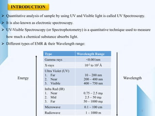
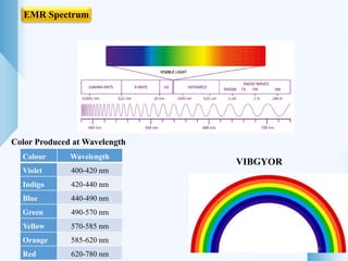
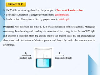
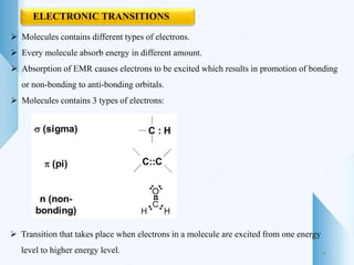
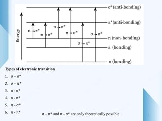
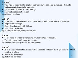
Spectral Shift
Solvent effect on λmax
5
Beers Lambert’s Law
Derivation, Deviation
6
Instrumentation
7
Applications
8
Introduction 1
Principle 2
Electronic
Transition
3
Chromophore,
Auxochrome
4
CONTENT
2
 Quantitative analysis of sample by using UV and Visible light is called UV Spectroscopy.
 It is also known as electronic spectroscopy.
 UV-Visible Spectroscopy (or Spectrophotometry) is a quantitative technique used to measure
how much a chemical substance absorbs light.
 Different types of EMR & their Wavelength range:
Type Wavelength Range
Gamma rays <0.001nm
X-rays 10-2 to 102 Å
Ultra Violet (UV)
1. Far
2. Near
3. Visible
10 – 200 nm
200 – 400 nm
400 – 750 nm
Infra Red (IR)
1. Near
2. Mid
3. Far
0.75 – 2.5 mµ
2.5 – 50 mµ
50 – 1000 mµ
Microwave 0.1 – 100 cm
Radiowave 1 – 1000 m
Energy Wavelength
INTRODUCTION
3
Color Produced at Wavelength
VIBGYOR
EMR Spectrum
4
Colour Wavelength
Violet 400-420 nm
Indigo 420-440 nm
Blue 440-490 nm
Green 490-570 nm
Yellow 570-585 nm
Orange 585-620 nm
Red 620-780 nm
 UV Visible spectroscopy based on the principle of Beers and Lamberts law.
 Beers law: Absorption is directly proportional to concentration.
 Lamberts law: Absorption is directly proportional to pathlength.
Principle: Any molecule has either n, π, σ or a combination of these electrons. Molecules
containing these bonding and bonding electrons absorb the energy in the form of UV light
and undergo a transition from the ground state to an excited state. By the characteristics
absorption peak, the nature of electron present and hence the molecular structure can be
determined.
I0 It
Incident light Transmitted light
PRINCIPLE
5
 Molecules contains different types of electrons.
 Every molecule absorb energy in different amount.
 Absorption of EMR causes electrons to be excited which results in promotion of bonding
or non-bonding to anti-bonding orbitals.
 Molecules contains 3 types of electrons:
 Transition that takes place when electrons in a molecule are excited from one energy
level to higher energy level.
ELECTRONIC TRANSITIONS
6
Types of electronic transition
1. σ – σ*
2. σ – π*
3. n - σ*
4. n - π*
5. π - σ*
6. π - π* σ – π* and π - σ* are only theoretically possible. 7
1. σ – σ*
 This type of transition takes place between lower occupied molecular orbitals to
higher occupied molecular orbitals.
 Such transition requires more energy.
 Shows absorbance at 125 nm.
e.g. Alkane – Methane
2. n - σ*
 Saturated compound containing 1 hetero atom with unshared pair of electrons.
 Requires less energy.
 Show absorbance at 150-250 nm.
 Sensitive to H-bonding.
e.g. Aldehyde, ketones, ether, alcohol, etc.
3. π - π*
 Takes place in aromatic compound or unsaturated compound.
 Transition occurs at longer wavelength.
e.g. alkenes, alkynes, cyanides, azo compounds
4. n - π*
 In this, an electrons of unshared pair of electrons on hetero atom get excited to π*anti-
bonding orbitals.
 It requires less energy.
e.g. Saturated aldehyde
8
 Molecules or parts of molecules that absorb light in UV visible region and
responsible for imparting color to the compounds.
 Covalently unsaturated group responsible for electronic transition.
e.g. C=C, C=O
 Chromogen: Comp. containing chromophores
Types of Chromophores:
1. Independent chromophore:
Single chromophore impart color to compounds.
e.g. Azo group, Nitro group (-NO)
2. Dependent chromophore
e.g. Acetone: 1 Ketonic group – Colorless
2 Ketonic group – Yellow
3 Ketonic group - Orange
CHROMOPHORE
9
AUXOCHROMES
 Saturated group with non-bonding electrons when attached to chromophore, it will alter
wavelength as well as absorption of substance.
 Group of atoms that get attached to chromophore and increases their color intensity.
Benzene Aniline
255 nm 280 nm
10
SPECTRAL SHIFTS
The position of λmax and absorption intensity can be modified in different ways by some
structural or solvent changes.
11
Bathochromic shift
 It is an effect in which absorption maximum is shifted towards longer wavelength due to
presence of auxochromes or by change of solvent.
 Also called as Red shift.
Hypsochromic Shift
 Absorption maximum is shifted toward shorter wavelength.
 Also called as Blue shift.
Hyperchromic Shift
It is an effect due to which intensity of absorption maximum increases.
Hypochromic shift
Intensity of absorption maximum decreases.
12
SOLVENT EFFECT ON ABSORPTION MAXIMA
 Choice of solvent used in UV is important because of solvent can change λmax .
 Most suitable solvent is that it does not absorb radiation in the region under
investigation.
 The position as well as intensity of absorption maximum get shifted for a particular
chromophore by changing the polarity of solvent.
 By increasing the polarity of solvent compound such as dienes and conjugated
hydrocarbons do not experience any appreciable shift.
 In general absorption maximum for non-polar compound is usually shifted with the
change in polarity of solvent.
Polarity Lambda max
e.g. Lambda max of Acetone in water and hexane:
Water: 264 nm
Hexane: 279 nm
Water > Methanol > Ethanol > Benzene > Hexane
Polarity
13
Solvent Wavelength of absorption
Water 205
Cyclohexane 210
Ethanol 210
Methanol 210
Chloroform 245
Benzene 280
Alpha, beta unsaturated compounds show different shift:
1. n - π*
 Absorption band moves to shorter wavelength by increasing the polarity of solvent.
 Less intense
2. π - π*
 Absorption band moves to longer wavelength by increasing the polarity of solvent.
 More intense
 Group is more polar in ground state than excited: Shorter wavelength & Vive versa.
 Increase in polarity of solvent shift n - π* and n - σ* band to shorter wavelength and
π - π* to longer wavelength 14
BEER AND LAMBERT’S LAW
 Beer’s Law
When monochromatic light passes through an absorbing medium its intensity decreases
exponentially as the concentration of the absorbing medium increases.
Absorbance is directly proportional to the Concentration.
A ∝ C
 Lambert’s Law
When monochromatic light passes through an absorbing medium its intensity decreases
exponentially as the length of the absorbing medium increases.
Absorbance is directly proportional to the Pathlength.
A ∝ l
 Beer’s Lambert’s law
When a monochromatic light is passed through a solution containing absorbing species,
decrease in intensity of light with Pathlength is proportional to Concentration of solution
and intensity of light.
15
DERIVATION
-
𝑑𝐼
𝑑𝑙
∝ I.C
-
𝑑𝐼
𝑑𝑙
= K.I.C
-
𝑑𝐼
𝐼
= K .C. dl
Add integration
- 𝐼=𝐼0
𝐼=𝐼 𝑑𝐼
𝐼
= 𝐾𝐶 𝑙=0
𝑙=𝑙
𝑑𝑙
- ln
𝐼
𝐼0
= KCl
Interchange
ln
𝐼0
𝐼
= KCl
2.303 × ln
𝐼0
𝐼
= 2.303 KCl
Log10
𝐼0
𝐼
= εCl ……………1)
16
I0 It
A > 0, T < 100%
T =
𝐼
𝐼0
Now, A = log10 (
1
𝑇
) ---------- 2), As A ∝
1
𝑇
Put the value of T into equation 2)
A = log10 (
1
𝐼/𝐼0
)
A = log10 (
𝐼0
𝐼
) ----------- 3)
According to equation 1) and 3)
A = ε Cl
17
DEVIATION
As per law A ∝ C
 Positive deviation: Less concentration, Increase in absorbance
 Negative deviation: Increase in concentration, Decrease in absorbance
18
Types of Deviation
1. Real deviation
 Occurs due to higher concentration of molecules in solution and also due to RI of
absorbing species.
 Only dilute solution follows the Beer’s law very closely.
2. Instrumental deviation
It happens mainly due to:
 Polychromatic light
 Stray radiation reaching the detector
 Sensitivity changes in detector employed
 Fluctuation in radiation source
 Defect in detector amplification system
3. Chemical deviation
 Due to chemical changes such as pH, association, dissociation, etc.
 If absorbing species react with solvent, they may produce more species in solution with
varying absorptivity value. 19
INSTRUMENTATION
UV visible spectrophotometer consist of following parts:
1. Radiation source
2. Wavelength selector
3. Sample cell
4. Detector
5. Read out system
20
1. RADIATION SOURCE
 It is a device that generates a beam of radiation with sufficient power of energy.
 Best source of light is that which is more stable, intense and gives range of spectrum
from 200-800 nm. (UV visible range).
Types of radiation sources:
A) Line sources: emit limited number of radiation
B) Continuous sources: Intensity of radiation changes with wavelength.
Commonly used radiation sources in UV visible spectrophotometer are:
Hydrogen discharge lamp
Duterium lamp
Xenon discharge lamp
Mercury arc lamp
21
Hydrogen discharge lamp
 Hydrogen gas is stored under high pressure.
 When electric discharge is passed through lamp, excited hydrogen
molecules will produced which emits UV radiations.
Deuterium lamp
 Similar to hydrogen lamp, but Deuterium is used.
 Offers 3-5 times more intensity than hydrogen lamp.
Xenon discharge lamp
 Xenon is filled under 10-30 atm pressure.
 Has 2 tungsten electrodes.
 Greater intensity than hydrogen lamp.
22
2. WAVELENGTH SELECTOR
 It consist of Slit, dispersive element.
 For the selection of particular wavelength, as light source produce light of different
wavelength.
 Used for selection of desired wavelength.
Monochromator
 Convert Polychromatic light to monochromatic
 Consist of 3 parts:
Entrance slit
Prism or Grating (Dispersive element)
23
Types:
1. Prism monochromator
a) Single pass monochromator
b) Double pass monochromator
2. Grating monochromator
Highly polished surface of glass, quartz or fused silica.
 Device which get the light from light source & passes the light of desired wavelength
which is required for sample analysis.
24
3. SAMPLE CELL
 These are the sample holder made from glass, quartz or fused silica.
 Cells, cuvettes or test tube are commonly used for holding the sample.
Reflection Absorption Transmittance
4. DETECTOR (PHOTOMETRIC DETECTOR)
 Device that converts light signal into electric signal.
 Light or intensity of transmitted radiation by a sample is collected on detector device.
 This is to measure the amount of transmitted radiation.
Ideal requirements:
 High sensitivity
 Low noise level
 Short response time
 Give quantitative response.
25
Types of detectors
1. Barrier layer cell (Photoholding cell)
2. Phototube (Photoemissive tube)
3. Photomultiplier tube
1
2
3
26
1. Phototube detector
 Composed of evacuated glass tube which consist of a photocathode (light signal) and
a collector anode (electric signal).
 Photocathode is coated with layer of Ca, K which liberate electron that goes to
anode.
 This flow of electrons towards anode produce a current which is proportional to
intensity of light radiation.
 It is more sensitive because of high degree of amplification is used.
27
2. Photomultiplier tube
 Principle: Multiplication of photoelectrons by secondary emission of electrons.
 It consist of photocathode and series of anode (dynode chain).
 Multiplication of secondary electrons: 4-5 times.
 It can detect very low signal.
 Finally large number of electrons arrive at anode.
 Number of electrons falling on the collector measures the intensity of light incident on
cathode surface.
 Response time: 10-9 Second.
28
3. Barrier layer cell
 Operates without use of battery.
 Made from base plate of metals (Iron or aluminium) that act as electrode on this thin
layer of semiconductor material.
 When light radiation falls on selenium layer, electrons are generated at selenium silver
interface.
 These electrons are collected by silver, which creates a electric voltage difference
between 2 electrodes i.e. silver and base cell and causes flow of current.
 This flow is directly proportional to incident light.
29
5. Signal processor & Read out system
 Used for amplification of electric signal & alteration of DC to AC.
 Generated electric signal is amplified & finally recorded in computerized system.
30
APPLICATIONS
To find extent of conjugation (Conjugation can shift λmax to longer wavelength).
 To distinguish between conjugated and non-conjugated compound (Wavelength
comparison).
 Identification of isomer.
 Identification of unknown compound (Comparing spectrum with known).
Detection of functional group (Presence or absence of chromophore).
 Examination of polynuclear hydrocarbons (Multiple aromatic rings).
 Structural elucidation.
 Identification of compound in different solvents.
Detection of impurities.
31
32
Quantitative analysis
1. Spectrophotometric titrations
 Process of determining quantity of sample by adding increment of titrant until end
point.
 After titration, plot the graph Absorbance vs Volume of titrant
 Titration follows Beer’s law.
2. Single component analysis
 If only drug molecule in a sample absorb radiations at λmax of drug.
 Method: Standard absorptivity value
By using Beer’s curve
3. Multicomponent analysis
 If sample has multiple drugs & all of them absorb radiations at same wavelength.
 Excipient also absorb radiations.
 Method: Simultaneous equation method
Differential spectrophotometry.
33
References
1. Ashutosh Kar (2005). Pharmaceutical Drug Analysis, New Age International
Publishers, Page no. 293-311.
2. Y. R. Sharma (2013). Elementary Organic Spectroscopy, S. Chand & Company
Pvt. Ltd., Page no. 11-72.
3. Gurdeep Chatwal (2019). Instrumental methods of chemical analysis,
Himalaya Publishing House, Page no. 156-175.
35
 Explain principle, instrumentation and applications of UV-Visible spectroscopy.
 State & derive Beer’s Lambert’s law along with its limitations.
 Describe spectral shift & solvent effect on absorption spectra in UV spectroscopy.
 Define: Chromophores, Auxochromes,
Absorbance, Transmittance
 Explain the detectors used in UV-Visible spectroscopy.
 Discuss the concept of EMR & explain electronic transitions in UV-Visible spectroscopy.
????????????????????????????????

UV visible Spectroscopy

  • 2.
    Spectral Shift Solvent effecton λmax 5 Beers Lambert’s Law Derivation, Deviation 6 Instrumentation 7 Applications 8 Introduction 1 Principle 2 Electronic Transition 3 Chromophore, Auxochrome 4 CONTENT 2
  • 3.
     Quantitative analysisof sample by using UV and Visible light is called UV Spectroscopy.  It is also known as electronic spectroscopy.  UV-Visible Spectroscopy (or Spectrophotometry) is a quantitative technique used to measure how much a chemical substance absorbs light.  Different types of EMR & their Wavelength range: Type Wavelength Range Gamma rays <0.001nm X-rays 10-2 to 102 Å Ultra Violet (UV) 1. Far 2. Near 3. Visible 10 – 200 nm 200 – 400 nm 400 – 750 nm Infra Red (IR) 1. Near 2. Mid 3. Far 0.75 – 2.5 mµ 2.5 – 50 mµ 50 – 1000 mµ Microwave 0.1 – 100 cm Radiowave 1 – 1000 m Energy Wavelength INTRODUCTION 3
  • 4.
    Color Produced atWavelength VIBGYOR EMR Spectrum 4 Colour Wavelength Violet 400-420 nm Indigo 420-440 nm Blue 440-490 nm Green 490-570 nm Yellow 570-585 nm Orange 585-620 nm Red 620-780 nm
  • 5.
     UV Visiblespectroscopy based on the principle of Beers and Lamberts law.  Beers law: Absorption is directly proportional to concentration.  Lamberts law: Absorption is directly proportional to pathlength. Principle: Any molecule has either n, π, σ or a combination of these electrons. Molecules containing these bonding and bonding electrons absorb the energy in the form of UV light and undergo a transition from the ground state to an excited state. By the characteristics absorption peak, the nature of electron present and hence the molecular structure can be determined. I0 It Incident light Transmitted light PRINCIPLE 5
  • 6.
     Molecules containsdifferent types of electrons.  Every molecule absorb energy in different amount.  Absorption of EMR causes electrons to be excited which results in promotion of bonding or non-bonding to anti-bonding orbitals.  Molecules contains 3 types of electrons:  Transition that takes place when electrons in a molecule are excited from one energy level to higher energy level. ELECTRONIC TRANSITIONS 6
  • 7.
    Types of electronictransition 1. σ – σ* 2. σ – π* 3. n - σ* 4. n - π* 5. π - σ* 6. π - π* σ – π* and π - σ* are only theoretically possible. 7
  • 8.
    1. σ –σ*  This type of transition takes place between lower occupied molecular orbitals to higher occupied molecular orbitals.  Such transition requires more energy.  Shows absorbance at 125 nm. e.g. Alkane – Methane 2. n - σ*  Saturated compound containing 1 hetero atom with unshared pair of electrons.  Requires less energy.  Show absorbance at 150-250 nm.  Sensitive to H-bonding. e.g. Aldehyde, ketones, ether, alcohol, etc. 3. π - π*  Takes place in aromatic compound or unsaturated compound.  Transition occurs at longer wavelength. e.g. alkenes, alkynes, cyanides, azo compounds 4. n - π*  In this, an electrons of unshared pair of electrons on hetero atom get excited to π*anti- bonding orbitals.  It requires less energy. e.g. Saturated aldehyde 8
  • 9.
     Molecules orparts of molecules that absorb light in UV visible region and responsible for imparting color to the compounds.  Covalently unsaturated group responsible for electronic transition. e.g. C=C, C=O  Chromogen: Comp. containing chromophores Types of Chromophores: 1. Independent chromophore: Single chromophore impart color to compounds. e.g. Azo group, Nitro group (-NO) 2. Dependent chromophore e.g. Acetone: 1 Ketonic group – Colorless 2 Ketonic group – Yellow 3 Ketonic group - Orange CHROMOPHORE 9
  • 10.
    AUXOCHROMES  Saturated groupwith non-bonding electrons when attached to chromophore, it will alter wavelength as well as absorption of substance.  Group of atoms that get attached to chromophore and increases their color intensity. Benzene Aniline 255 nm 280 nm 10
  • 11.
    SPECTRAL SHIFTS The positionof λmax and absorption intensity can be modified in different ways by some structural or solvent changes. 11
  • 12.
    Bathochromic shift  Itis an effect in which absorption maximum is shifted towards longer wavelength due to presence of auxochromes or by change of solvent.  Also called as Red shift. Hypsochromic Shift  Absorption maximum is shifted toward shorter wavelength.  Also called as Blue shift. Hyperchromic Shift It is an effect due to which intensity of absorption maximum increases. Hypochromic shift Intensity of absorption maximum decreases. 12
  • 13.
    SOLVENT EFFECT ONABSORPTION MAXIMA  Choice of solvent used in UV is important because of solvent can change λmax .  Most suitable solvent is that it does not absorb radiation in the region under investigation.  The position as well as intensity of absorption maximum get shifted for a particular chromophore by changing the polarity of solvent.  By increasing the polarity of solvent compound such as dienes and conjugated hydrocarbons do not experience any appreciable shift.  In general absorption maximum for non-polar compound is usually shifted with the change in polarity of solvent. Polarity Lambda max e.g. Lambda max of Acetone in water and hexane: Water: 264 nm Hexane: 279 nm Water > Methanol > Ethanol > Benzene > Hexane Polarity 13
  • 14.
    Solvent Wavelength ofabsorption Water 205 Cyclohexane 210 Ethanol 210 Methanol 210 Chloroform 245 Benzene 280 Alpha, beta unsaturated compounds show different shift: 1. n - π*  Absorption band moves to shorter wavelength by increasing the polarity of solvent.  Less intense 2. π - π*  Absorption band moves to longer wavelength by increasing the polarity of solvent.  More intense  Group is more polar in ground state than excited: Shorter wavelength & Vive versa.  Increase in polarity of solvent shift n - π* and n - σ* band to shorter wavelength and π - π* to longer wavelength 14
  • 15.
    BEER AND LAMBERT’SLAW  Beer’s Law When monochromatic light passes through an absorbing medium its intensity decreases exponentially as the concentration of the absorbing medium increases. Absorbance is directly proportional to the Concentration. A ∝ C  Lambert’s Law When monochromatic light passes through an absorbing medium its intensity decreases exponentially as the length of the absorbing medium increases. Absorbance is directly proportional to the Pathlength. A ∝ l  Beer’s Lambert’s law When a monochromatic light is passed through a solution containing absorbing species, decrease in intensity of light with Pathlength is proportional to Concentration of solution and intensity of light. 15
  • 16.
    DERIVATION - 𝑑𝐼 𝑑𝑙 ∝ I.C - 𝑑𝐼 𝑑𝑙 = K.I.C - 𝑑𝐼 𝐼 =K .C. dl Add integration - 𝐼=𝐼0 𝐼=𝐼 𝑑𝐼 𝐼 = 𝐾𝐶 𝑙=0 𝑙=𝑙 𝑑𝑙 - ln 𝐼 𝐼0 = KCl Interchange ln 𝐼0 𝐼 = KCl 2.303 × ln 𝐼0 𝐼 = 2.303 KCl Log10 𝐼0 𝐼 = εCl ……………1) 16
  • 17.
    I0 It A >0, T < 100% T = 𝐼 𝐼0 Now, A = log10 ( 1 𝑇 ) ---------- 2), As A ∝ 1 𝑇 Put the value of T into equation 2) A = log10 ( 1 𝐼/𝐼0 ) A = log10 ( 𝐼0 𝐼 ) ----------- 3) According to equation 1) and 3) A = ε Cl 17
  • 18.
    DEVIATION As per lawA ∝ C  Positive deviation: Less concentration, Increase in absorbance  Negative deviation: Increase in concentration, Decrease in absorbance 18
  • 19.
    Types of Deviation 1.Real deviation  Occurs due to higher concentration of molecules in solution and also due to RI of absorbing species.  Only dilute solution follows the Beer’s law very closely. 2. Instrumental deviation It happens mainly due to:  Polychromatic light  Stray radiation reaching the detector  Sensitivity changes in detector employed  Fluctuation in radiation source  Defect in detector amplification system 3. Chemical deviation  Due to chemical changes such as pH, association, dissociation, etc.  If absorbing species react with solvent, they may produce more species in solution with varying absorptivity value. 19
  • 20.
    INSTRUMENTATION UV visible spectrophotometerconsist of following parts: 1. Radiation source 2. Wavelength selector 3. Sample cell 4. Detector 5. Read out system 20
  • 21.
    1. RADIATION SOURCE It is a device that generates a beam of radiation with sufficient power of energy.  Best source of light is that which is more stable, intense and gives range of spectrum from 200-800 nm. (UV visible range). Types of radiation sources: A) Line sources: emit limited number of radiation B) Continuous sources: Intensity of radiation changes with wavelength. Commonly used radiation sources in UV visible spectrophotometer are: Hydrogen discharge lamp Duterium lamp Xenon discharge lamp Mercury arc lamp 21
  • 22.
    Hydrogen discharge lamp Hydrogen gas is stored under high pressure.  When electric discharge is passed through lamp, excited hydrogen molecules will produced which emits UV radiations. Deuterium lamp  Similar to hydrogen lamp, but Deuterium is used.  Offers 3-5 times more intensity than hydrogen lamp. Xenon discharge lamp  Xenon is filled under 10-30 atm pressure.  Has 2 tungsten electrodes.  Greater intensity than hydrogen lamp. 22
  • 23.
    2. WAVELENGTH SELECTOR It consist of Slit, dispersive element.  For the selection of particular wavelength, as light source produce light of different wavelength.  Used for selection of desired wavelength. Monochromator  Convert Polychromatic light to monochromatic  Consist of 3 parts: Entrance slit Prism or Grating (Dispersive element) 23
  • 24.
    Types: 1. Prism monochromator a)Single pass monochromator b) Double pass monochromator 2. Grating monochromator Highly polished surface of glass, quartz or fused silica.  Device which get the light from light source & passes the light of desired wavelength which is required for sample analysis. 24
  • 25.
    3. SAMPLE CELL These are the sample holder made from glass, quartz or fused silica.  Cells, cuvettes or test tube are commonly used for holding the sample. Reflection Absorption Transmittance 4. DETECTOR (PHOTOMETRIC DETECTOR)  Device that converts light signal into electric signal.  Light or intensity of transmitted radiation by a sample is collected on detector device.  This is to measure the amount of transmitted radiation. Ideal requirements:  High sensitivity  Low noise level  Short response time  Give quantitative response. 25
  • 26.
    Types of detectors 1.Barrier layer cell (Photoholding cell) 2. Phototube (Photoemissive tube) 3. Photomultiplier tube 1 2 3 26
  • 27.
    1. Phototube detector Composed of evacuated glass tube which consist of a photocathode (light signal) and a collector anode (electric signal).  Photocathode is coated with layer of Ca, K which liberate electron that goes to anode.  This flow of electrons towards anode produce a current which is proportional to intensity of light radiation.  It is more sensitive because of high degree of amplification is used. 27
  • 28.
    2. Photomultiplier tube Principle: Multiplication of photoelectrons by secondary emission of electrons.  It consist of photocathode and series of anode (dynode chain).  Multiplication of secondary electrons: 4-5 times.  It can detect very low signal.  Finally large number of electrons arrive at anode.  Number of electrons falling on the collector measures the intensity of light incident on cathode surface.  Response time: 10-9 Second. 28
  • 29.
    3. Barrier layercell  Operates without use of battery.  Made from base plate of metals (Iron or aluminium) that act as electrode on this thin layer of semiconductor material.  When light radiation falls on selenium layer, electrons are generated at selenium silver interface.  These electrons are collected by silver, which creates a electric voltage difference between 2 electrodes i.e. silver and base cell and causes flow of current.  This flow is directly proportional to incident light. 29
  • 30.
    5. Signal processor& Read out system  Used for amplification of electric signal & alteration of DC to AC.  Generated electric signal is amplified & finally recorded in computerized system. 30
  • 31.
    APPLICATIONS To find extentof conjugation (Conjugation can shift λmax to longer wavelength).  To distinguish between conjugated and non-conjugated compound (Wavelength comparison).  Identification of isomer.  Identification of unknown compound (Comparing spectrum with known). Detection of functional group (Presence or absence of chromophore).  Examination of polynuclear hydrocarbons (Multiple aromatic rings).  Structural elucidation.  Identification of compound in different solvents. Detection of impurities. 31
  • 32.
    32 Quantitative analysis 1. Spectrophotometrictitrations  Process of determining quantity of sample by adding increment of titrant until end point.  After titration, plot the graph Absorbance vs Volume of titrant  Titration follows Beer’s law. 2. Single component analysis  If only drug molecule in a sample absorb radiations at λmax of drug.  Method: Standard absorptivity value By using Beer’s curve 3. Multicomponent analysis  If sample has multiple drugs & all of them absorb radiations at same wavelength.  Excipient also absorb radiations.  Method: Simultaneous equation method Differential spectrophotometry.
  • 33.
    33 References 1. Ashutosh Kar(2005). Pharmaceutical Drug Analysis, New Age International Publishers, Page no. 293-311. 2. Y. R. Sharma (2013). Elementary Organic Spectroscopy, S. Chand & Company Pvt. Ltd., Page no. 11-72. 3. Gurdeep Chatwal (2019). Instrumental methods of chemical analysis, Himalaya Publishing House, Page no. 156-175.
  • 35.
    35  Explain principle,instrumentation and applications of UV-Visible spectroscopy.  State & derive Beer’s Lambert’s law along with its limitations.  Describe spectral shift & solvent effect on absorption spectra in UV spectroscopy.  Define: Chromophores, Auxochromes, Absorbance, Transmittance  Explain the detectors used in UV-Visible spectroscopy.  Discuss the concept of EMR & explain electronic transitions in UV-Visible spectroscopy. ????????????????????????????????