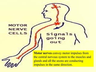
Our  nervous system  allows us to sense and respond to our external environment and also allows us to control the internal activities of our bodies to maintain homeostasis.
There is only one  nervous system .  However, anatomists and physiologists have found it useful to partition it into various divisions.  Structurally , it can be divided into the  central nervous system (CNS) and the peripheral nervous system (PNS).
The  peripheral nervous system  (PNS) includes the  cranial nerves  that arise from the brain and the  spinal nerves  that arise from the spinal cord.  Also included are  ganglia,  which are clusters of nerve cell bodies located outside the CNS.
Together the  CNS and PNS   collect information  with the help of receptors in the PNS,  process and evaluate sensory input  so the CNS can determine the appropriate response, and then  respond  by sending impulses via the PNS to effectors (muscles and glands).


Sympathetic division , which typically  speeds up  body activities Parasympathetic division , which typically  slows down  body activities
 
IF YOU HAD PAINFUL BLOATING OF YOUR STOMACH,  WHAT FUNCTIONAL DIVISION OF YOUR NERVOUS SYSTEM   WOULD BE RESPONSIBLE FOR SENDING IMPULSES  TO  YOUR CENTRAL NERVOUS SYSTEM (CNS)? A VISCERAL SENSORY B AUTONOMIC MOTOR C SOMATIC SENSORY D SOMATIC MOTOR E SYMPATHETIC DIVISION
There are  two distinct cell types  that form nervous tissue: Neurons , which are excitable Glial cells , which are supportive Nervous tissue cells of the  CNS
Neurons  are the basic structural unit of the nervous system and conduct impulses from on part of the body to another.  They have a high metabolic rate, they have extreme longevity, and most cease mitosis after fetal development.
Network of neurons Read  clinical view  about the possibility of new neurons in adults.
Dendrites  conduct impulses towards the  cell body  while  axons  conduct impulses away from the cell body.
 
Neurons  are classified into three types:  Sensory  (afferent) neurons,  motor  (efferent) neurons, and  interneurons  (association neurons).  It is estimated that 99% of all neurons are interneurons.
Glial cells  (neuroglial cells) are capable of mitosis, do support, nourish, and protect neurons, and collectively account for one-half the volume of the nervous system.  There are  four types  found in the CNS (shown above) and  two types  found in the PNS. Glial cells of the CNS
The most abundant type of glial cells are the  astrocytes , so named because of the cytoplasmic projections that give the cells a star-like shape.
Astrocytes  envelop the brain capillaries and reduce capillary permeability.  This helps create the  blood-brain barrier .
 
Ependymal cells  line the internal cavities (ventricles) of the brain and the central canal of the spinal cord (CNS).  These cells, and nearby capillaries, form a network called the  choroid plexus , which produces cerebrospinal fluid (CSF).
Choroid plexi  are formed by the ependymal cells that line the ventricles and by nearby blood vessels.  CSF is essentially a filtrate of plasma. In lateral ventricles of brain
Microglial cells  are wandering phagocytic cells that ingest and remove debris throughout the CNS.
Oligodendrocytes  are cells that ensheathe portions of axons in the CNS by repeatedly wrapping around an axon to create a  myelin sheath .
Oligodendrocytes  creating a  myelin sheath .
Satellite cells  are flattened cells that arrange around neuronal cell bodies in ganglia of the PNS and help regulate the movement of nutrients and waste products
Note numerous dark  satellite cells  arranged around pink neuronal cell bodies in a ganglion of the PNS.  They regulate the movement of nutrients and wastes. Satellite cells Cell bodies of a ganglion
Schwann cells  (neurolemmocytes) are responsible for forming the myelin sheath around axons in the PNS.
Schwann cells  (neurolemmocytes) in action in the PNS
Schwann cells  (neurolemmocytes) finishing up the job of myelination in the PNS.
Read about  tumors of the CNS  in the  clinical view  in the text.
ACCORDING TO THE CLINICAL VIEW ON TUMORS OF THE CNS, WHICH OF THE FOLLOWING IS  MOST LIKELY  TO DEVELOP INTO A NEOPLASM (TUMOR) IN THE CNS? A INTERNEURONS B AXONS C MITOCHONDRIA IN NERVE CELL BODIES D DENDRITES E ASTROCYTES
A  nerve  is a cable-like bundle of parallel axons.  While a single axon tends to be microscopic, nerves are larger and tend to be visible to the naked eye.
Sensory nerves  convey sensory information  to the central nervous system  and all axons are conducting impulses in the same direction.
Motor nerves  convey motor impulses from the central nervous system to the muscles and glands and all the axons are conducting impulses in the same direction.
Mixed nerves  carry both types of information and some axons are transmitting impulses in one direction, while other axons are transmitting impulses in the opposite direction.
Read about  nerve regeneration and spinal cord injuries  in the  clinical view  in your text.
Read about  nervous system disorders  in the clinical view in the text.
Nervous tissue development begins in the embryo during the third week of development.  A  neural groove  appears and by the end of the third week the sides of this groove come together and fuse along the midline to form a  neural tube .
The  neural tube  has an opening near the future head and an opening near the future buttocks.
If the openings in the neural tube do not close, the developing human will have a  neural tube defect .
Read about  neural tube defects  in the clinical view in the text.
The cranial part of the neural tube expands to form the  brain. The caudal part of the neural tube expands to form the  spinal cord .
ACCORDING TO THE CLINICAL VIEW ON NEURAL TUBE DEFECTS (NTDs), WHICH OF THE FOLLOWING IS  CORRECT ? A ANENCEPHALY CAN BE DETECTED WITH  ULTRASOUND B SPINA BIFIDA OFTEN CAUSES PARALYSIS C SPINA BIFIDA OCCULTA MAY BE  ASYMPTOMATIC D VITAMIN B 12  CAN REDUCE INCIDENCE OF  NTDs E ALL OF THE ABOVE
EVERYTHING PAST THIS POINT IS EXTRA OR FOR USE IN EXAMS
Neuroglial cells creating a  myelin sheath
Glial cell wrapped around neuron
 
Figure 14.co
Neurology  is the study of the nervous system


A stimulus , if sufficiently strong, will generate a nerve impulse (action potential)
Figure 14.01
The  central nervous system (CNS)  consists of the brain and spinal cord.  The  peripheral nervous system (PNS)  consists of the cranial nerves arising from the brain and the spinal nerves that arise from the spinal cord.
The  autonomic motor component  has  two subdivisions
Structural  division of the nervous system. Functional  division of the nervous system
The nervous system conveys information rapidly and enables us to respond to stimuli.
The nervous system enables us to orient our bodies to changes in the internal and external environment.
The nervous system allows coordination of body activities and assimilation of experiences for memory, learning, and intelligence.
The nervous system provides for  instinctual behavior , such as rooting and sucking to facilitate breast feeding.
The nervous system is composed of conductive  neurons  and supportive  neuroglia  (glial) cells
Neuroglial cells wrap a  myelin sheath  around the axons and dendrites of some nerve cells.  This sheath  is interrupted by Nodes of Ranvier .
Nerve impulse rapidly jumping from node to node.
Neurons can be  sensory  (afferent),  motor  (efferent), or i nterneurons  (found only in CNS) which connect the sensory and motor neurons
A  nerve  is a collection of nerve fibers (neurons) outside the CNS


Figure 14.04
Figure 14.09
Figure 14.03b


13 Nervous Tissue

  • 1.
    Our nervoussystem allows us to sense and respond to our external environment and also allows us to control the internal activities of our bodies to maintain homeostasis.
  • 2.
    There is onlyone nervous system . However, anatomists and physiologists have found it useful to partition it into various divisions. Structurally , it can be divided into the central nervous system (CNS) and the peripheral nervous system (PNS).
  • 3.
    The peripheralnervous system (PNS) includes the cranial nerves that arise from the brain and the spinal nerves that arise from the spinal cord. Also included are ganglia, which are clusters of nerve cell bodies located outside the CNS.
  • 4.
    Together the CNS and PNS collect information with the help of receptors in the PNS, process and evaluate sensory input so the CNS can determine the appropriate response, and then respond by sending impulses via the PNS to effectors (muscles and glands).
  • 5.
  • 6.
  • 7.
  • 8.
    Sympathetic division ,which typically speeds up body activities Parasympathetic division , which typically slows down body activities
  • 9.
  • 10.
    IF YOU HADPAINFUL BLOATING OF YOUR STOMACH, WHAT FUNCTIONAL DIVISION OF YOUR NERVOUS SYSTEM WOULD BE RESPONSIBLE FOR SENDING IMPULSES TO YOUR CENTRAL NERVOUS SYSTEM (CNS)? A VISCERAL SENSORY B AUTONOMIC MOTOR C SOMATIC SENSORY D SOMATIC MOTOR E SYMPATHETIC DIVISION
  • 11.
    There are two distinct cell types that form nervous tissue: Neurons , which are excitable Glial cells , which are supportive Nervous tissue cells of the CNS
  • 12.
    Neurons arethe basic structural unit of the nervous system and conduct impulses from on part of the body to another. They have a high metabolic rate, they have extreme longevity, and most cease mitosis after fetal development.
  • 13.
    Network of neuronsRead clinical view about the possibility of new neurons in adults.
  • 14.
    Dendrites conductimpulses towards the cell body while axons conduct impulses away from the cell body.
  • 15.
  • 16.
    Neurons areclassified into three types: Sensory (afferent) neurons, motor (efferent) neurons, and interneurons (association neurons). It is estimated that 99% of all neurons are interneurons.
  • 17.
    Glial cells (neuroglial cells) are capable of mitosis, do support, nourish, and protect neurons, and collectively account for one-half the volume of the nervous system. There are four types found in the CNS (shown above) and two types found in the PNS. Glial cells of the CNS
  • 18.
    The most abundanttype of glial cells are the astrocytes , so named because of the cytoplasmic projections that give the cells a star-like shape.
  • 19.
    Astrocytes envelopthe brain capillaries and reduce capillary permeability. This helps create the blood-brain barrier .
  • 20.
  • 21.
    Ependymal cells line the internal cavities (ventricles) of the brain and the central canal of the spinal cord (CNS). These cells, and nearby capillaries, form a network called the choroid plexus , which produces cerebrospinal fluid (CSF).
  • 22.
    Choroid plexi are formed by the ependymal cells that line the ventricles and by nearby blood vessels. CSF is essentially a filtrate of plasma. In lateral ventricles of brain
  • 23.
    Microglial cells are wandering phagocytic cells that ingest and remove debris throughout the CNS.
  • 24.
    Oligodendrocytes arecells that ensheathe portions of axons in the CNS by repeatedly wrapping around an axon to create a myelin sheath .
  • 25.
    Oligodendrocytes creatinga myelin sheath .
  • 26.
    Satellite cells are flattened cells that arrange around neuronal cell bodies in ganglia of the PNS and help regulate the movement of nutrients and waste products
  • 27.
    Note numerous dark satellite cells arranged around pink neuronal cell bodies in a ganglion of the PNS. They regulate the movement of nutrients and wastes. Satellite cells Cell bodies of a ganglion
  • 28.
    Schwann cells (neurolemmocytes) are responsible for forming the myelin sheath around axons in the PNS.
  • 29.
    Schwann cells (neurolemmocytes) in action in the PNS
  • 30.
    Schwann cells (neurolemmocytes) finishing up the job of myelination in the PNS.
  • 31.
    Read about tumors of the CNS in the clinical view in the text.
  • 32.
    ACCORDING TO THECLINICAL VIEW ON TUMORS OF THE CNS, WHICH OF THE FOLLOWING IS MOST LIKELY TO DEVELOP INTO A NEOPLASM (TUMOR) IN THE CNS? A INTERNEURONS B AXONS C MITOCHONDRIA IN NERVE CELL BODIES D DENDRITES E ASTROCYTES
  • 33.
    A nerve is a cable-like bundle of parallel axons. While a single axon tends to be microscopic, nerves are larger and tend to be visible to the naked eye.
  • 34.
    Sensory nerves convey sensory information to the central nervous system and all axons are conducting impulses in the same direction.
  • 35.
    Motor nerves convey motor impulses from the central nervous system to the muscles and glands and all the axons are conducting impulses in the same direction.
  • 36.
    Mixed nerves carry both types of information and some axons are transmitting impulses in one direction, while other axons are transmitting impulses in the opposite direction.
  • 37.
    Read about nerve regeneration and spinal cord injuries in the clinical view in your text.
  • 38.
    Read about nervous system disorders in the clinical view in the text.
  • 39.
    Nervous tissue developmentbegins in the embryo during the third week of development. A neural groove appears and by the end of the third week the sides of this groove come together and fuse along the midline to form a neural tube .
  • 40.
    The neuraltube has an opening near the future head and an opening near the future buttocks.
  • 41.
    If the openingsin the neural tube do not close, the developing human will have a neural tube defect .
  • 42.
    Read about neural tube defects in the clinical view in the text.
  • 43.
    The cranial partof the neural tube expands to form the brain. The caudal part of the neural tube expands to form the spinal cord .
  • 44.
    ACCORDING TO THECLINICAL VIEW ON NEURAL TUBE DEFECTS (NTDs), WHICH OF THE FOLLOWING IS CORRECT ? A ANENCEPHALY CAN BE DETECTED WITH ULTRASOUND B SPINA BIFIDA OFTEN CAUSES PARALYSIS C SPINA BIFIDA OCCULTA MAY BE ASYMPTOMATIC D VITAMIN B 12 CAN REDUCE INCIDENCE OF NTDs E ALL OF THE ABOVE
  • 45.
    EVERYTHING PAST THISPOINT IS EXTRA OR FOR USE IN EXAMS
  • 46.
  • 47.
    Glial cell wrappedaround neuron
  • 48.
  • 49.
  • 50.
    Neurology isthe study of the nervous system
  • 51.
  • 52.
  • 53.
  • 54.
  • 55.
    A stimulus ,if sufficiently strong, will generate a nerve impulse (action potential)
  • 56.
  • 57.
    The centralnervous system (CNS) consists of the brain and spinal cord. The peripheral nervous system (PNS) consists of the cranial nerves arising from the brain and the spinal nerves that arise from the spinal cord.
  • 58.
    The autonomicmotor component has two subdivisions
  • 59.
    Structural divisionof the nervous system. Functional division of the nervous system
  • 60.
    The nervous systemconveys information rapidly and enables us to respond to stimuli.
  • 61.
    The nervous systemenables us to orient our bodies to changes in the internal and external environment.
  • 62.
    The nervous systemallows coordination of body activities and assimilation of experiences for memory, learning, and intelligence.
  • 63.
    The nervous systemprovides for instinctual behavior , such as rooting and sucking to facilitate breast feeding.
  • 64.
    The nervous systemis composed of conductive neurons and supportive neuroglia (glial) cells
  • 65.
    Neuroglial cells wrapa myelin sheath around the axons and dendrites of some nerve cells. This sheath is interrupted by Nodes of Ranvier .
  • 66.
    Nerve impulse rapidlyjumping from node to node.
  • 67.
    Neurons can be sensory (afferent), motor (efferent), or i nterneurons (found only in CNS) which connect the sensory and motor neurons
  • 68.
    A nerve is a collection of nerve fibers (neurons) outside the CNS
  • 69.
  • 70.
  • 71.
  • 72.
  • 73.
  • 74.
  • 75.
  • 76.
  • 77.
  • 78.
  • 79.
  • 80.
  • 81.
  • 82.
  • 83.