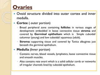
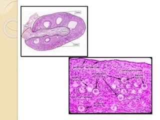
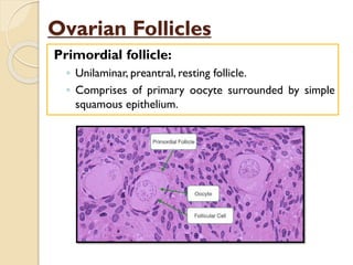
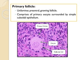
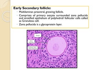
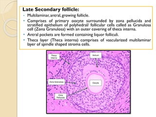
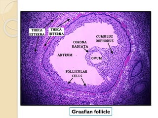
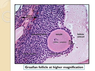
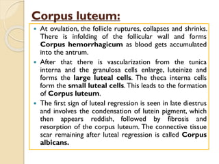
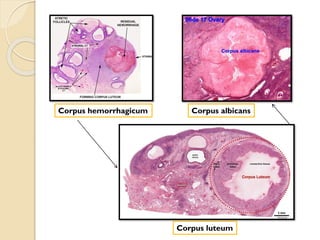
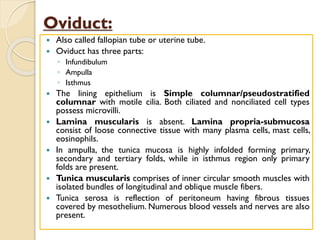
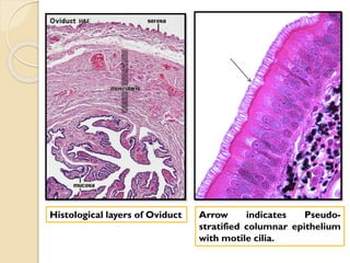
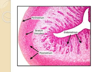
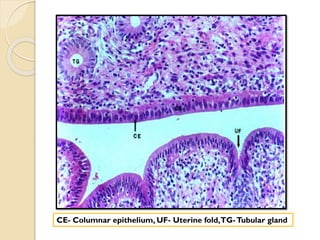
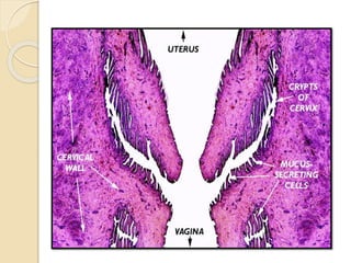
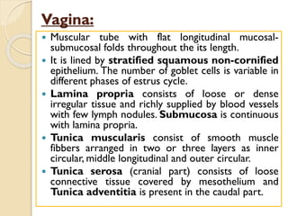
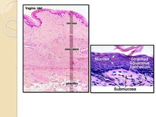
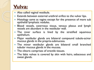
SlideShare a Scribd company logo
1 of 26
Download to read offline
Histology of Female
Reproductive System
Dr. Rajesh Ranjan
Assistant Professor
Deptt. ofVeterinary Anatomy
C.V.Sc., Rewa
Female Reproductive System
▪Ovaries
▪Oviducts
▪Uterus
▪Vagina
▪Vulva
Ovaries
 Ovoid structure divided into outer cortex and inner
medulla.
 Cortex ( outer portion)
◦ Broad peripheral zone containing follicles in various stages of
development embedded in loose connective tissue stroma and
covered by Germinal epithelium which is Simple cuboidal/
columnar (young) and low cuboidal/ squamous (adult).
◦ Stroma: supporting tissue and covered by Tunica albuginea just
beneath the germinal epithelium.
 Medulla (Inner portion)
◦ Contains nerves, blood vessels, lymphatics, loose connective tissue
and smooth muscles.
◦ Also contains rete ovarii which is a solid cellular cords or networks
of irregular channels lined by cuboidal epithelium.
Ovarian Follicles
Primordial follicle:
◦ Unilaminar, preantral, resting follicle.
◦ Comprises of primary oocyte surrounded by simple
squamous epithelium.
Primary follicle:
◦ Unilaminar, preantral, growing follicle.
◦ Comprises of primary oocyte surrounded by simple
cuboidal epithelium.
Early Secondary follicle:
◦ Multilaminar, preantral, growing follicle.
◦ Comprises of primary oocyte surrounded zona pellucida
and stratified epithelium of polyhedral/ follicular cells called
as Granulosa cell.
◦ Zona pellucida is a glycoprotein layer.
Late Secondary follicle:
• Multilaminar, antral, growing follicle.
• Comprises of primary oocyte surrounded by zona pellucida and
stratified epithelium of polyhedral/ follicular cells called as Granulosa
cell (Zona Granulosa) with an outer covering of theca interna.
• Antral pockets are formed containing liquor folliculi.
• Theca layer (Theca interna) comprises of vascularized multilaminar
layer of spindle shaped stroma cells.
Graafian follicle:
 Also calledVesicular/Tertiary follicle.
 Multilaminar, antral, growing follicle.
 Comprises of primary oocyte surrounded by Zona
pellucida, Granulosa cells (Stratum granulosum) with
Antrum andTheca layers.
 Primary oocyte is spherical with centrally located
nucleus having sparse chromatin network and prominent
nucleolus.
 The primary oocyte lies in the accumulation of granulosa
cells known as Cumulus oophorus.
 The innermost cell layer of cumulus oophorus
immediately surrounding the oocyte becomes columnar
and radially disposed called Corona radiata.
 Antrum is filled with liquor folliculi.
 The granulosa cells form a parietal follicular lining, the
Stratum granulosum.
◦ It comprises of polyhedral cells (basal layer may be
columnar).
 Some of the granulosa cells may contain large PAS-positive
inclusions called the Call-Exner bodies which represent
intracellular precursors of liquor folliculi.
 Theca layers: comprises of Theca interna (inner vascular
layer) and Theca externa (outer fibrous layer).
 Cells in the theca interna are spindle shaped with delicate
reticular fiber network.
 Theca externa is loose connective tissue layer with fibrocytes
arranged concentrically around the theca interna.
Graafian follicle
Graafian follicle at higher magnification
Corpus luteum:
 At ovulation, the follicle ruptures, collapses and shrinks.
There is infolding of the follicular wall and forms
Corpus hemorrhagicum as blood gets accumulated
into the antrum.
 After that there is vascularization from the tunica
interna and the granulosa cells enlarge, luteinize and
forms the large luteal cells. The theca interna cells
form the small luteal cells. This leads to the formation
of Corpus luteum.
 The first sign of luteal regression is seen in late diestrus
and involves the condensation of lutein pigment, which
then appears reddish, followed by fibrosis and
resorption of the corpus luteum. The connective tissue
scar remaining after luteal regression is called Corpus
albicans.
Corpus hemorrhagicum
Corpus luteum
Corpus albicans
Oviduct:
 Also called fallopian tube or uterine tube.
 Oviduct has three parts:
◦ Infundibulum
◦ Ampulla
◦ Isthmus
 The lining epithelium is Simple columnar/pseudostratified
columnar with motile cilia. Both ciliated and nonciliated cell types
possess microvilli.
 Lamina muscularis is absent. Lamina propria-submucosa
consist of loose connective tissue with many plasma cells, mast cells,
eosinophils.
 In ampulla, the tunica mucosa is highly infolded forming primary,
secondary and tertiary folds, while in isthmus region only primary
folds are present.
 Tunica muscularis comprises of inner circular smooth muscles with
isolated bundles of longitudinal and oblique muscle fibers.
 Tunica serosa is reflection of peritoneum having fibrous tissues
covered by mesothelium. Numerous blood vessels and nerves are also
present.
Histological layers of Oviduct Arrow indicates Pseudo-
stratified columnar epithelium
with motile cilia.
Uterus:
 Uterus has three parts: Two horns or cornua, Body or corpus and Neck
or cervix
 Histological structure of cornua and corpus:
 Endometrium (Tunica mucosa and submucosa)
 The lining epithelium is
◦ Simple columnar in mare, bitch, queen.
◦ Simple columnar/ pseudostratified columnar in sow, ruminants.
 Lamina propria- submucosa is divided into sub-epithelial functional zone
that comprises of richly vascular L.C.T. with many fibrocytes, macrophages,
mast cells, etc and a deep part comprising of L.C.T. and simple coiled
branched tubular glands. These glands are absent in caruncular areas in
ruminants. Uterine glands are lined by ciliated and non-ciliated simple
columnar epithelium.
 Myometrium (Tunica muscularis): Consists of thick inner circular layer
and outer longitudinal layer of smooth muscles. In between these two layers
stratum vasculare is present.
 Perimetrium (Tunica serosa): Consists of L.C.T. covered by mesothelium,
few smooth muscle cells, lymphatics, blood vessels and nerves.
CE- Columnar epithelium, UF- Uterine fold,TG-Tubular gland
Cervix (Neck of uterus):
 Thick walled, muscular tube rich in elastic fibers.
 The mucosa-submucosa forms high primary, secondary and
tertiary folds. The lining epithelium is simple columnar with
many mucigenous cells including goblet cells.
 Lamina propria-submucosa contains dense irregular
connective tissue with collagenous and elastic fibers. Intra-
epithelial and simple tubular glands are present in ruminants.
Uterine glands do not extend into the cervix.
 Tunica muscularis consists of Inner circular and outer
longitudinal muscle layers. Elastic fibers are prominent in the
circular layer.
 Tunica serosa contains loose connective tissue covered by
mesothelium.
Vagina:
 Muscular tube with flat longitudinal mucosal-
submucosal folds throughout the its length.
 It is lined by stratified squamous non-cornified
epithelium. The number of goblet cells is variable in
different phases of estrus cycle.
 Lamina propria consists of loose or dense
irregular tissue and richly supplied by blood vessels
with few lymph nodules. Submucosa is continuous
with lamina propria.
 Tunica muscularis consist of smooth muscle
fibbers arranged in two or three layers as inner
circular, middle longitudinal and outer circular.
 Tunica serosa (cranial part) consists of loose
connective tissue covered by mesothelium and
Tunica adventitia is present in the caudal part.
Vulva:
 Also called vaginal vestibule.
 Extends between external urethral orifice to the vulvar lips.
 Histology same as vagina except for the presence of more sub
epithelial lymphatic nodules.
 Blood vessels, cavernous tissue, venous plexus and lymph
vessels are abundant in the vestibular wall.
 The inner surface is lined by the stratified squamous
epithelium.
 Major vestibular glands are bilateral compound tubulo-acinar
mucous glands in the propria-submucosa.
 The minor vestibular glands are bilateral small branched
tubular mucous glands in the mucosa.
 The clitoris comprises of erectile tissues.
 The labia vulvae is covered by skin with hairs, sebaceous and
sweat glands.
Assignment
 Well labelled histological representation
of female genitalia of different species of
animals in practical notebook.
Thanks!

More Related Content

What's hot

Histology of stomach
Histology of stomachHistology of stomach
Histology of stomachTehseen Anwar
 
Histology slides with Identification Points 2nd yr mbbs Nishtar by Umer Salman
Histology slides with Identification Points 2nd yr mbbs Nishtar by Umer SalmanHistology slides with Identification Points 2nd yr mbbs Nishtar by Umer Salman
Histology slides with Identification Points 2nd yr mbbs Nishtar by Umer Salmanumersalman
 
12.08.08: Histology of the Urinary Tract
12.08.08: Histology of the Urinary Tract12.08.08: Histology of the Urinary Tract
12.08.08: Histology of the Urinary TractOpen.Michigan
 
Histology (histology of upper urinary tract)
Histology (histology of upper urinary tract)Histology (histology of upper urinary tract)
Histology (histology of upper urinary tract)Osama Al-Zahrani
 
Histological structure of pancreas
Histological structure of pancreasHistological structure of pancreas
Histological structure of pancreaschet08
 
Gross anatomy & histology of ileum, jejunum
Gross anatomy & histology of ileum, jejunumGross anatomy & histology of ileum, jejunum
Gross anatomy & histology of ileum, jejunumAbdul Ansari
 
histology of the tongue and salivary gland
histology of the tongue and salivary glandhistology of the tongue and salivary gland
histology of the tongue and salivary glandGbenga Pita Oderinde
 
Histology of testes & epididymis
Histology of testes & epididymisHistology of testes & epididymis
Histology of testes & epididymisRohit Paswan
 
General histology
General  histologyGeneral  histology
General histologyKemUnited
 
Adrenal gland Anatomy and histology
Adrenal gland  Anatomy and histologyAdrenal gland  Anatomy and histology
Adrenal gland Anatomy and histologyDrsurajkc
 
Histology of cns.dk.2014
Histology of cns.dk.2014Histology of cns.dk.2014
Histology of cns.dk.2014Deepak Khedekar
 
Histology of Gall bladder
Histology of Gall bladderHistology of Gall bladder
Histology of Gall bladderNahry Omer
 
Histology of respiratory system
Histology of respiratory systemHistology of respiratory system
Histology of respiratory systemAddisu Alemu
 

What's hot (20)

Histology of stomach
Histology of stomachHistology of stomach
Histology of stomach
 
Histology slides with Identification Points 2nd yr mbbs Nishtar by Umer Salman
Histology slides with Identification Points 2nd yr mbbs Nishtar by Umer SalmanHistology slides with Identification Points 2nd yr mbbs Nishtar by Umer Salman
Histology slides with Identification Points 2nd yr mbbs Nishtar by Umer Salman
 
12.08.08: Histology of the Urinary Tract
12.08.08: Histology of the Urinary Tract12.08.08: Histology of the Urinary Tract
12.08.08: Histology of the Urinary Tract
 
Histology (histology of upper urinary tract)
Histology (histology of upper urinary tract)Histology (histology of upper urinary tract)
Histology (histology of upper urinary tract)
 
Histological structure of pancreas
Histological structure of pancreasHistological structure of pancreas
Histological structure of pancreas
 
histology of digestive system (1)
histology of digestive system  (1)histology of digestive system  (1)
histology of digestive system (1)
 
Gross anatomy & histology of ileum, jejunum
Gross anatomy & histology of ileum, jejunumGross anatomy & histology of ileum, jejunum
Gross anatomy & histology of ileum, jejunum
 
Histology of Stomach
Histology of StomachHistology of Stomach
Histology of Stomach
 
histology of the tongue and salivary gland
histology of the tongue and salivary glandhistology of the tongue and salivary gland
histology of the tongue and salivary gland
 
Histology of testes & epididymis
Histology of testes & epididymisHistology of testes & epididymis
Histology of testes & epididymis
 
Histology of digestive system 2
Histology of digestive system 2Histology of digestive system 2
Histology of digestive system 2
 
Histology of lymphatic system
Histology of lymphatic systemHistology of lymphatic system
Histology of lymphatic system
 
General histology
General  histologyGeneral  histology
General histology
 
Adrenal gland Anatomy and histology
Adrenal gland  Anatomy and histologyAdrenal gland  Anatomy and histology
Adrenal gland Anatomy and histology
 
Histology of cns.dk.2014
Histology of cns.dk.2014Histology of cns.dk.2014
Histology of cns.dk.2014
 
Histology of liver ppt
Histology of liver pptHistology of liver ppt
Histology of liver ppt
 
Histology of Lung
Histology of LungHistology of Lung
Histology of Lung
 
Histology of Gall bladder
Histology of Gall bladderHistology of Gall bladder
Histology of Gall bladder
 
Histology 1st year medschool
Histology 1st year medschoolHistology 1st year medschool
Histology 1st year medschool
 
Histology of respiratory system
Histology of respiratory systemHistology of respiratory system
Histology of respiratory system
 

Similar to histology-of-female-reproductive-system.pdf

Female reproductive system
Female  reproductive systemFemale  reproductive system
Female reproductive systemReach Na
 
Histology Of Female Genital Tract
Histology Of Female Genital TractHistology Of Female Genital Tract
Histology Of Female Genital Tractdreyngerous
 
Urinary system
Urinary systemUrinary system
Urinary systemReach Na
 
Digestiv tube.english histology
Digestiv tube.english histologyDigestiv tube.english histology
Digestiv tube.english histologyHasan Sultan
 
Histology-of-urinary-system.pdf
Histology-of-urinary-system.pdfHistology-of-urinary-system.pdf
Histology-of-urinary-system.pdfHema752685
 
female reproductive system.pptx
female reproductive system.pptxfemale reproductive system.pptx
female reproductive system.pptxKalanaDenuwan
 
STPM: Specialization of animal cells
STPM: Specialization of animal cellsSTPM: Specialization of animal cells
STPM: Specialization of animal cellsShaleni Kavirajan
 
HAP 8 semester 2 PCI FIRST YEAR BPHARMACY
HAP 8 semester 2 PCI FIRST YEAR BPHARMACYHAP 8 semester 2 PCI FIRST YEAR BPHARMACY
HAP 8 semester 2 PCI FIRST YEAR BPHARMACYchristinajohn24
 
GROUP 11 FINAL 2023.pptx
GROUP 11 FINAL 2023.pptxGROUP 11 FINAL 2023.pptx
GROUP 11 FINAL 2023.pptxBoas4
 
914909 female-reproductive
914909 female-reproductive914909 female-reproductive
914909 female-reproductiveabctutor
 
914909 female-reproductive
914909 female-reproductive914909 female-reproductive
914909 female-reproductiveYoAmoNYC
 
Lect. 3 Epithelial Tissues
Lect. 3 Epithelial TissuesLect. 3 Epithelial Tissues
Lect. 3 Epithelial TissuesHara O.
 
Eithelial tissue,connective tissue by Dr Jyoti Prakash Pani Associate Profess...
Eithelial tissue,connective tissue by Dr Jyoti Prakash Pani Associate Profess...Eithelial tissue,connective tissue by Dr Jyoti Prakash Pani Associate Profess...
Eithelial tissue,connective tissue by Dr Jyoti Prakash Pani Associate Profess...phupu
 
Histology slide guide
Histology slide guideHistology slide guide
Histology slide guideMUBOSScz
 

Similar to histology-of-female-reproductive-system.pdf (20)

Female reproductive system
Female  reproductive systemFemale  reproductive system
Female reproductive system
 
3.tissues of body
3.tissues of body3.tissues of body
3.tissues of body
 
Histology Of Female Genital Tract
Histology Of Female Genital TractHistology Of Female Genital Tract
Histology Of Female Genital Tract
 
Urinary system
Urinary systemUrinary system
Urinary system
 
Digestiv tube.english histology
Digestiv tube.english histologyDigestiv tube.english histology
Digestiv tube.english histology
 
Histology-of-urinary-system.pdf
Histology-of-urinary-system.pdfHistology-of-urinary-system.pdf
Histology-of-urinary-system.pdf
 
female reproductive system.pptx
female reproductive system.pptxfemale reproductive system.pptx
female reproductive system.pptx
 
Tissues
TissuesTissues
Tissues
 
STPM: Specialization of animal cells
STPM: Specialization of animal cellsSTPM: Specialization of animal cells
STPM: Specialization of animal cells
 
HAP 8 semester 2 PCI FIRST YEAR BPHARMACY
HAP 8 semester 2 PCI FIRST YEAR BPHARMACYHAP 8 semester 2 PCI FIRST YEAR BPHARMACY
HAP 8 semester 2 PCI FIRST YEAR BPHARMACY
 
Female2
Female2Female2
Female2
 
GROUP 11 FINAL 2023.pptx
GROUP 11 FINAL 2023.pptxGROUP 11 FINAL 2023.pptx
GROUP 11 FINAL 2023.pptx
 
Animal tissues epithelial & connective
Animal tissues epithelial & connectiveAnimal tissues epithelial & connective
Animal tissues epithelial & connective
 
Animal tissues epithelial & connective
Animal tissues epithelial & connectiveAnimal tissues epithelial & connective
Animal tissues epithelial & connective
 
914909 female-reproductive
914909 female-reproductive914909 female-reproductive
914909 female-reproductive
 
914909 female-reproductive
914909 female-reproductive914909 female-reproductive
914909 female-reproductive
 
5.TISSUES.pdf
5.TISSUES.pdf5.TISSUES.pdf
5.TISSUES.pdf
 
Lect. 3 Epithelial Tissues
Lect. 3 Epithelial TissuesLect. 3 Epithelial Tissues
Lect. 3 Epithelial Tissues
 
Eithelial tissue,connective tissue by Dr Jyoti Prakash Pani Associate Profess...
Eithelial tissue,connective tissue by Dr Jyoti Prakash Pani Associate Profess...Eithelial tissue,connective tissue by Dr Jyoti Prakash Pani Associate Profess...
Eithelial tissue,connective tissue by Dr Jyoti Prakash Pani Associate Profess...
 
Histology slide guide
Histology slide guideHistology slide guide
Histology slide guide
 

Recently uploaded

BEST ✨ Call Girls In Park Plaza Faridabad ✔️ 9871031762 ✔️ Escorts Service In...
BEST ✨ Call Girls In Park Plaza Faridabad ✔️ 9871031762 ✔️ Escorts Service In...BEST ✨ Call Girls In Park Plaza Faridabad ✔️ 9871031762 ✔️ Escorts Service In...
BEST ✨ Call Girls In Park Plaza Faridabad ✔️ 9871031762 ✔️ Escorts Service In...noida100girls
 
BEST ✨Call Girls In Park Plaza Gurugram ✔️9773824855✔️ Escorts Service In Del...
BEST ✨Call Girls In Park Plaza Gurugram ✔️9773824855✔️ Escorts Service In Del...BEST ✨Call Girls In Park Plaza Gurugram ✔️9773824855✔️ Escorts Service In Del...
BEST ✨Call Girls In Park Plaza Gurugram ✔️9773824855✔️ Escorts Service In Del...noida100girls
 
Call Girls In Hauz Khas Delhi 9654467111 Independent Escorts Service
Call Girls In Hauz Khas Delhi 9654467111 Independent Escorts ServiceCall Girls In Hauz Khas Delhi 9654467111 Independent Escorts Service
Call Girls In Hauz Khas Delhi 9654467111 Independent Escorts ServiceSapana Sha
 
HI-Profiles Call girls in Hyatt Residency Delhi | 8377087607
HI-Profiles Call girls in Hyatt Residency Delhi | 8377087607HI-Profiles Call girls in Hyatt Residency Delhi | 8377087607
HI-Profiles Call girls in Hyatt Residency Delhi | 8377087607dollysharma2066
 
BEST ✨ Call Girls In MG Road Gurgaon ✔️ 9871031762 ✔️ Escorts Service In De...
BEST ✨ Call Girls In  MG Road Gurgaon  ✔️ 9871031762 ✔️ Escorts Service In De...BEST ✨ Call Girls In  MG Road Gurgaon  ✔️ 9871031762 ✔️ Escorts Service In De...
BEST ✨ Call Girls In MG Road Gurgaon ✔️ 9871031762 ✔️ Escorts Service In De...noida100girls
 
办理学位证(USC文凭证书)南加州大学毕业证成绩单原版一模一样
办理学位证(USC文凭证书)南加州大学毕业证成绩单原版一模一样办理学位证(USC文凭证书)南加州大学毕业证成绩单原版一模一样
办理学位证(USC文凭证书)南加州大学毕业证成绩单原版一模一样7pn7zv3i
 
NIGHT DREAN Genuine Call girls in Vasant Vihar Delhi | 83778 77756
NIGHT DREAN Genuine Call girls in Vasant Vihar Delhi | 83778 77756NIGHT DREAN Genuine Call girls in Vasant Vihar Delhi | 83778 77756
NIGHT DREAN Genuine Call girls in Vasant Vihar Delhi | 83778 77756dollysharma2066
 
BEST ✨ Call Girls In Greater Noida ✔️ 9871031762 ✔️ Escorts Service In Delhi ...
BEST ✨ Call Girls In Greater Noida ✔️ 9871031762 ✔️ Escorts Service In Delhi ...BEST ✨ Call Girls In Greater Noida ✔️ 9871031762 ✔️ Escorts Service In Delhi ...
BEST ✨ Call Girls In Greater Noida ✔️ 9871031762 ✔️ Escorts Service In Delhi ...noida100girls
 

Recently uploaded (9)

BEST ✨ Call Girls In Park Plaza Faridabad ✔️ 9871031762 ✔️ Escorts Service In...
BEST ✨ Call Girls In Park Plaza Faridabad ✔️ 9871031762 ✔️ Escorts Service In...BEST ✨ Call Girls In Park Plaza Faridabad ✔️ 9871031762 ✔️ Escorts Service In...
BEST ✨ Call Girls In Park Plaza Faridabad ✔️ 9871031762 ✔️ Escorts Service In...
 
BEST ✨Call Girls In Park Plaza Gurugram ✔️9773824855✔️ Escorts Service In Del...
BEST ✨Call Girls In Park Plaza Gurugram ✔️9773824855✔️ Escorts Service In Del...BEST ✨Call Girls In Park Plaza Gurugram ✔️9773824855✔️ Escorts Service In Del...
BEST ✨Call Girls In Park Plaza Gurugram ✔️9773824855✔️ Escorts Service In Del...
 
Call Girls In Hauz Khas Delhi 9654467111 Independent Escorts Service
Call Girls In Hauz Khas Delhi 9654467111 Independent Escorts ServiceCall Girls In Hauz Khas Delhi 9654467111 Independent Escorts Service
Call Girls In Hauz Khas Delhi 9654467111 Independent Escorts Service
 
Cheap Rate ➥8448380779 ▻Call Girls In Sector 57 Gurgaon
Cheap Rate ➥8448380779 ▻Call Girls In Sector 57 GurgaonCheap Rate ➥8448380779 ▻Call Girls In Sector 57 Gurgaon
Cheap Rate ➥8448380779 ▻Call Girls In Sector 57 Gurgaon
 
HI-Profiles Call girls in Hyatt Residency Delhi | 8377087607
HI-Profiles Call girls in Hyatt Residency Delhi | 8377087607HI-Profiles Call girls in Hyatt Residency Delhi | 8377087607
HI-Profiles Call girls in Hyatt Residency Delhi | 8377087607
 
BEST ✨ Call Girls In MG Road Gurgaon ✔️ 9871031762 ✔️ Escorts Service In De...
BEST ✨ Call Girls In  MG Road Gurgaon  ✔️ 9871031762 ✔️ Escorts Service In De...BEST ✨ Call Girls In  MG Road Gurgaon  ✔️ 9871031762 ✔️ Escorts Service In De...
BEST ✨ Call Girls In MG Road Gurgaon ✔️ 9871031762 ✔️ Escorts Service In De...
 
办理学位证(USC文凭证书)南加州大学毕业证成绩单原版一模一样
办理学位证(USC文凭证书)南加州大学毕业证成绩单原版一模一样办理学位证(USC文凭证书)南加州大学毕业证成绩单原版一模一样
办理学位证(USC文凭证书)南加州大学毕业证成绩单原版一模一样
 
NIGHT DREAN Genuine Call girls in Vasant Vihar Delhi | 83778 77756
NIGHT DREAN Genuine Call girls in Vasant Vihar Delhi | 83778 77756NIGHT DREAN Genuine Call girls in Vasant Vihar Delhi | 83778 77756
NIGHT DREAN Genuine Call girls in Vasant Vihar Delhi | 83778 77756
 
BEST ✨ Call Girls In Greater Noida ✔️ 9871031762 ✔️ Escorts Service In Delhi ...
BEST ✨ Call Girls In Greater Noida ✔️ 9871031762 ✔️ Escorts Service In Delhi ...BEST ✨ Call Girls In Greater Noida ✔️ 9871031762 ✔️ Escorts Service In Delhi ...
BEST ✨ Call Girls In Greater Noida ✔️ 9871031762 ✔️ Escorts Service In Delhi ...
 

histology-of-female-reproductive-system.pdf

  • 1. Histology of Female Reproductive System Dr. Rajesh Ranjan Assistant Professor Deptt. ofVeterinary Anatomy C.V.Sc., Rewa
  • 3. Ovaries  Ovoid structure divided into outer cortex and inner medulla.  Cortex ( outer portion) ◦ Broad peripheral zone containing follicles in various stages of development embedded in loose connective tissue stroma and covered by Germinal epithelium which is Simple cuboidal/ columnar (young) and low cuboidal/ squamous (adult). ◦ Stroma: supporting tissue and covered by Tunica albuginea just beneath the germinal epithelium.  Medulla (Inner portion) ◦ Contains nerves, blood vessels, lymphatics, loose connective tissue and smooth muscles. ◦ Also contains rete ovarii which is a solid cellular cords or networks of irregular channels lined by cuboidal epithelium.
  • 4.
  • 5. Ovarian Follicles Primordial follicle: ◦ Unilaminar, preantral, resting follicle. ◦ Comprises of primary oocyte surrounded by simple squamous epithelium.
  • 6. Primary follicle: ◦ Unilaminar, preantral, growing follicle. ◦ Comprises of primary oocyte surrounded by simple cuboidal epithelium.
  • 7. Early Secondary follicle: ◦ Multilaminar, preantral, growing follicle. ◦ Comprises of primary oocyte surrounded zona pellucida and stratified epithelium of polyhedral/ follicular cells called as Granulosa cell. ◦ Zona pellucida is a glycoprotein layer.
  • 8. Late Secondary follicle: • Multilaminar, antral, growing follicle. • Comprises of primary oocyte surrounded by zona pellucida and stratified epithelium of polyhedral/ follicular cells called as Granulosa cell (Zona Granulosa) with an outer covering of theca interna. • Antral pockets are formed containing liquor folliculi. • Theca layer (Theca interna) comprises of vascularized multilaminar layer of spindle shaped stroma cells.
  • 9. Graafian follicle:  Also calledVesicular/Tertiary follicle.  Multilaminar, antral, growing follicle.  Comprises of primary oocyte surrounded by Zona pellucida, Granulosa cells (Stratum granulosum) with Antrum andTheca layers.  Primary oocyte is spherical with centrally located nucleus having sparse chromatin network and prominent nucleolus.  The primary oocyte lies in the accumulation of granulosa cells known as Cumulus oophorus.  The innermost cell layer of cumulus oophorus immediately surrounding the oocyte becomes columnar and radially disposed called Corona radiata.
  • 10.  Antrum is filled with liquor folliculi.  The granulosa cells form a parietal follicular lining, the Stratum granulosum. ◦ It comprises of polyhedral cells (basal layer may be columnar).  Some of the granulosa cells may contain large PAS-positive inclusions called the Call-Exner bodies which represent intracellular precursors of liquor folliculi.  Theca layers: comprises of Theca interna (inner vascular layer) and Theca externa (outer fibrous layer).  Cells in the theca interna are spindle shaped with delicate reticular fiber network.  Theca externa is loose connective tissue layer with fibrocytes arranged concentrically around the theca interna.
  • 12. Graafian follicle at higher magnification
  • 13. Corpus luteum:  At ovulation, the follicle ruptures, collapses and shrinks. There is infolding of the follicular wall and forms Corpus hemorrhagicum as blood gets accumulated into the antrum.  After that there is vascularization from the tunica interna and the granulosa cells enlarge, luteinize and forms the large luteal cells. The theca interna cells form the small luteal cells. This leads to the formation of Corpus luteum.  The first sign of luteal regression is seen in late diestrus and involves the condensation of lutein pigment, which then appears reddish, followed by fibrosis and resorption of the corpus luteum. The connective tissue scar remaining after luteal regression is called Corpus albicans.
  • 15. Oviduct:  Also called fallopian tube or uterine tube.  Oviduct has three parts: ◦ Infundibulum ◦ Ampulla ◦ Isthmus  The lining epithelium is Simple columnar/pseudostratified columnar with motile cilia. Both ciliated and nonciliated cell types possess microvilli.  Lamina muscularis is absent. Lamina propria-submucosa consist of loose connective tissue with many plasma cells, mast cells, eosinophils.  In ampulla, the tunica mucosa is highly infolded forming primary, secondary and tertiary folds, while in isthmus region only primary folds are present.  Tunica muscularis comprises of inner circular smooth muscles with isolated bundles of longitudinal and oblique muscle fibers.  Tunica serosa is reflection of peritoneum having fibrous tissues covered by mesothelium. Numerous blood vessels and nerves are also present.
  • 16. Histological layers of Oviduct Arrow indicates Pseudo- stratified columnar epithelium with motile cilia.
  • 17. Uterus:  Uterus has three parts: Two horns or cornua, Body or corpus and Neck or cervix  Histological structure of cornua and corpus:  Endometrium (Tunica mucosa and submucosa)  The lining epithelium is ◦ Simple columnar in mare, bitch, queen. ◦ Simple columnar/ pseudostratified columnar in sow, ruminants.  Lamina propria- submucosa is divided into sub-epithelial functional zone that comprises of richly vascular L.C.T. with many fibrocytes, macrophages, mast cells, etc and a deep part comprising of L.C.T. and simple coiled branched tubular glands. These glands are absent in caruncular areas in ruminants. Uterine glands are lined by ciliated and non-ciliated simple columnar epithelium.  Myometrium (Tunica muscularis): Consists of thick inner circular layer and outer longitudinal layer of smooth muscles. In between these two layers stratum vasculare is present.  Perimetrium (Tunica serosa): Consists of L.C.T. covered by mesothelium, few smooth muscle cells, lymphatics, blood vessels and nerves.
  • 18.
  • 19. CE- Columnar epithelium, UF- Uterine fold,TG-Tubular gland
  • 20. Cervix (Neck of uterus):  Thick walled, muscular tube rich in elastic fibers.  The mucosa-submucosa forms high primary, secondary and tertiary folds. The lining epithelium is simple columnar with many mucigenous cells including goblet cells.  Lamina propria-submucosa contains dense irregular connective tissue with collagenous and elastic fibers. Intra- epithelial and simple tubular glands are present in ruminants. Uterine glands do not extend into the cervix.  Tunica muscularis consists of Inner circular and outer longitudinal muscle layers. Elastic fibers are prominent in the circular layer.  Tunica serosa contains loose connective tissue covered by mesothelium.
  • 21.
  • 22. Vagina:  Muscular tube with flat longitudinal mucosal- submucosal folds throughout the its length.  It is lined by stratified squamous non-cornified epithelium. The number of goblet cells is variable in different phases of estrus cycle.  Lamina propria consists of loose or dense irregular tissue and richly supplied by blood vessels with few lymph nodules. Submucosa is continuous with lamina propria.  Tunica muscularis consist of smooth muscle fibbers arranged in two or three layers as inner circular, middle longitudinal and outer circular.  Tunica serosa (cranial part) consists of loose connective tissue covered by mesothelium and Tunica adventitia is present in the caudal part.
  • 23.
  • 24. Vulva:  Also called vaginal vestibule.  Extends between external urethral orifice to the vulvar lips.  Histology same as vagina except for the presence of more sub epithelial lymphatic nodules.  Blood vessels, cavernous tissue, venous plexus and lymph vessels are abundant in the vestibular wall.  The inner surface is lined by the stratified squamous epithelium.  Major vestibular glands are bilateral compound tubulo-acinar mucous glands in the propria-submucosa.  The minor vestibular glands are bilateral small branched tubular mucous glands in the mucosa.  The clitoris comprises of erectile tissues.  The labia vulvae is covered by skin with hairs, sebaceous and sweat glands.
  • 25. Assignment  Well labelled histological representation of female genitalia of different species of animals in practical notebook.