The document describes the nervous system and its divisions. It includes:
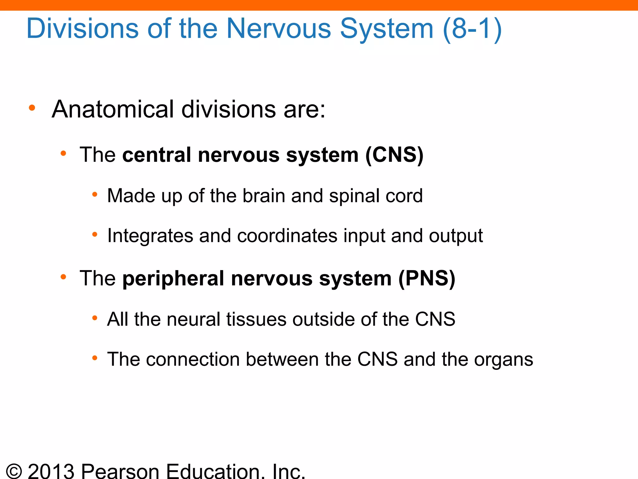
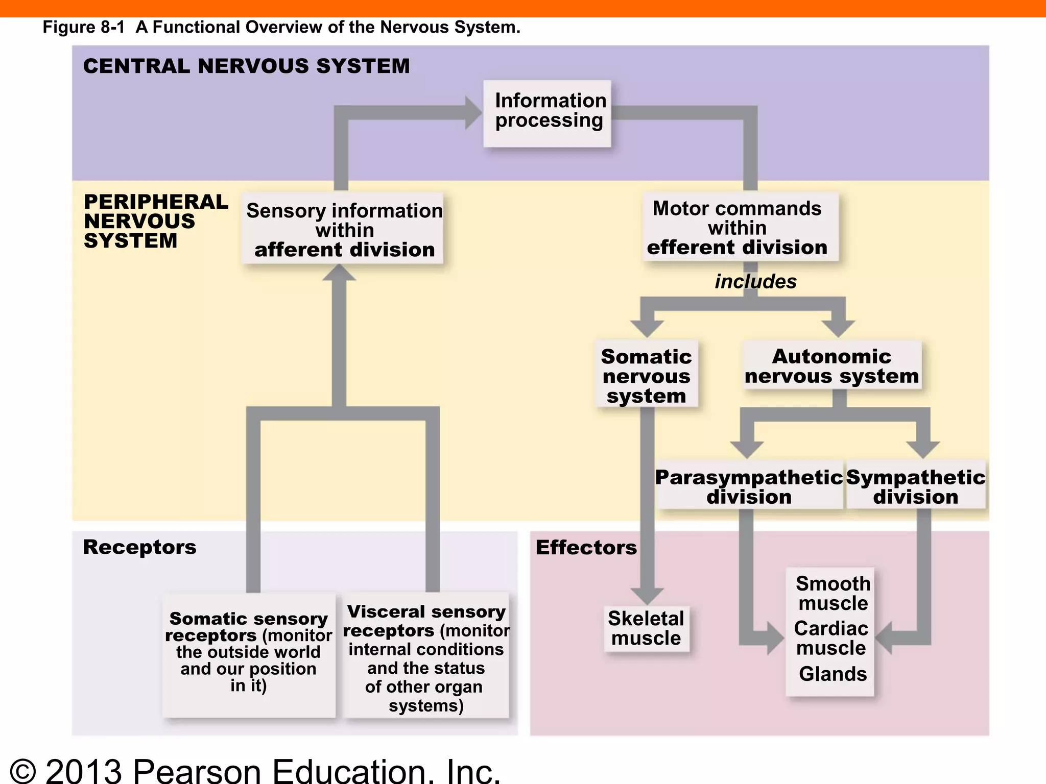
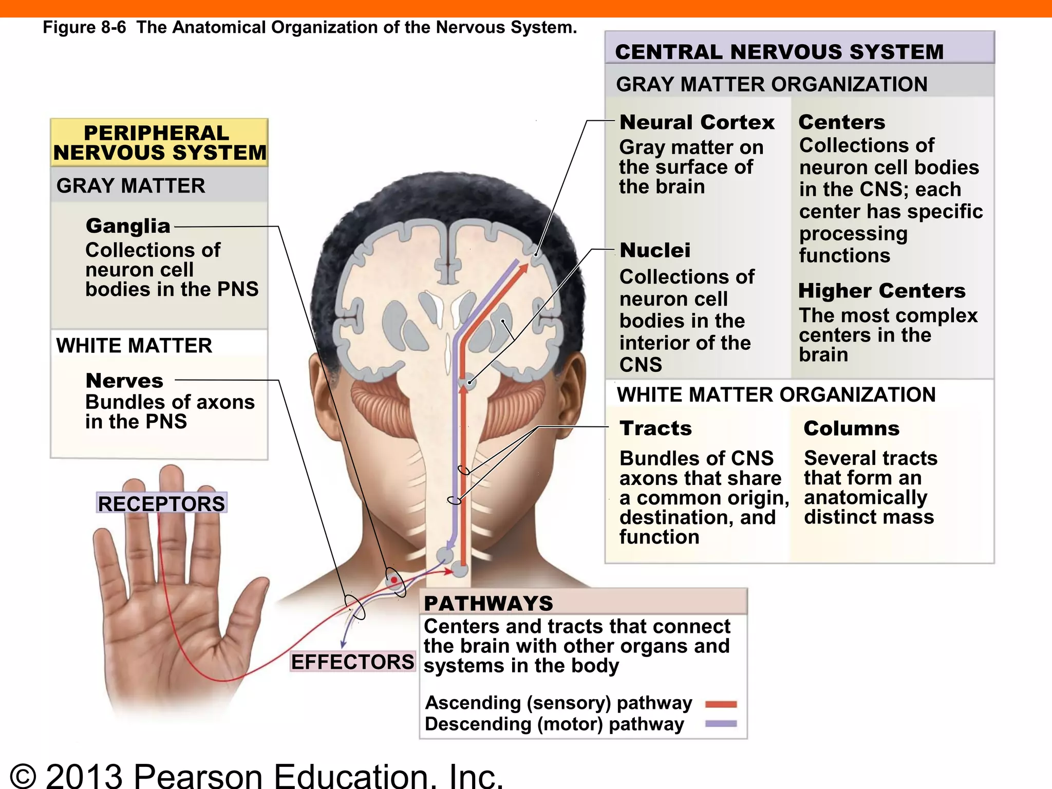
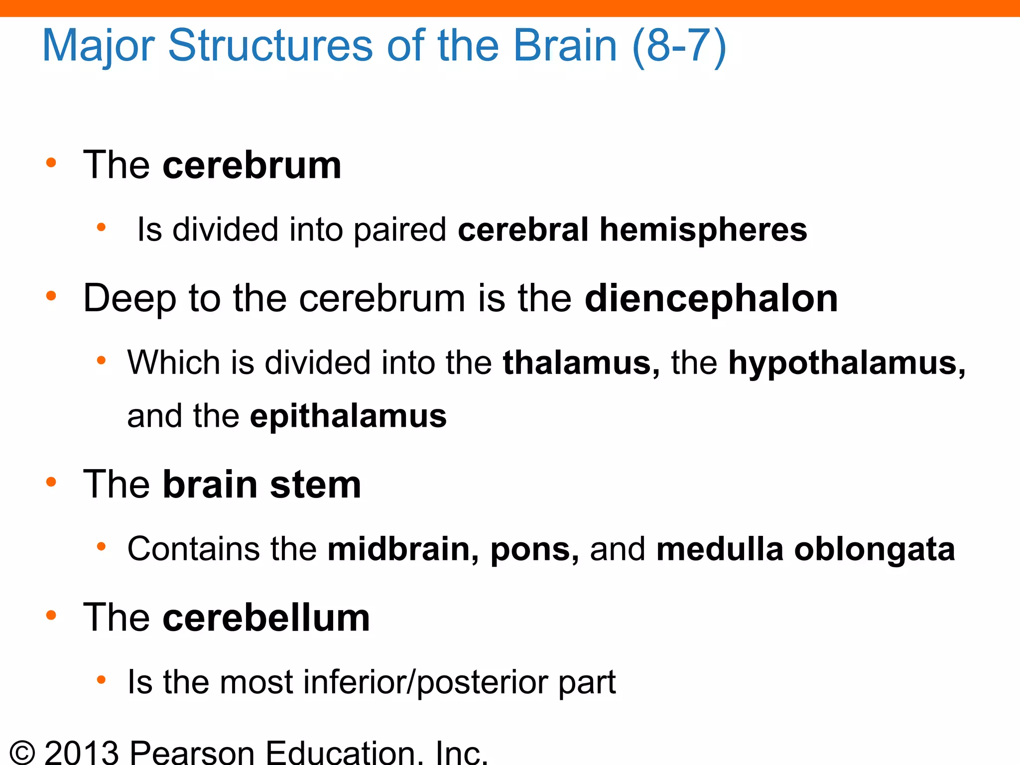
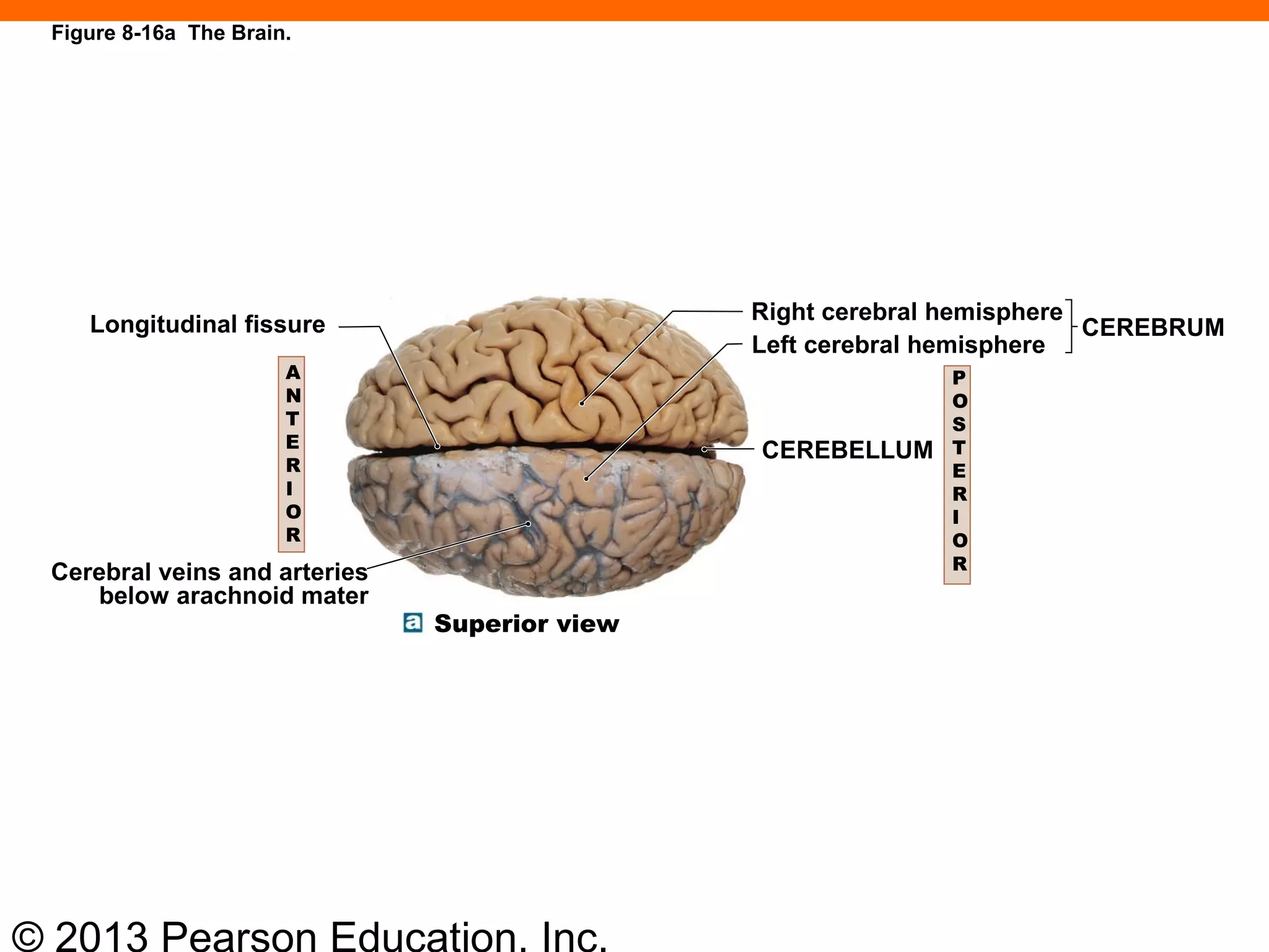
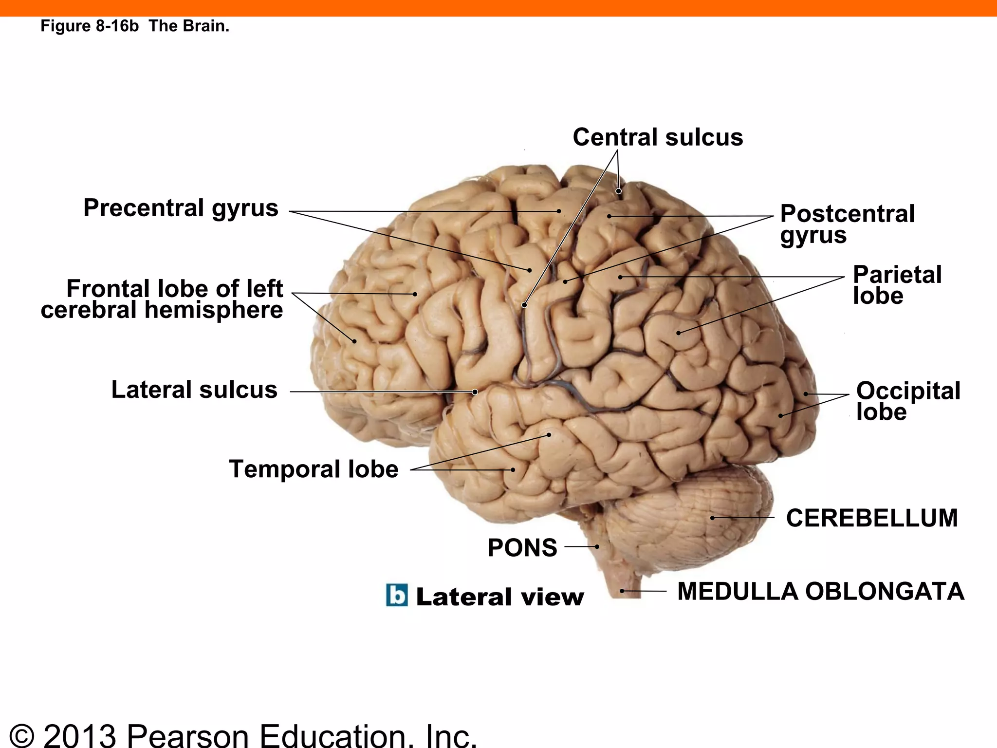
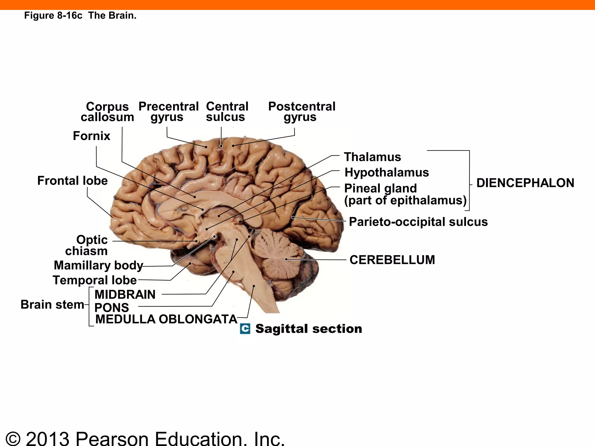
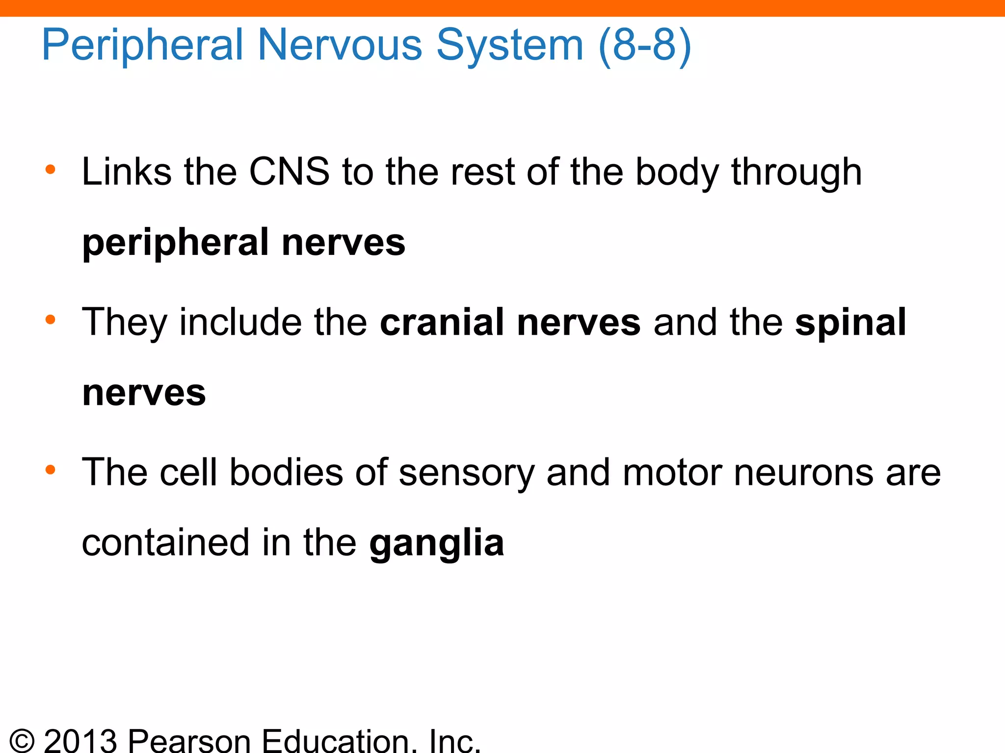
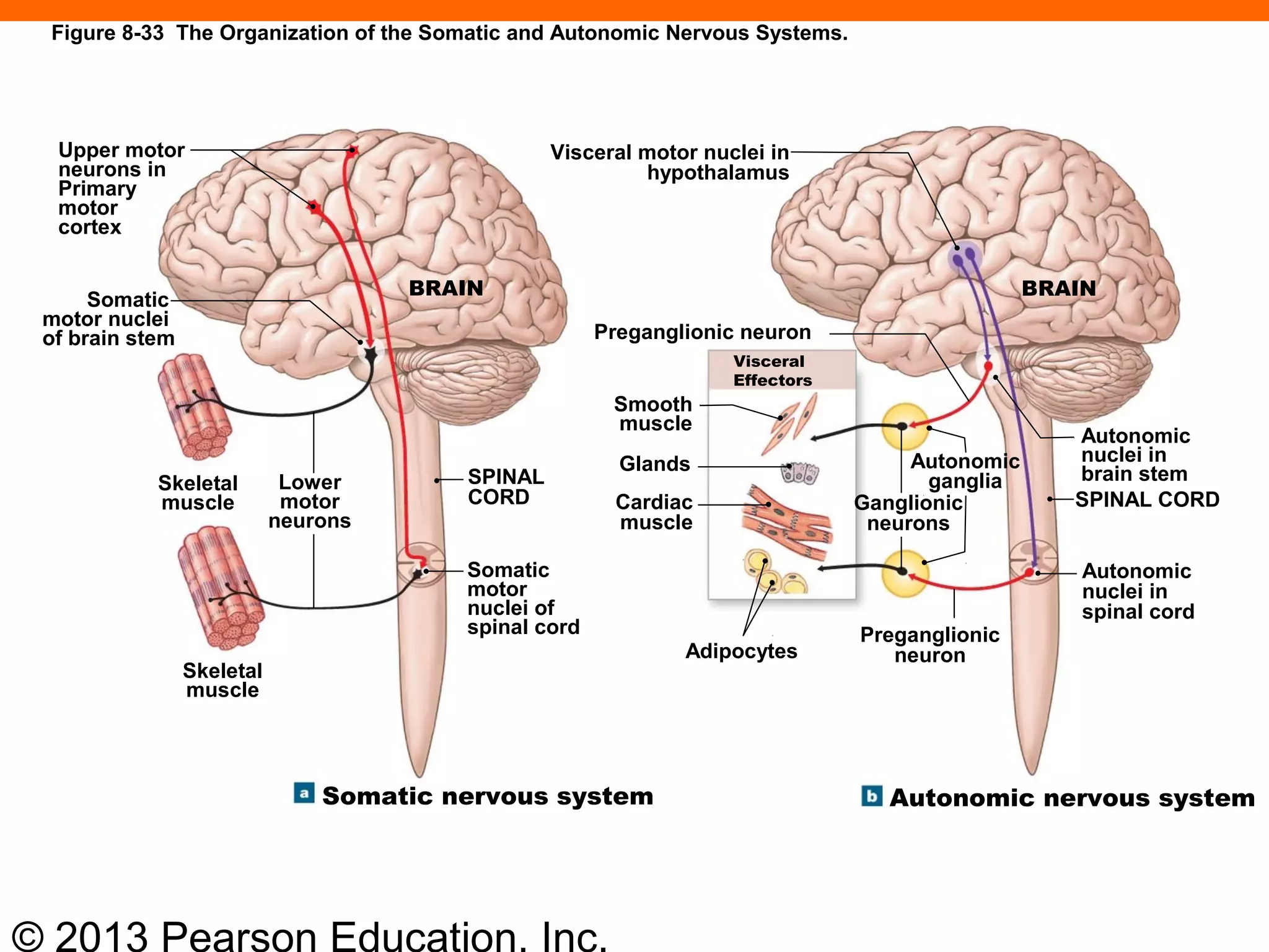
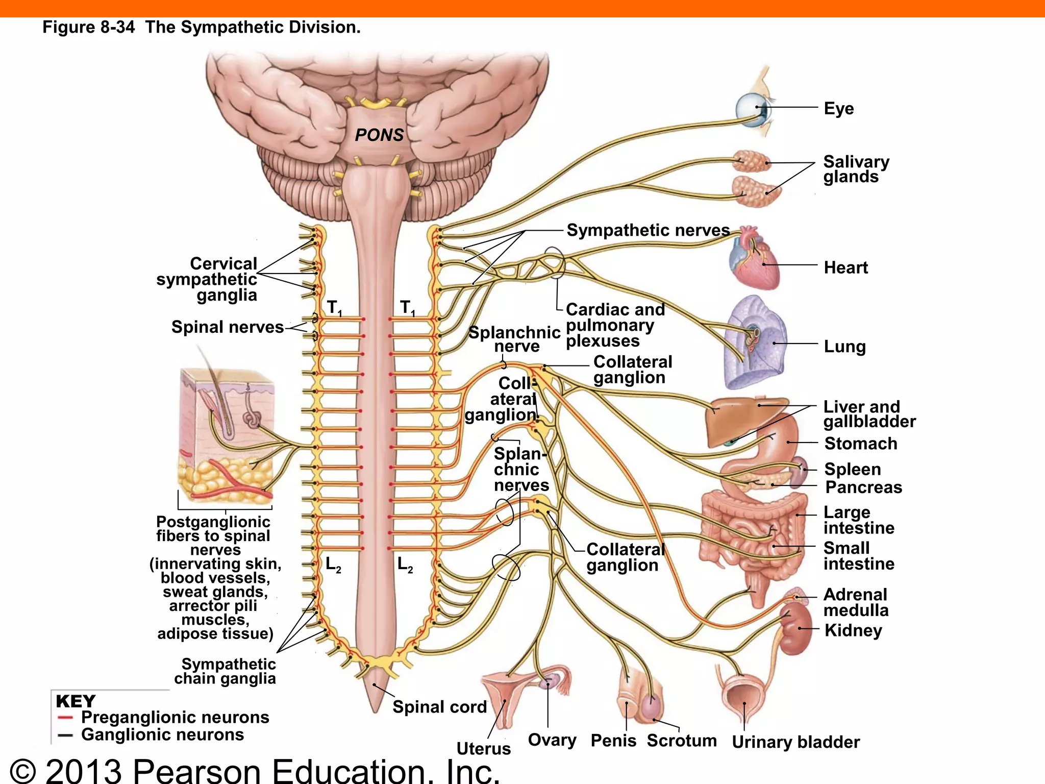
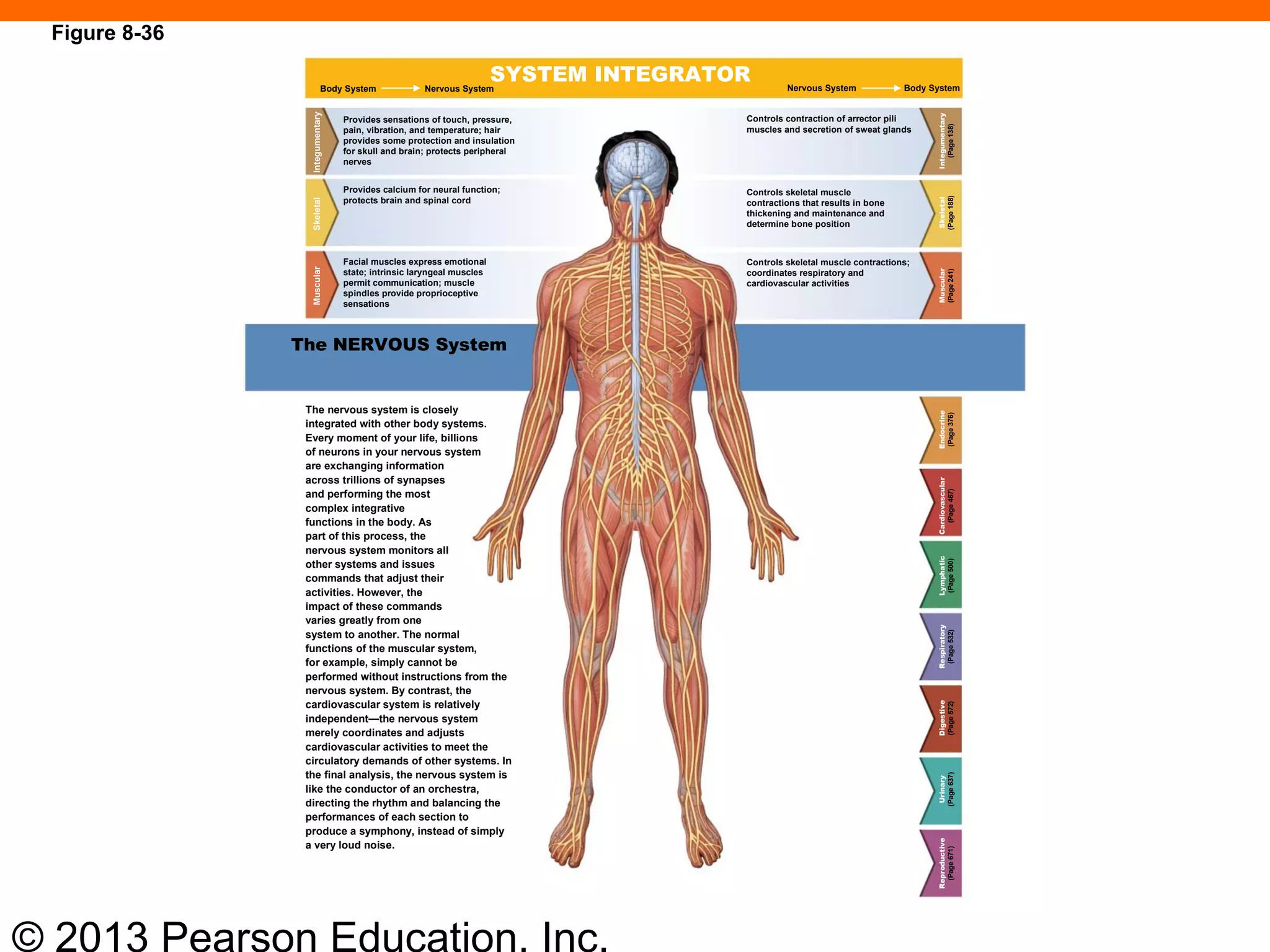
1) The nervous system has two main divisions - the central nervous system (CNS) made up of the brain and spinal cord, and the peripheral nervous system (PNS) made up of nerves outside the CNS.
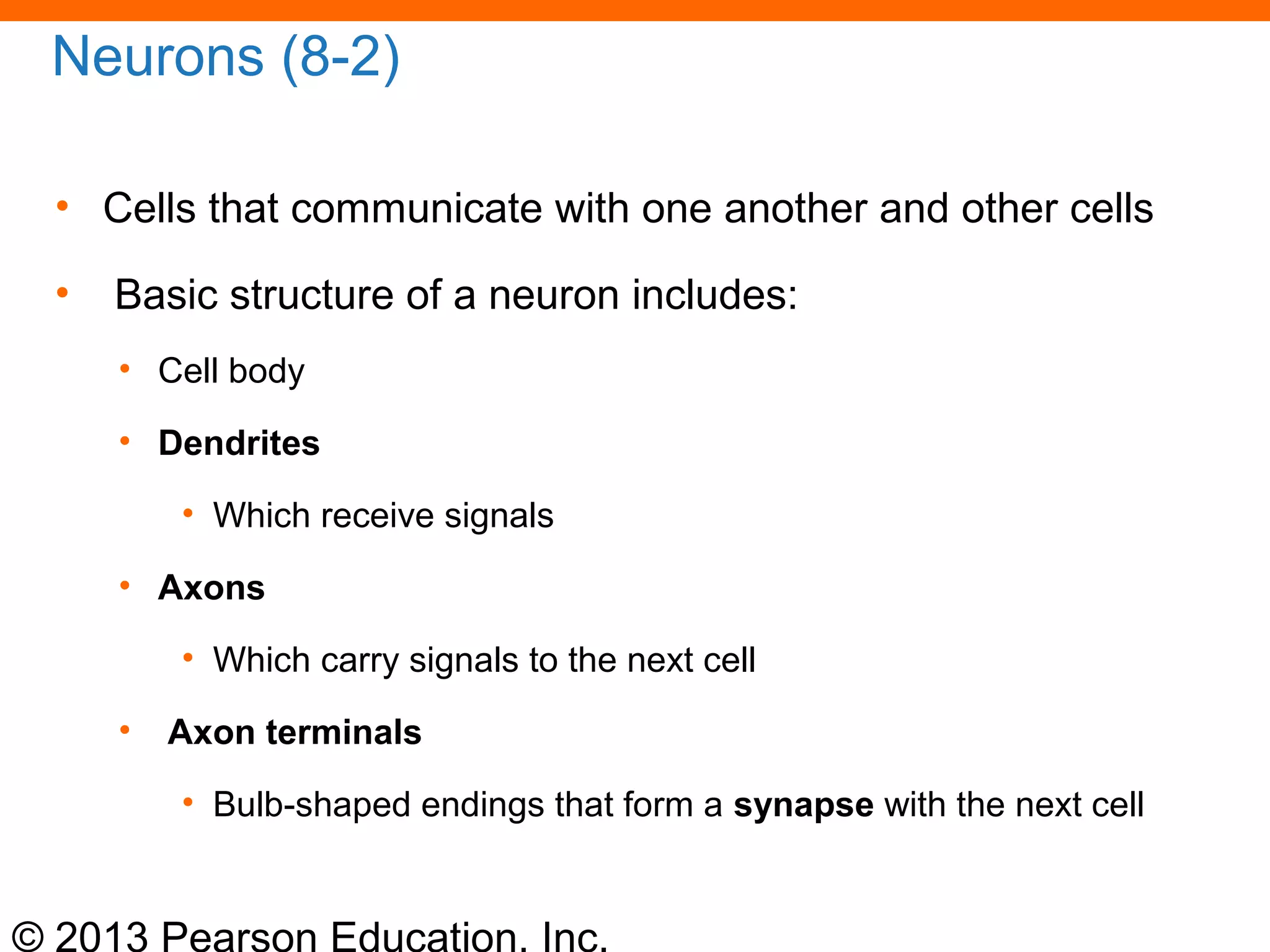
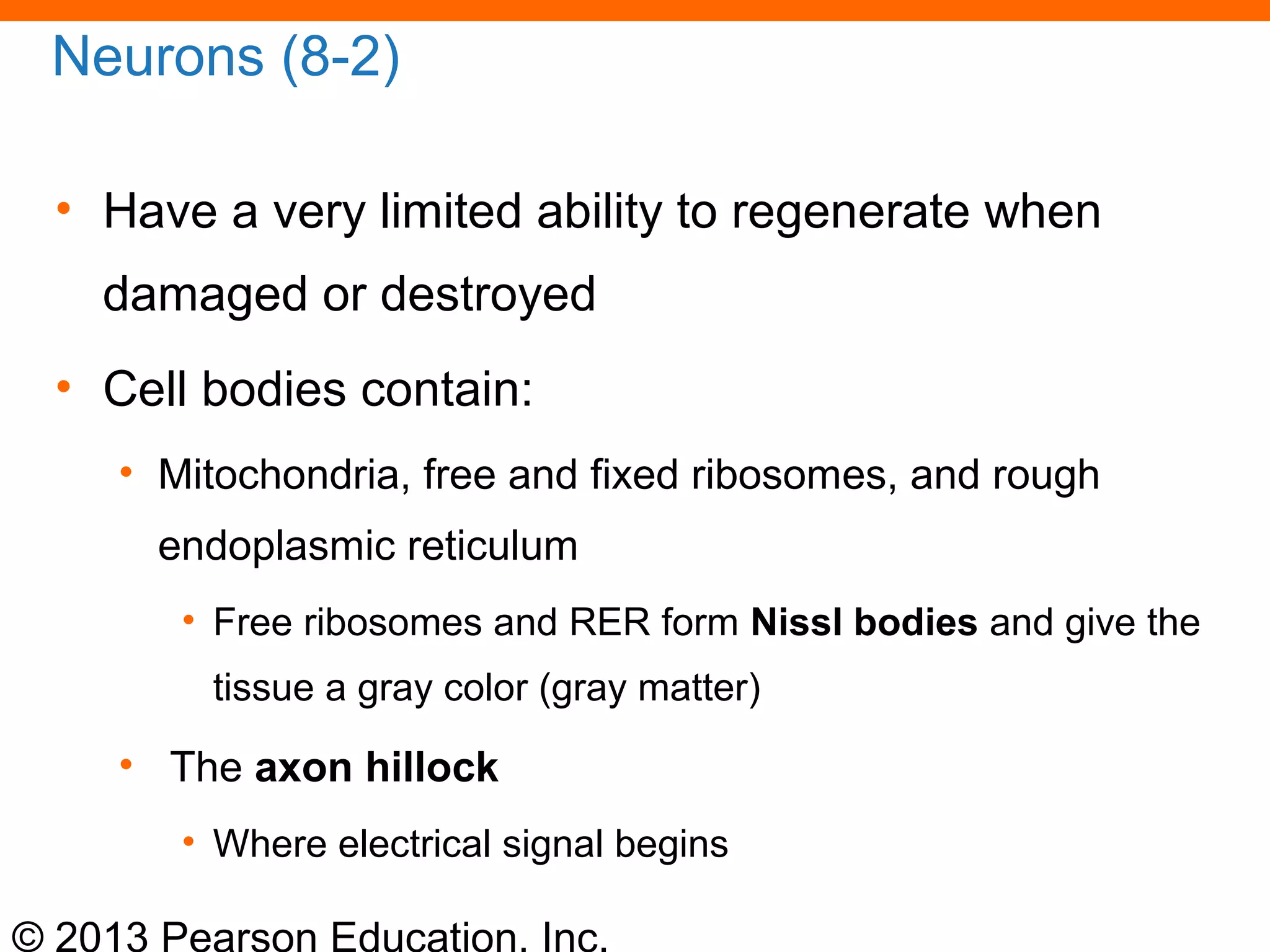
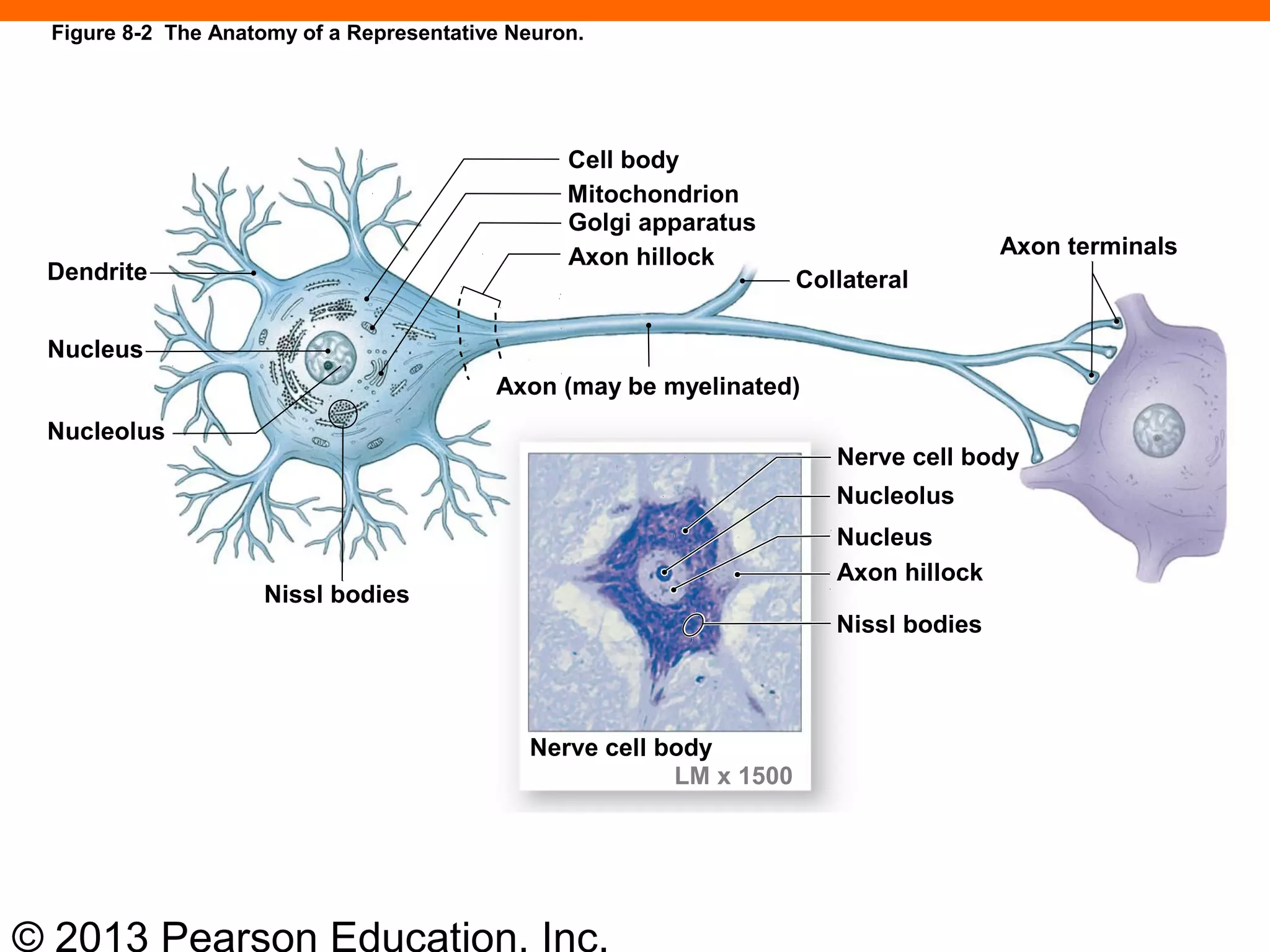
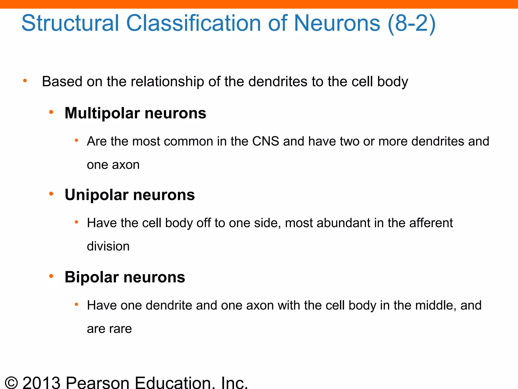
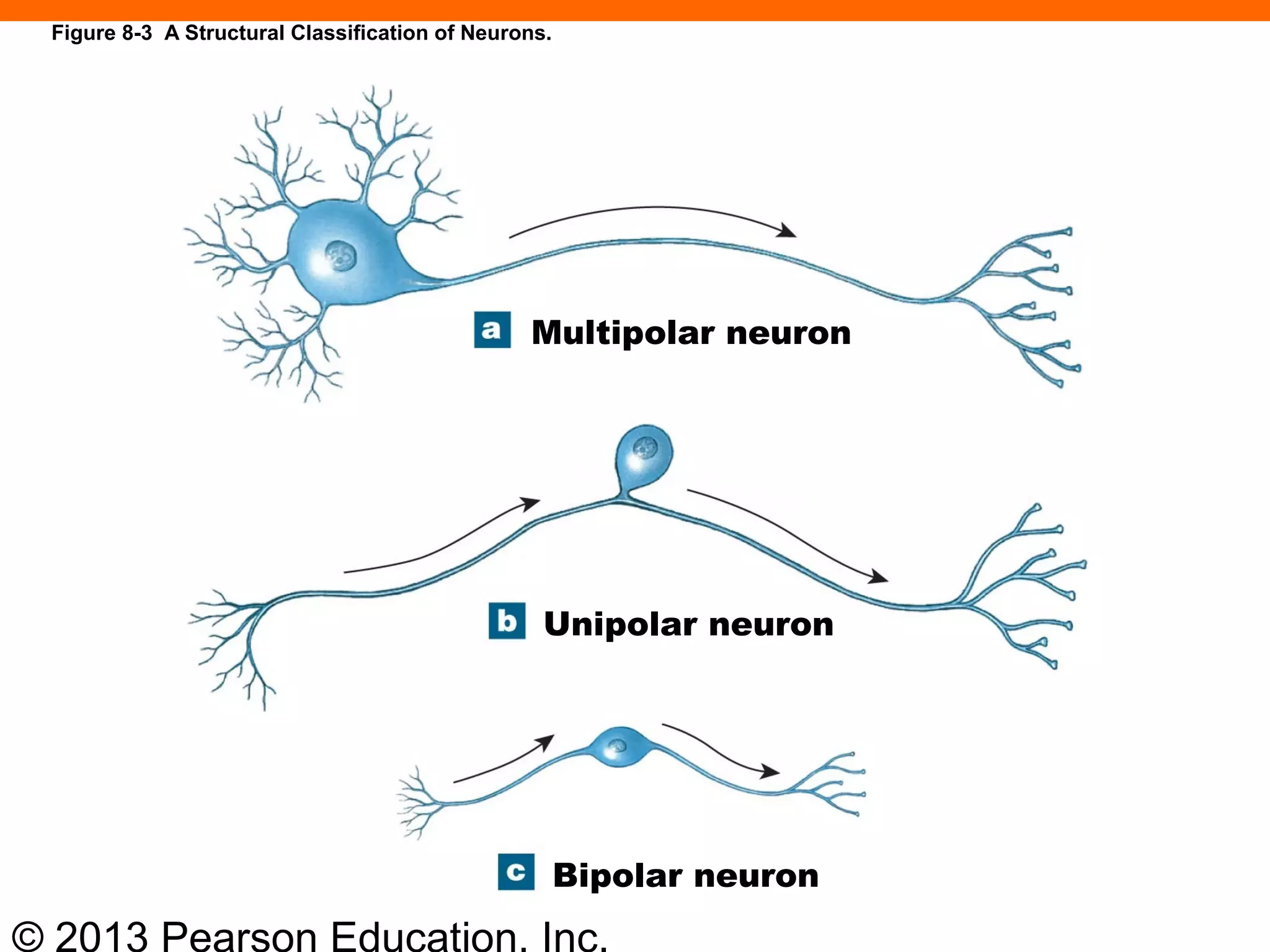
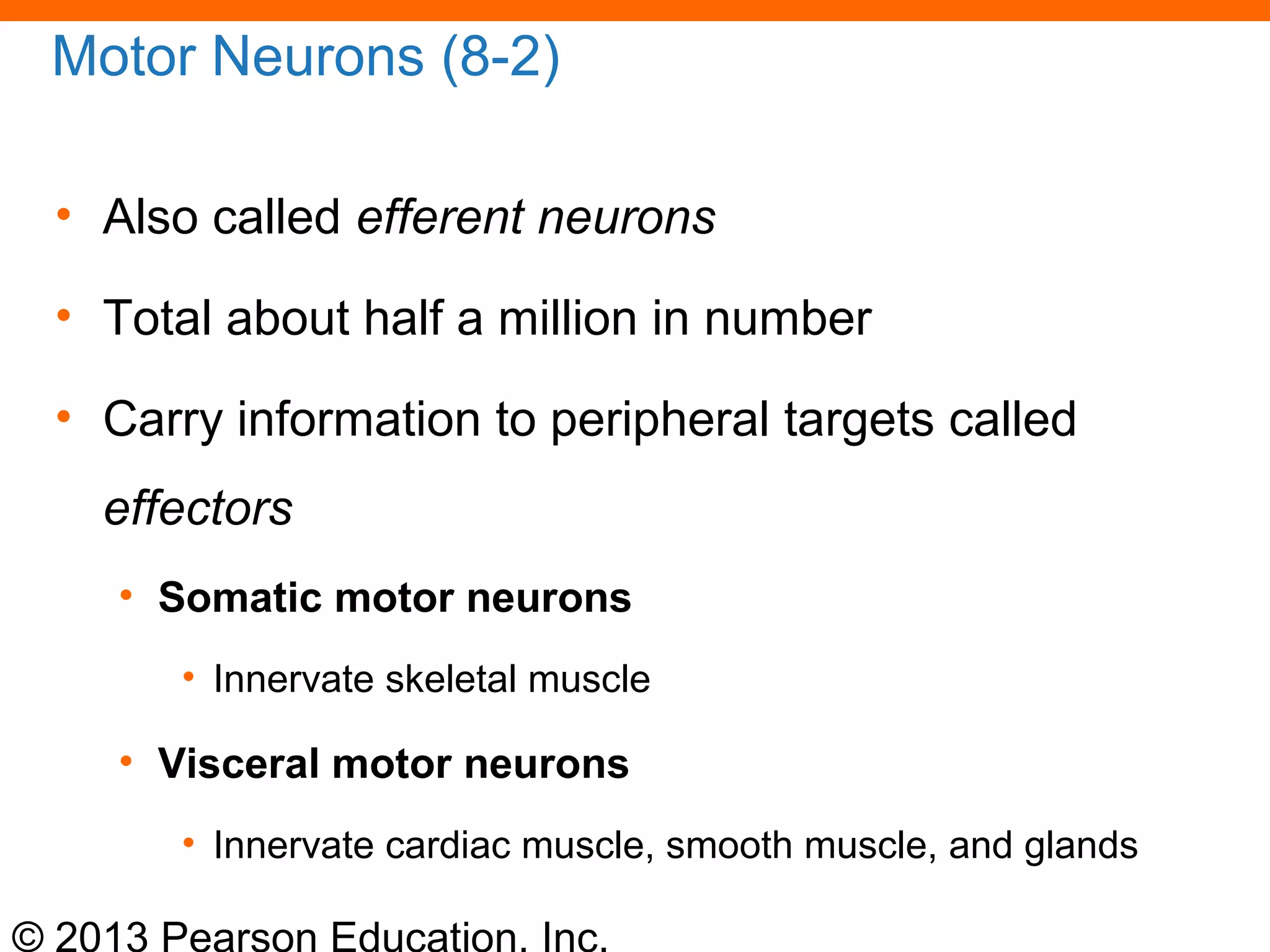
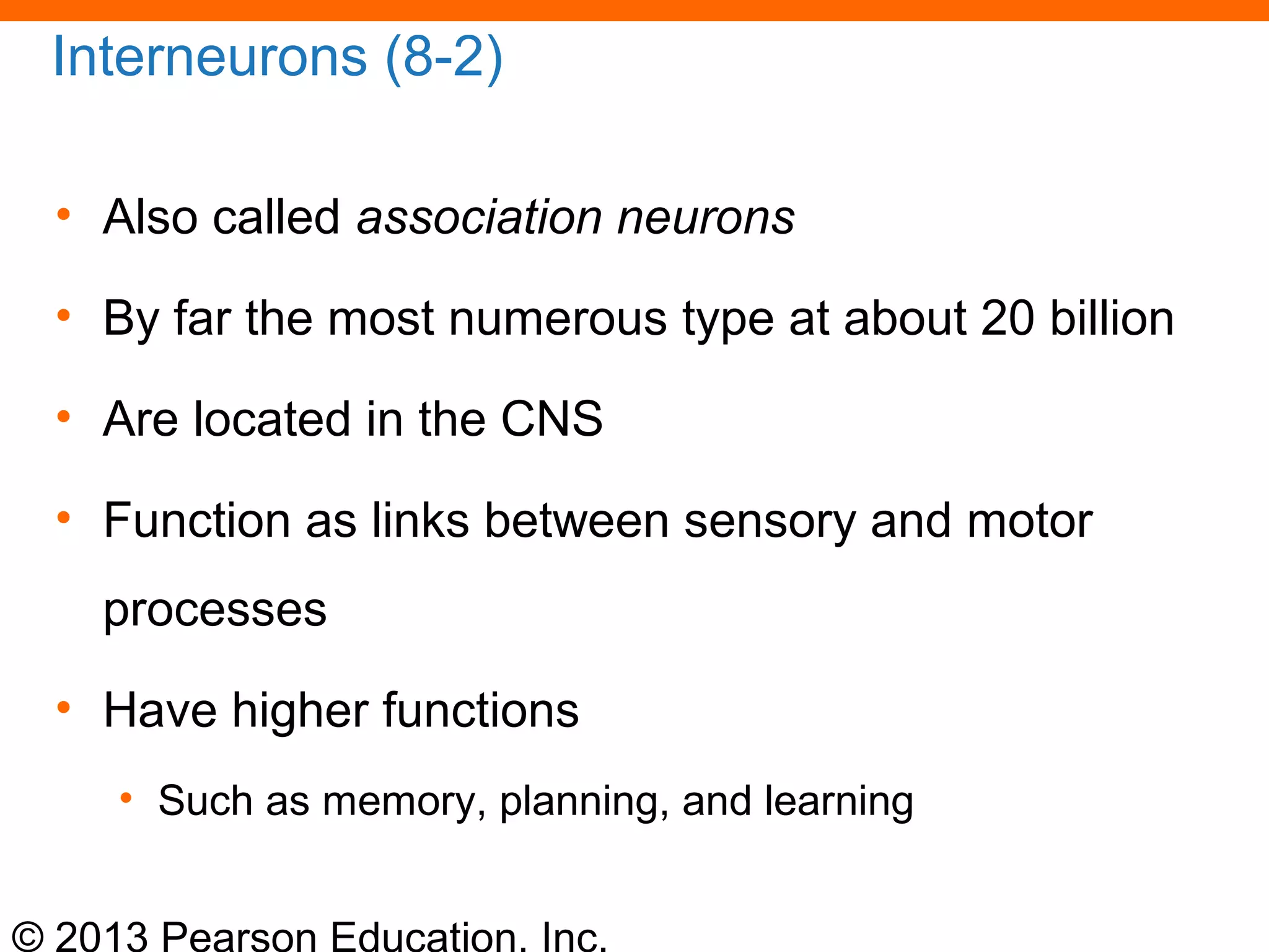
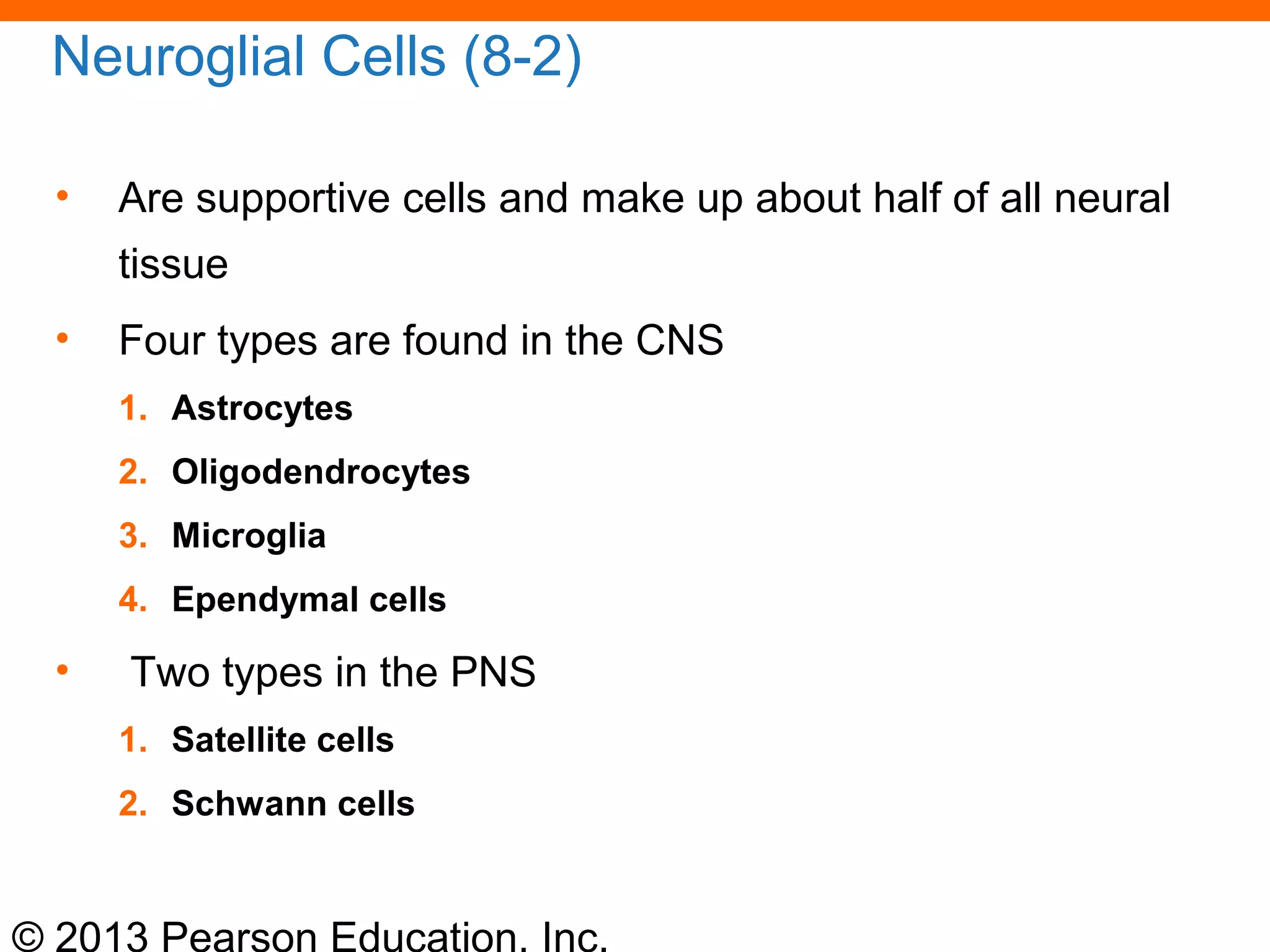
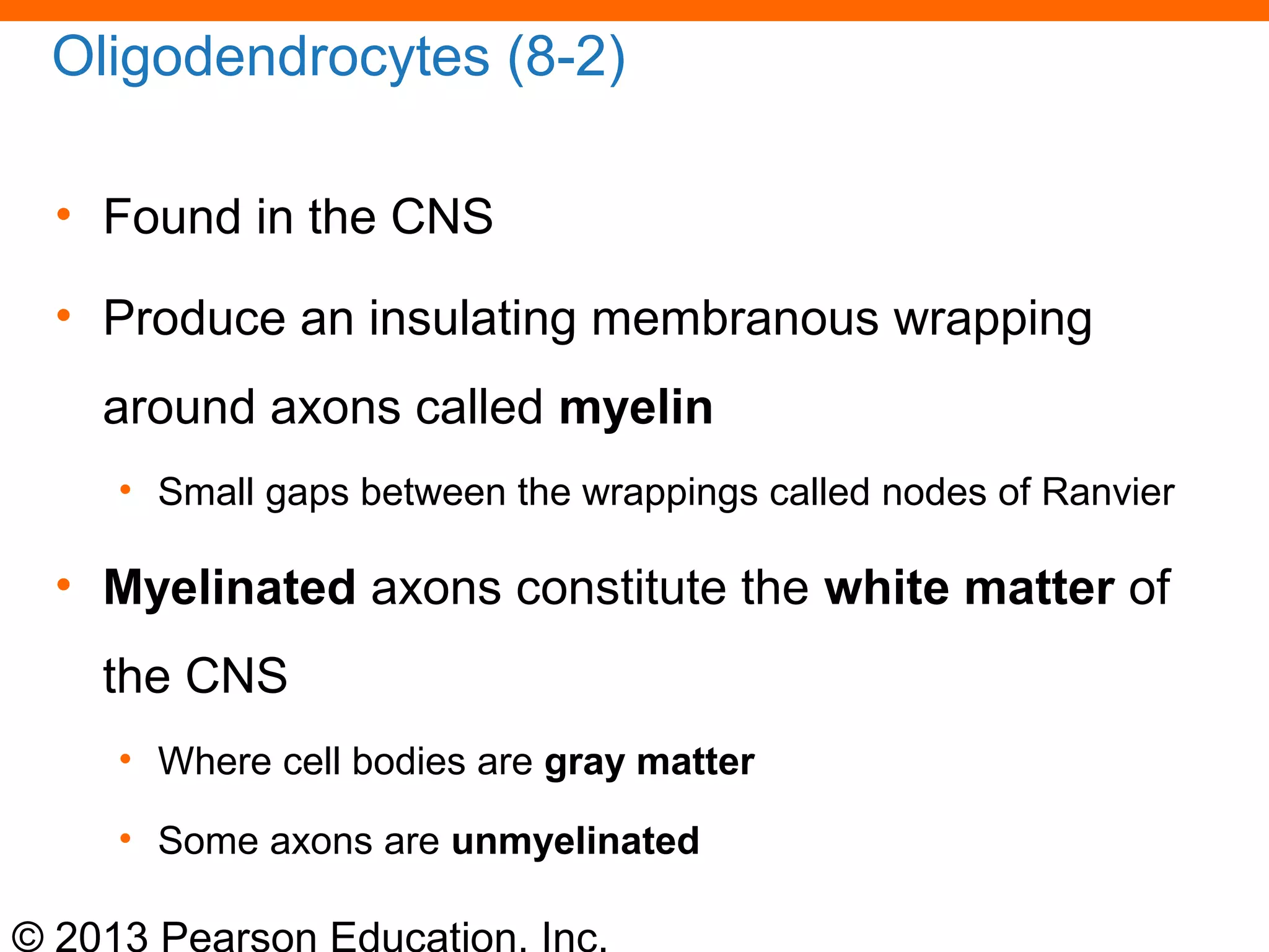
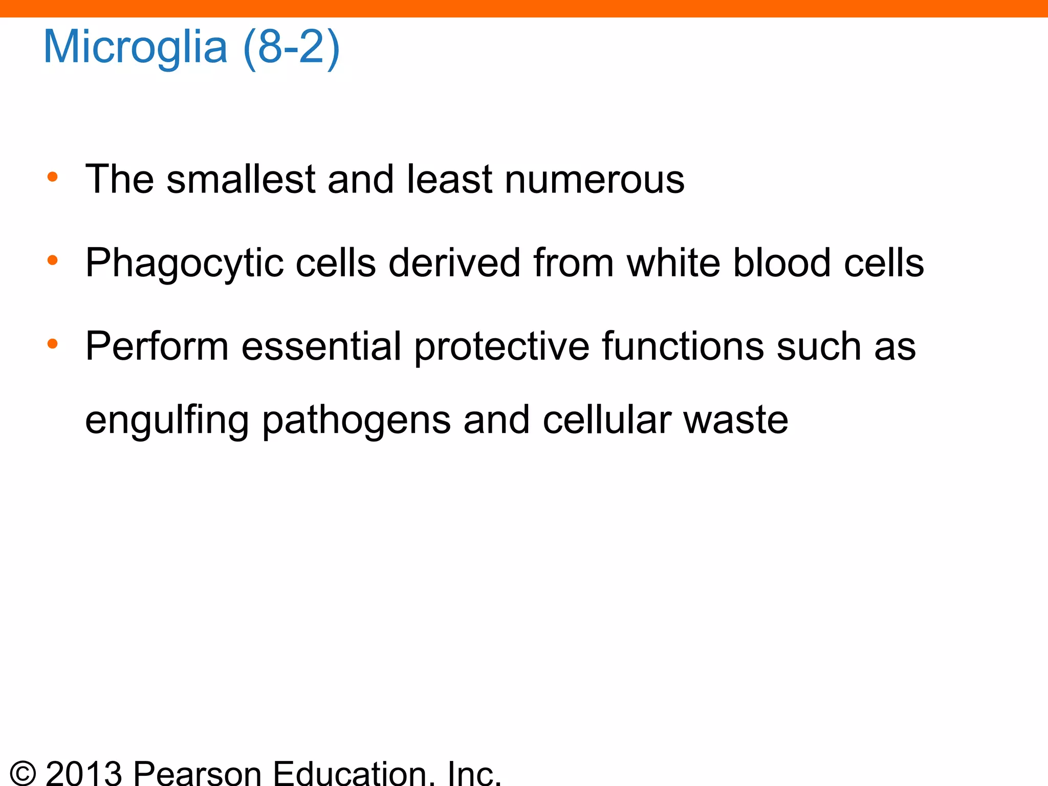
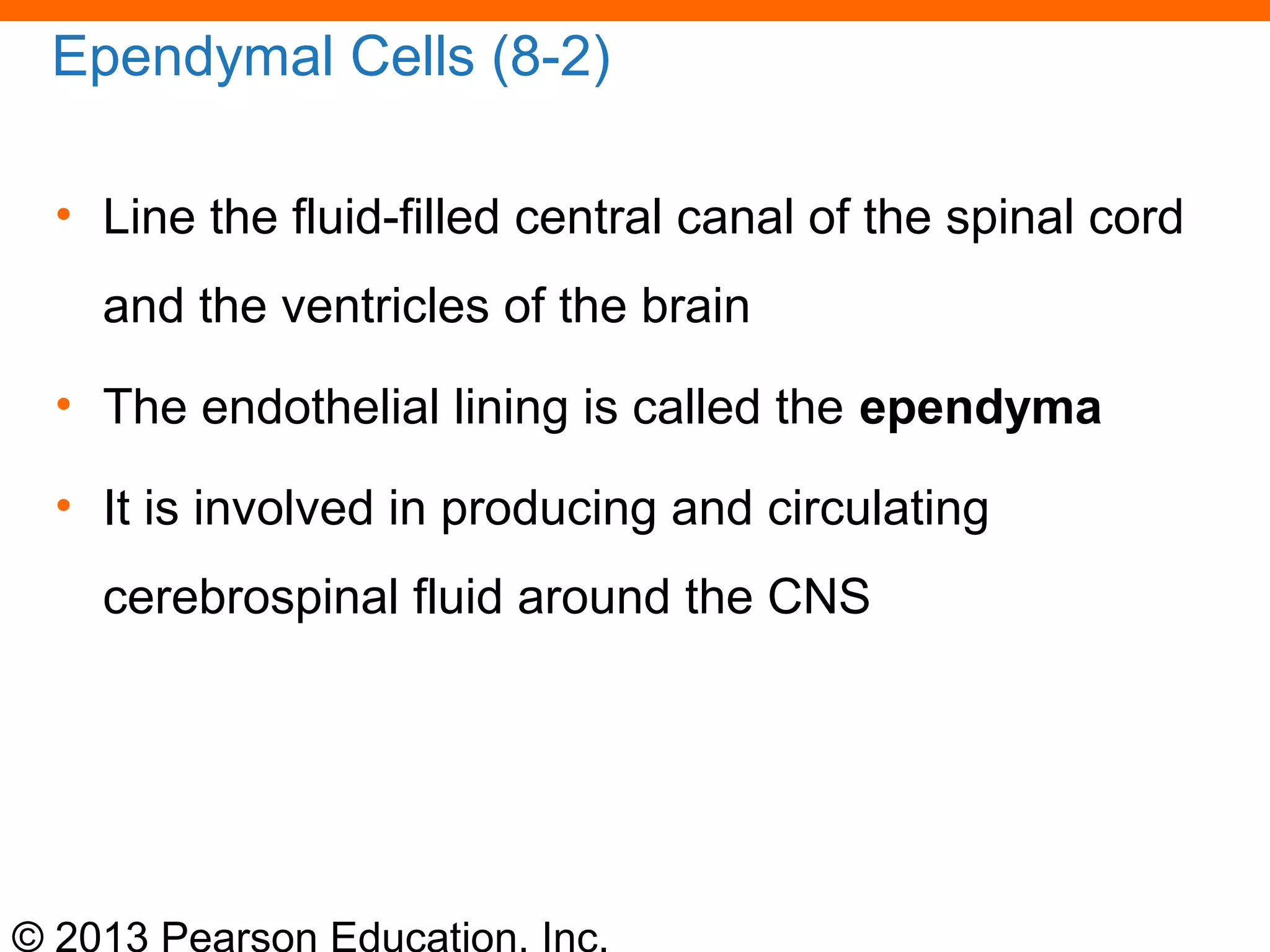
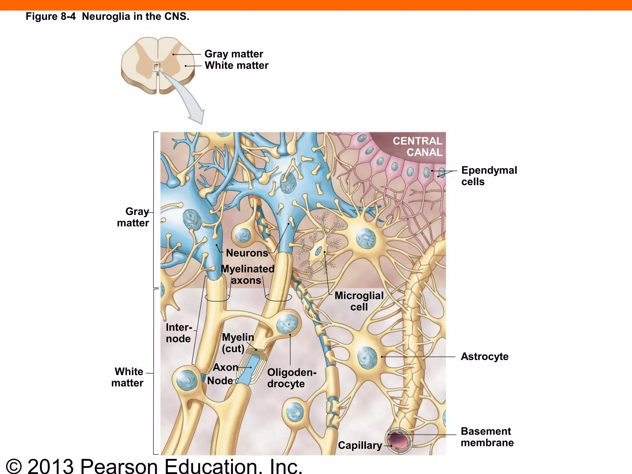
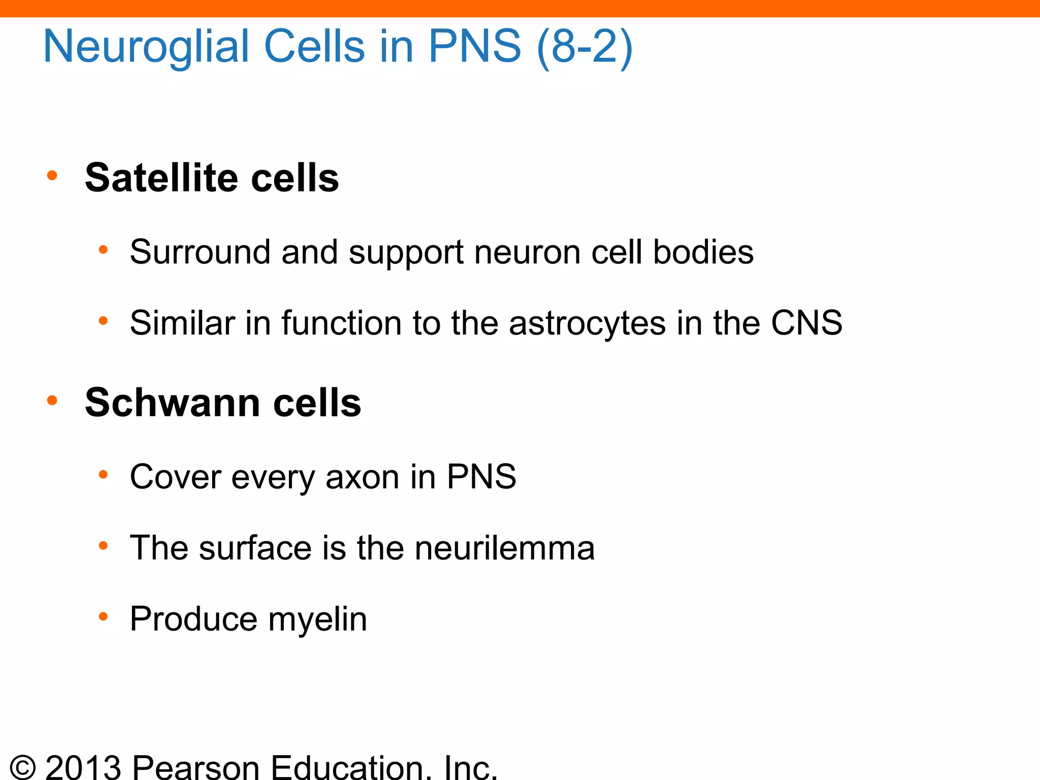
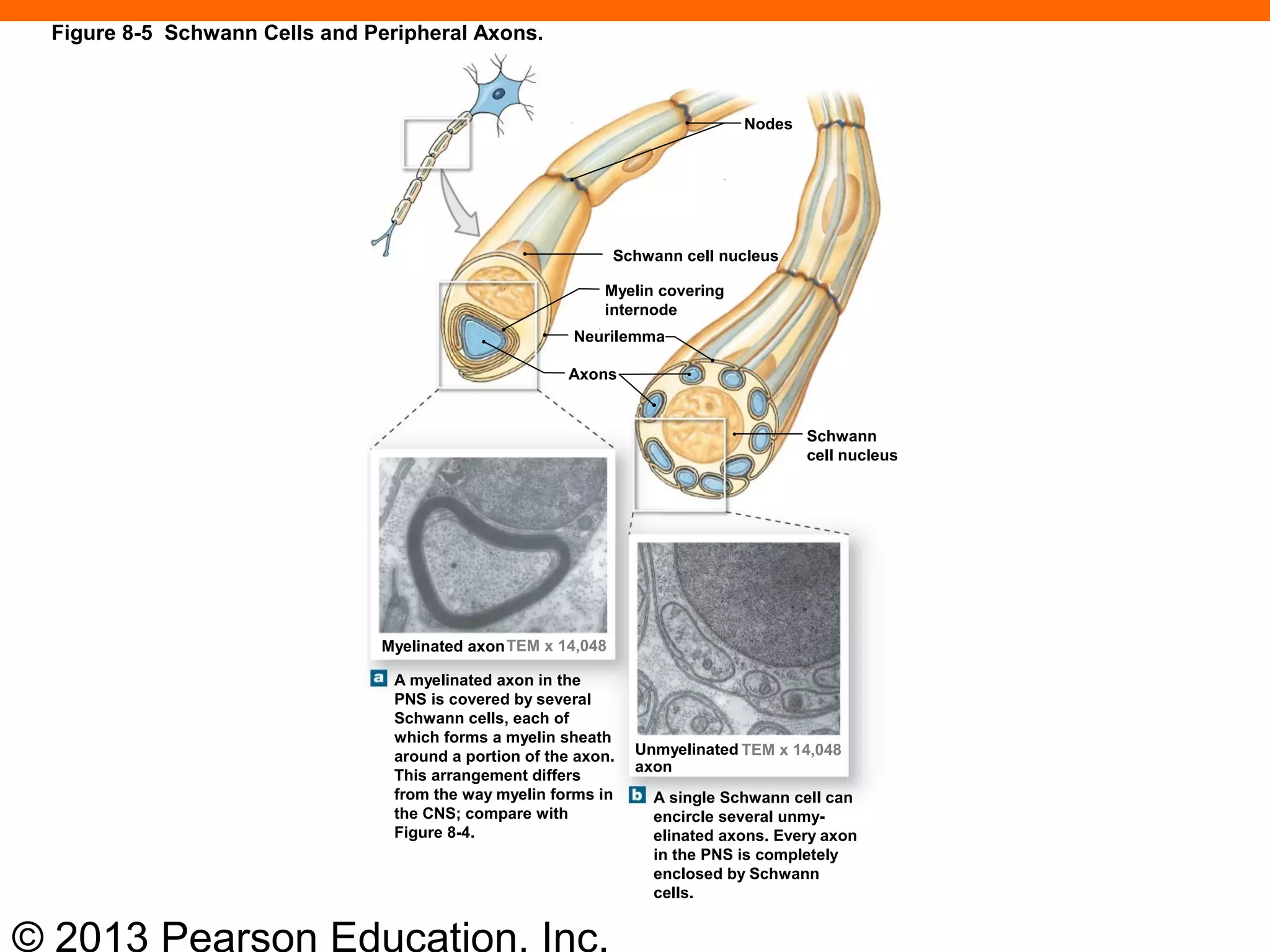
2) Neurons are the basic functional units and come in sensory, motor, and interneuron types. Neuroglia are supportive glial cells like astrocytes and oligodendrocytes.
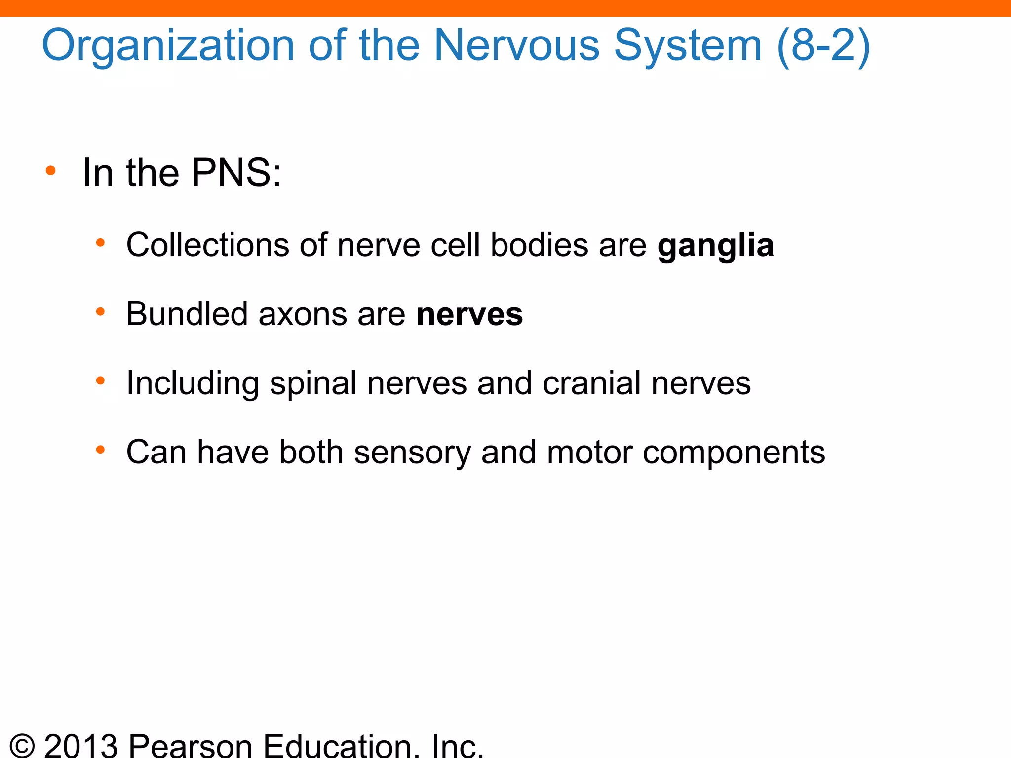
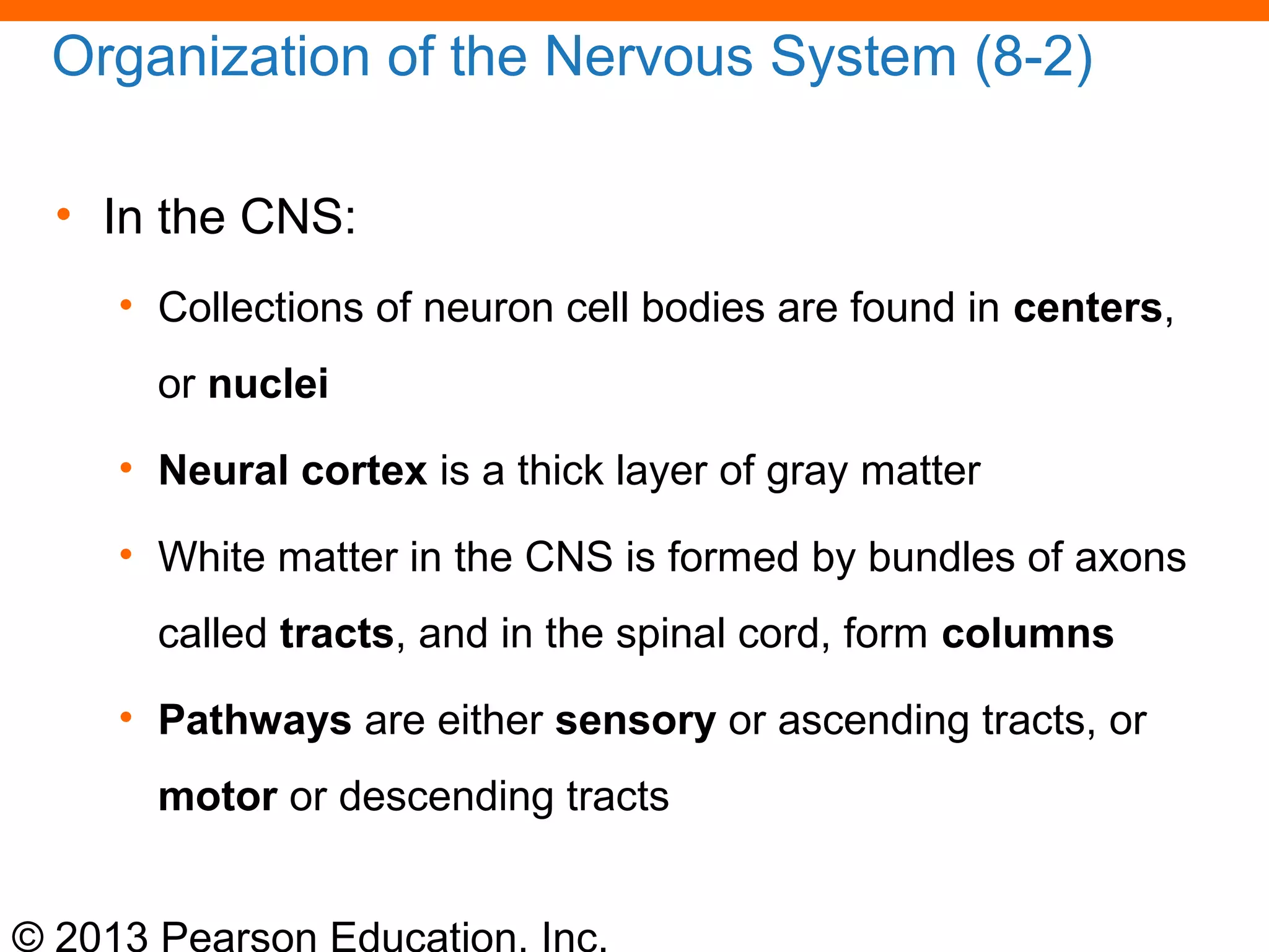
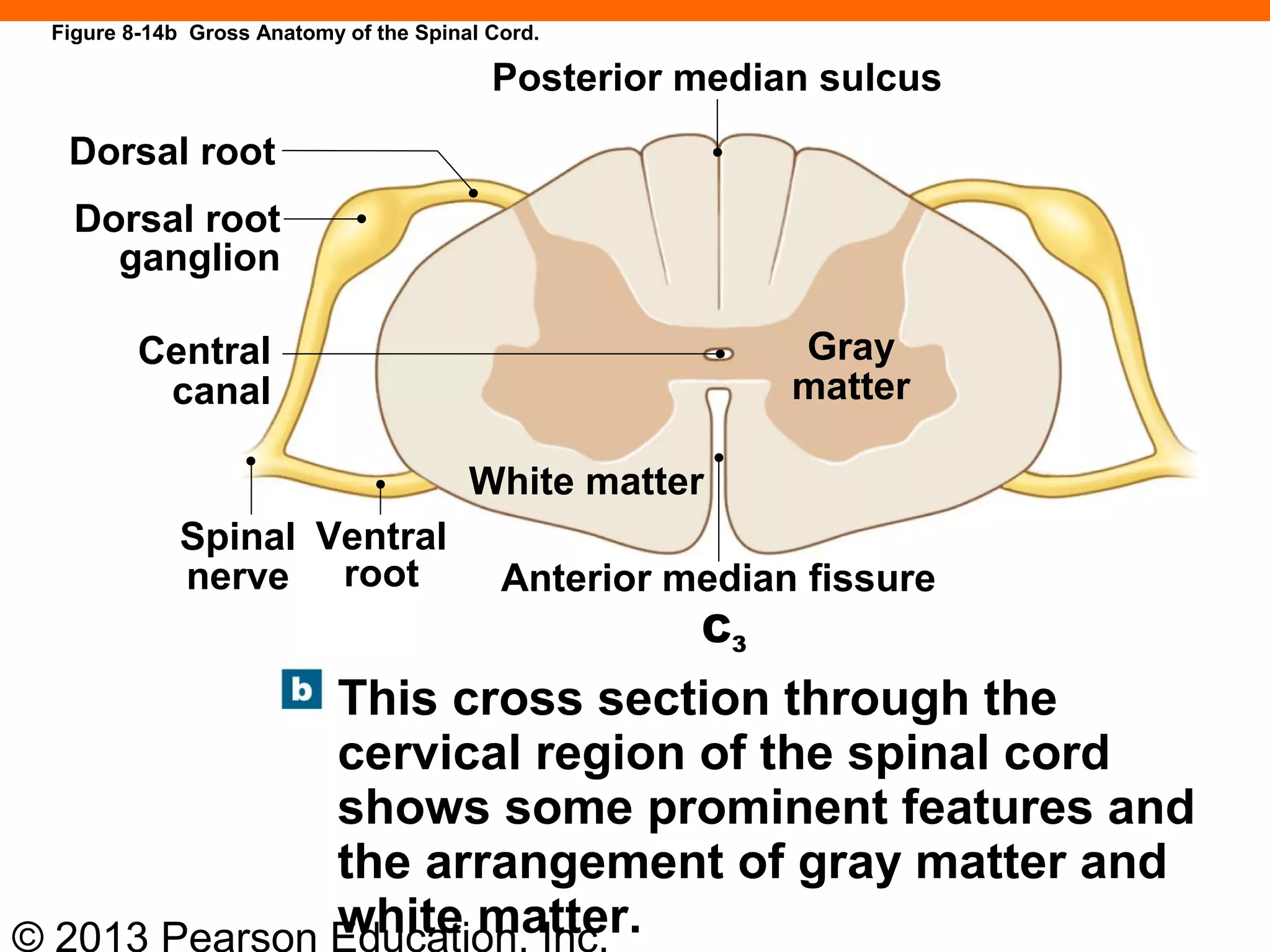
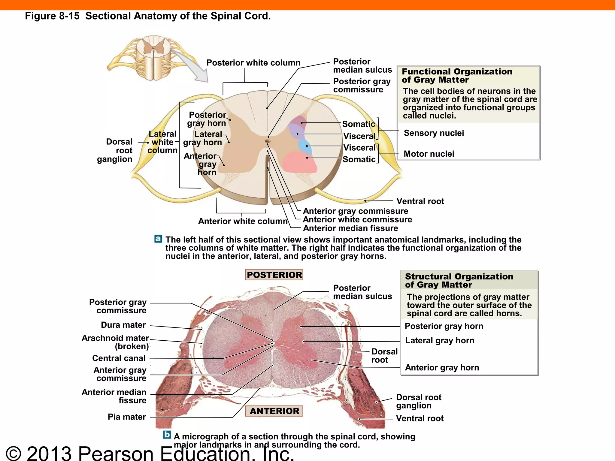
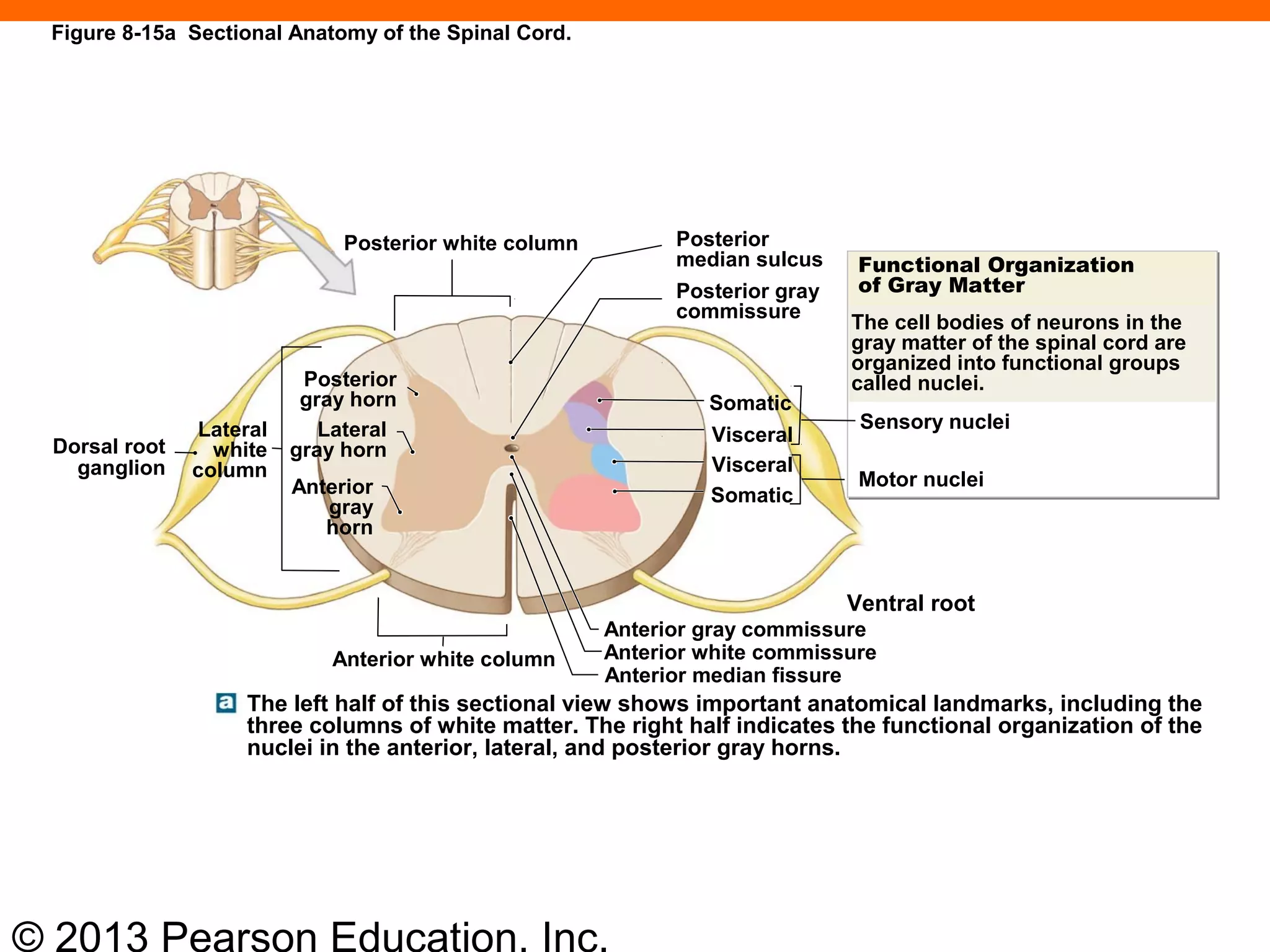
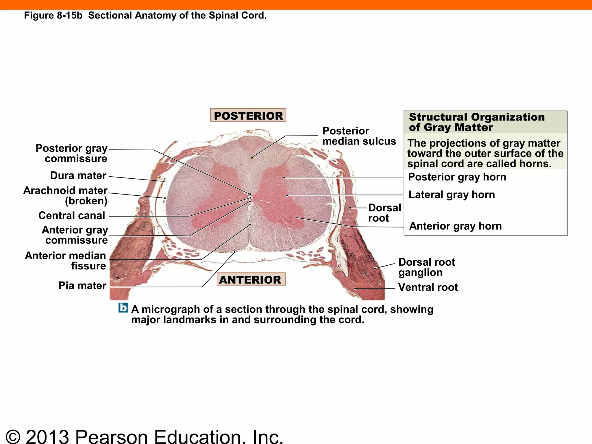
3) The CNS contains gray matter with neuron cell bodies and white matter with myelinated axon tracts. The PNS contains ganglia with neuron cell bodies and nerves with bundled axons.


![04 [chapter 4 the tissue level of organization][11e]](https://cdn.slidesharecdn.com/ss_thumbnails/04chapter4thetissueleveloforganization11e-170828035609-thumbnail.jpg?width=640&height=640&fit=bounds)


![07 [chapter 7 the skeletal system the axial skeleton]](https://cdn.slidesharecdn.com/ss_thumbnails/07chapter7theskeletalsystem-theaxialskeleton-170828035650-thumbnail.jpg?width=640&height=640&fit=bounds)


![Vibe Coding vs. Spec-Driven Development [Free Meetup]](https://cdn.slidesharecdn.com/ss_thumbnails/vibecodingvsspecdrivendevelopment-251209105622-43f455e7-thumbnail.jpg?width=640&height=640&fit=bounds)


