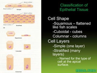
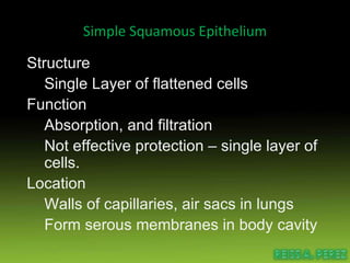
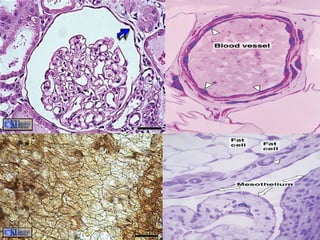
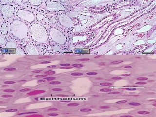
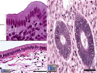
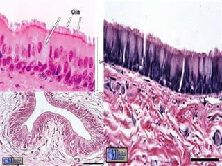
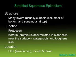
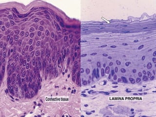
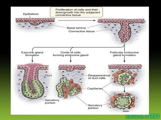
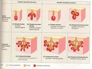
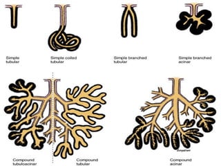
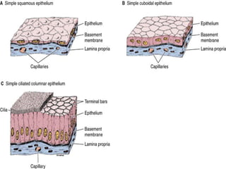
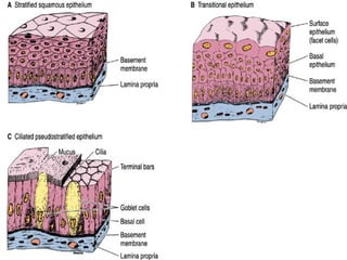
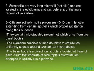
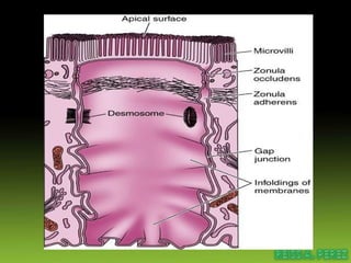
Epithelial Tissue  :-D
Tissues   are groups of cells that are similar in structure and function. * Epithelium Coverings Linings of surfaces * Connective Support Bone, ligaments, fat * Muscle Movement * Nervous Control Brain, nerves, spinal cord
Epithelial Tissue Function Protection  Skin protects from sunlight & bacteria & physical damage. Absorption Lining of small intestine, absorbing nutrients into blood Filtration Lining of Kidney tubules filtering wastes from blood plasma Secretion Different glands produce perspiration, oil, digestive enzymes and mucus
Classification of Epithelial Tissue Cell Shape -Squamous – flattened like fish scales -Cuboidal - cubes Columnar - columns Cell Layers -Simple (one layer) -Stratified (many layers) - Named for the type of cell at the apical surface.
Simple Squamous Epithelium Structure Single Layer of flattened cells Function Absorption, and filtration Not effective protection – single layer of cells. Location Walls of capillaries, air sacs in lungs Form serous membranes in body cavity
 
Simple Cuboidal Epithelium Structure Single layer of cube shaped cells Function Secretion and transportation in glands, filtration in kidneys Location Glands and ducts (pancreas & salivary), kidney tubules, covers ovaries
 
Simple Columnar Epithelium Structure Elongated layer of cells with nuclei at same level Function Absorption, Protection & Secretion When open to body cavities – called mucous membranes Special Features Microvill i , bumpy extension of apical surface, increase surface area and absorption rate. Goblet cells , single cell glands, produce protective mucus. Location Linings of entire digestive tract
 
Pseudostratified Epithelium Structure Irregularly shaped cells with nuclei at different levels – appear stratified, but aren’t. All cells reach basement membrane Function Absorption and Secretion Goblet cells produce mucus Cilia (larger than microvilli) sweep mucus  Location Respiratory Linings & Reproductive tract
 
Stratified Squamous Epithelium Structure Many layers (usually cubodial/columnar at bottom and squamous at top) Function Protection Keratin (protein) is accumulated in older cells near the surface – waterproofs and toughens skin. Location Skin (keratinized), mouth & throat
 
Transitional Epithelium  Structure Many layers Very specialized – cells at base are cuboidal or columnar, at surface will vary. Change between stratified & simple as tissue is stretched out. Function Allows stretching (change size) Location Urinary bladder, ureters & urethra
 
Glands One or more cells that make and secrete a product. Secretion = protein in aqueous solution: hormones, acids, oils. Endocrine glands No duct, release secretion into blood vessels  Often hormones Thyroid, adrenal and pituitary glands Exocrine glands Contain ducts, empty onto epithelial surface Sweat, Oil glands, Salivary glands, Mammary  glands.
 
Shapes of Exocrine glands Branching Simple – single, unbranched duct Compound – branched. Shape: tubular or alveolar Tubular – shaped like a tube Alveolar – shaped like flasks or sacs Tubuloalveolar – has both tubes and sacs in gland


Apical epithelial surfaces  These surfaces may possess specialized structures such as  microvilli ,  stereocilia , and  cilia .   1- Microvilli are finger like projections of epithelia (1µm length) extend into a lumen to increase the surface area - Glycocalyx is present on their surfaces - composed of actin filaments run longitudinally inserted in the terminal web - they constitute the brush and striated borders
2- Stereocilia are very long microvilli (not cilia) and are located in the epididymis and vas deferens of the male reproductive system 3- Cilia are actively motile processes (5-10 µm in length) extending from certain epithelia which propel substance along their surfaces They contain microtubules (axoneme) which arise from the basal bodies The axoneme consists of nine doublets microtubules uniformly spaced around two central microtubules The basal body is a cylindrical structure located at base of each cilium that consists of nine triplets microtubules arranged in radially like a pinwheel
 
THANK YOU MAM ALMOND :-D

Epithelial Tissue

  • 1.
  • 2.
  • 3.
    Tissues are groups of cells that are similar in structure and function. * Epithelium Coverings Linings of surfaces * Connective Support Bone, ligaments, fat * Muscle Movement * Nervous Control Brain, nerves, spinal cord
  • 4.
    Epithelial Tissue FunctionProtection Skin protects from sunlight & bacteria & physical damage. Absorption Lining of small intestine, absorbing nutrients into blood Filtration Lining of Kidney tubules filtering wastes from blood plasma Secretion Different glands produce perspiration, oil, digestive enzymes and mucus
  • 5.
    Classification of EpithelialTissue Cell Shape -Squamous – flattened like fish scales -Cuboidal - cubes Columnar - columns Cell Layers -Simple (one layer) -Stratified (many layers) - Named for the type of cell at the apical surface.
  • 6.
    Simple Squamous EpitheliumStructure Single Layer of flattened cells Function Absorption, and filtration Not effective protection – single layer of cells. Location Walls of capillaries, air sacs in lungs Form serous membranes in body cavity
  • 7.
  • 8.
    Simple Cuboidal EpitheliumStructure Single layer of cube shaped cells Function Secretion and transportation in glands, filtration in kidneys Location Glands and ducts (pancreas & salivary), kidney tubules, covers ovaries
  • 9.
  • 10.
    Simple Columnar EpitheliumStructure Elongated layer of cells with nuclei at same level Function Absorption, Protection & Secretion When open to body cavities – called mucous membranes Special Features Microvill i , bumpy extension of apical surface, increase surface area and absorption rate. Goblet cells , single cell glands, produce protective mucus. Location Linings of entire digestive tract
  • 11.
  • 12.
    Pseudostratified Epithelium StructureIrregularly shaped cells with nuclei at different levels – appear stratified, but aren’t. All cells reach basement membrane Function Absorption and Secretion Goblet cells produce mucus Cilia (larger than microvilli) sweep mucus Location Respiratory Linings & Reproductive tract
  • 13.
  • 14.
    Stratified Squamous EpitheliumStructure Many layers (usually cubodial/columnar at bottom and squamous at top) Function Protection Keratin (protein) is accumulated in older cells near the surface – waterproofs and toughens skin. Location Skin (keratinized), mouth & throat
  • 15.
  • 16.
    Transitional Epithelium Structure Many layers Very specialized – cells at base are cuboidal or columnar, at surface will vary. Change between stratified & simple as tissue is stretched out. Function Allows stretching (change size) Location Urinary bladder, ureters & urethra
  • 17.
  • 18.
    Glands One ormore cells that make and secrete a product. Secretion = protein in aqueous solution: hormones, acids, oils. Endocrine glands No duct, release secretion into blood vessels Often hormones Thyroid, adrenal and pituitary glands Exocrine glands Contain ducts, empty onto epithelial surface Sweat, Oil glands, Salivary glands, Mammary glands.
  • 19.
  • 20.
    Shapes of Exocrineglands Branching Simple – single, unbranched duct Compound – branched. Shape: tubular or alveolar Tubular – shaped like a tube Alveolar – shaped like flasks or sacs Tubuloalveolar – has both tubes and sacs in gland
  • 21.
  • 22.
  • 23.
  • 24.
  • 25.
    Apical epithelial surfaces These surfaces may possess specialized structures such as microvilli , stereocilia , and cilia . 1- Microvilli are finger like projections of epithelia (1µm length) extend into a lumen to increase the surface area - Glycocalyx is present on their surfaces - composed of actin filaments run longitudinally inserted in the terminal web - they constitute the brush and striated borders
  • 26.
    2- Stereocilia arevery long microvilli (not cilia) and are located in the epididymis and vas deferens of the male reproductive system 3- Cilia are actively motile processes (5-10 µm in length) extending from certain epithelia which propel substance along their surfaces They contain microtubules (axoneme) which arise from the basal bodies The axoneme consists of nine doublets microtubules uniformly spaced around two central microtubules The basal body is a cylindrical structure located at base of each cilium that consists of nine triplets microtubules arranged in radially like a pinwheel
  • 27.
  • 28.
    THANK YOU MAMALMOND :-D