SlideShare a Scribd company logo
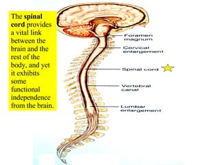
The  spinal cord  provides a vital link between the brain and the rest of the body, and yet  it exhibits some functional independence from the brain.
The  adult spinal cord  travels from the foramen magnum and terminates within the vertebral foramen of the  first lumbar vertebra (L1) in adults.
The  spinal cord  can be subdivided into five regions:  cervical region, thoracic region, lumbar region, sacral region, and coccygeal region  (which has only one pair of nerves). Don’t  be confused and think that the sacral “region” of the spinal cord is surrounded by sacral vertebrae.  It is NOT!
The diameter of the spinal cord is the largest in the  cervical region  and there is a larger proportion of white matter compared to gray matter.
The diameter of the  sacral region  of the spinal cord (which is surrounded by the T12/L1 vertebrae) is the smallest  and the proportion of gray matter is largest in the spinal cord.
The  cervical enlargement  contains the neurons that innervate the  upper  limbs The  lumbar enlargement  contains the neurons that innervate the  lower  limbs.
The tapering end of the spinal cord is called the  conus medullaris . The conus medullaris is surrounded by  L1 in and adult and L2 in a child .
The  adult  spinal cord terminates at the level of the first lumbar vertebra (L1) In a developing  child , the spinal cord can extend to the level of the second lumbar vertebra (L2)
The  cauda equina  (horse’s tail) is composed of nerves that arise from the conus medullaris and extend inferiorly.
The  filum terminale , which is composed of pia mater, extends from the conus medullaris to the coccyx. Note the  subarachnoid space  also continues for some distance.
There are  31 pairs of spinal nerves  that serve  defined segments  of the human body.
There are  8 pairs of cervical spinal nerves.   This is possible because the first pair (C1 spinal nerves) exits the spinal column  between  the occipital bone and the atlas (C1).  The remaining 7 pairs (C2-C8 spinal nerves) exit  below  each of the 7 cervical vertebrae via the intervertebral foramina.  All the spinal nerves are mixed nerves.
The spinal cord is surrounded by the  dura, arachnoid, and pia maters  (the meninges)
WHAT IS THE NAME OF THE NERVE  THAT EXITS VIA THE INTERVERTEBRAL FORAMEN BETWEEN THE ATLAS AND THE AXIS? A VAGUS NERVE B FIRST CERVICAL SPINAL NERVE C ACCESSORY NERVE D LONG THORACIC NERVE E SPINAL NERVE C2
The  epidural space  is between the vertebra and the dura mater
Epidural anesthesia
Epidural anesthesia
Effect of  epidural anesthesia
A person who needs an epidural!
The  dura mater  extends along the entire length of the vertebral canal and surrounds the spinal cord. It also extends along the initial portion of the radiating spinal nerves
The  subarachnoid space  is a real space filled with CSF
In this midsagittal picture #3 is the dura mater, #5 is the spinal cord, # 4 is the epidural space, and  #6 is the subarachnoid space  where CSF is located (#1 is an intervertebral disc and #2 is the body of a vertebrae).
Needle for spinal tap
Spinal taps  are done between the third and fourth lumbar vertebrae because there is no spinal cord at that location
The tip of the needle is inserted into the subarachnoid space  outside the cauda equina and spinal fluid is removed for testing.
The entering pressure can be determined  when the needle is inserted into the subarachnoid space during a spinal tap.
Spinal fluid is normally crystal clear like water.  Cloudy spinal fluid, like the specimen shown, is a sign of white blood cells (pus).   The most common cause for white blood cells in the spinal fluid is viral or bacterial  meningitis.
The  pia mater  directly adheres to the spinal cord
WHICH OF THE FOLLOWING  IS  TYPICALLY PENETRATED DURING A ROUTINE SPINAL TAP? A PIA MATER B NUCLEUS PULPOSUS C ANULUS FIBROSUS D SPINAL CORD E NONE OF THE ABOVE
The cross-sectional view shows that  the gray matter is central and the white matter is peripheral
The  peripheral white matter  contains ascending and descending tracts of nerves traveling to and from the brain.  The  central gray matter  serves as a center for spinal reflexes.
The  central canal  runs the entire length of the spinal cord, is contiguous with the brain and contains cerebrospinal fluid (CSF)
The spinal cord develops as 31 segments , each of which gives rise to a pair of spinal nerves that emerge from the cord through the  intervertebral foraminae
Nerves  can be sensory, motor, or mixed (sensory and motor)
Mixed nerves  carry both types of information and some axons are transmitting impulses in one direction, while other axons are transmitting impulses in the opposite direction.  All spinal nerves are mixed nerves.
There are  8 pairs of cervical spinal nerves.   This is possible because the first pair (C1 spinal nerves) exits the spinal column  between  the occipital bone and the atlas (C1).  The remaining 7 pairs (C2-C8 spinal nerves) exit  below  each of the 7 cervical vertebrae via the intervertebral foramina.  All the spinal nerves are mixed nerves.
Most of the spinal nerves are associated with specific  dermatomes  (an area of skin innervated by all the cutaneous neurons of a certain spinal or cranial nerve).
Dermatome  map.  Note the  trigeminal nerve  has dermatomes on the face. trigeminal
Dermatomes of the  trigeminal nerve  (cranial nerve V) are seen on the face
Note that the trigeminal nerve has dermatomes on the face (see white area) and that the first pair of cervical spinal nerves (C1 spinal nerves) are not represented on the surface at all.
 
Chickenpox  (varicella) virus is acquired by the respiratory route and causes a  head-to-toe rash in children . The chickenpox virus can invade the ganglia along the spinal cord and remain latent until adulthood.  It can then be activated by suppression of the immune system.  It will then travel through sensory axons of a single dermatome and  erupt onto the skin in a single dermatome  on one side of the body (unilateral eruption)
Shingles  is a reactivation of latent chickenpox from childhood that travels to the surface via a single nerve on one side of the body.
Shingles  involving the first (ophthalmic) division of the trigeminal nerve (cranial nerve V) on face.
Explanation of referred pain .  Numerous cutaneous and visceral sensory neurons share the same ascending tracts.
Body diagram of where  referred pain  may be felt.
WHICH OF THE FOLLOWING  IS CORRECT  ABOUT SHINGLES? A ADULTS WITH THIS CONDITION CAN CAUSE A  HEAD-TO-TOE RASH IN CHILDREN B IT TYPICALLY OCCURS BILATERALLY C IT IS TRIGGERED BY ETHYL ALCOHOL AND  PROLONGED NERVE COMPRESSION D IT IS MOST COMMON IN PERSONS UNDER 50 E ALL OF THE ABOVE
The majority of the spinal nerves combine and then split again as networks of nerves referred to as  plexuses.  The  exceptions  are T2-T12 and S5-Co1, which do NOT form plexuses
The  cervical plexus  is formed primarily by spinal nerves C1-C4 (C5 is  not  considered part of this plexus, even though it contributes some axons)
The cervical plexus, and particularly spinal nerves C3, C4, and part of C5, give rise to the  phrenic nerve  which  innervates the diaphragm .  Injury above C3 would lead to death by suffocation.
The  brachial plexus  is formed primarily by spinal nerves C5-C8, and T1.  This plexus gives rise to five nerves that serve the arm or hand.
There are  five nerves  that arise from the brachial plexus
The  axillary nerve  innervates the  teres minor  muscle and the  deltoid  muscle. It receives sensory information from the superolateral part of the arm and skin.
The  median nerve  innervates muscles in the antebrachium and manus.  It receives sensory information from the palmar side of fingers #1, #2, #3, and the lateral one-half of finger #4 and from the dorsal tips of these same fingers.
Median nerve dermatome
(Flexor retinaculum)
Note adduction of thumb
Note adducted thumbs
When draping my arm around my daughter’s neck for prolonged periods I would develop  anesthesia of fingers #1-#4 only  from pressure on my median nerve.
The  musculocutaneous nerve  innervates the  biceps brachii  muscle and several other muscles. It receives sensory input from the lateral surface of the forearm.
The  radial nerve  innervates the  triceps brachii  and numerous muscles of the antebrachium. It receives sensory input from the posterior arm and forearm surface and the dorsolateral side of the hand.
The  radial nerve  receives sensory input from the posterior arm and forearm surface and the  dorsolateral surface of the hand .
The  ulnar nerve , which passes near the medial epicondyle of the humerus, is the  “funny bone”.   It innervates muscles in the antebrachium and manus. It receives sensory input from the skin of the dorsal and palmar aspects of fingers #5, and the medial half of finger #4.
The  ulnar nerve  receives sensations from the  skin of the dorsal and palmar surfaces of fingers #5 and medial half of fingers #4.
Atrophy of the arm muscles caused by a  brachial plexus injury  in adulthood. Read about  brachial plexus injuries  in the  clinical view  in your text.
A PATIENT HAS SUFFERED A FRACTURE OF THE SURGICAL NECK OF THE FEMUR THAT IS SUCCESSFULLY REPAIRED WITH A MEDULLARY ROD THAT BRIDGES THE FRACTURE LINE.  HOWEVER, AS THE WEEKS PASS, THE PATIENT IS  UNABLE TO ABDUCT THE ARM AND SUFFERS FROM ANESTHESIA ALONG THE SUPEROLATERAL SKIN OF THE ARM .  WHAT HAS BEEN DAMAGED? A MEDIAN NERVE B MUSCULOCUTANEOUS NERVE C AXILLARY NERVE D ULNAR NERVE E RADIAL NERVE
The  lumbar plexus  is formed by spinal nerves L1-L4.  It gives rise to two major nerves: the femoral nerve and the obturator nerve
Note the two  major  nerves that arise from the  lumbar plexus
The  femoral nerve  innervates the  quadriceps femoris muscles  on the anterior of the thigh to help extend the knee.  It also innervates the  sartorius  muscle and several other muscles. It receives sensory input from the anterior and inferomedial thigh as well as the medial aspect of the leg.
The  obturator nerve  innervates the  gracilis  and several other muscles. It receives sensory information from the superomedial skin of the thigh
The  sacral plexus  is formed by spinal nerves from L4, L5, and S1-S4.  It gives rise to the  sciatic nerve , which is actually composed of two nerves: the  tibial nerve  and the  common fibular  (common peroneal)  nerve.
The  sacral plexus  gives rise to two principal nerves: the  tibial nerve  and the  common fibular (peroneal) nerve.   The  sciatic nerve  is composed of these two nerves wrapped in a common connective sheath.  These two nerves separate just above the popliteal fossa.
The  sciatic nerve  splits into its separate components [tibial nerve and common fibular (peroneal) nerve] just superior to the popliteal fossa.
A tight  piriformis muscle  can compress the sciatic nerve
Don’t stick needles in the sciatic nerve!
The  tibial nerve  innervates  the hamstrings, the gastrocnemius, the soleus,   and several other muscles.  It receives sensory input from the skin on the plantar surface of the foot.
The  common fibular nerve  (common peroneal nerve) innervates  the peroneus (fibularis) longus, the tibialis anterior , and several other muscles of the leg and foot. This is the branch of the sciatic nerve that caused me problems! It receives sensory input from the anterolateral part of the leg, the toes, and dorsum of the foot.
Read about sacral plexus injuries and sciatica in the  clinical view  in your text.
Reflexes  are rapid, automatic, involuntary reactions of muscles or glands to a stimulus
An example occurs when you accidentally touch a hot object. You remove your hand even before you are completely aware of the heat.  A reflex can precede sensation  (a reflex does not need to involve the brain)
A typical  reflex arc
Generally, there are five steps involved in a  neural reflex : stimulus, transmission via a sensory neuron, processing in CNS, transmission via a motor neuron, and an effector (muscle or gland) responds.
Read in your text about different types of reflexes for your own information  if  you desire.
The cranial (superior) portion of the  neural tube  expands and develops into the brain, while the caudal (inferior) part of the neural tube forms the spinal cord.  The hollow  neural canal  develops into the central canal of the spinal cord. Neural tube
The bony vertebral column (dark line shown) grows faster than the spinal cord .  A  newborn’s  spinal cord extends to about the level of L 3 .  A  child’s  spinal cord may extend to the level of L 2 .  An  adult’s  spinal cord typically terminates at the level of L 1 . Bony vertebral column
The inner delicate  spinal cord  terminates in an adult, as the conus medullaris, at the level of the L 1 vertebra.
Damage to the spinal cord can lead to paralysis or death
Severing the spinal cord  above C3 typically leads to death  by asphyxiation because the victim cannot use the  spinal nerves  to contract the intercostal muscles and cannot utilize the  phrenic nerve  to contract the diaphragm.
Christopher Reeve  as Superman
In his fall, he crushed both the atlas (C1) and the axis (C2).  He would have died by asphyxiation, but his accident was witnessed and rescue breathing was done.  He became a very famous respirator-dependent quadriplegic .
Christopher Reeve  after his fall.
Christopher Reeeve shortly before his death  from an infected bed sore (decubitus ulcer).  He did a great deal to advance research on spinal trauma.
 
Damage  below  C3 also results in  quadriplegia , but the person can still utilize their diaphragm for breathing via their  intact phrenic nerves .
 
 
WHICH OF THE FOLLOWING  IS CORRECT ? A “FOOT DROP” IS TYPICALLY CAUSED BY  DAMAGE TO THE COMMON PERONEAL NERVE B THE OBTURATOR NERVE STIMULATES  ADDUCTION OF THE THIGH C THE EMBRYONIC NEURAL CANAL BECOMES THE  CENTRAL CANAL OF THE SPINAL CORD D THE PHRENIC NERVE IS FUNCTIONAL  FOLLOWING TRANSECTIONS OF THE SPINAL  CORD AT C 5 /C 6 E ALL OF THE ABOVE

More Related Content

What's hot

Clinical anatomy of the lower limb
Clinical anatomy of the lower limbClinical anatomy of the lower limb
Clinical anatomy of the lower limb
Muhammad Ramzan Ul Rehman
 
Anatomy of diencephalon
Anatomy of diencephalonAnatomy of diencephalon
Anatomy of diencephalon
Dr. Mohammad Mahmoud
 
Ulnar nerve ppt
Ulnar nerve pptUlnar nerve ppt
Ulnar nerve ppt
Dr.vidya Sagar
 
Fourth ventricle
Fourth ventricleFourth ventricle
Fourth ventricle
Dr. Noura El Tahawy
 
Slideshow: Hand
Slideshow: Hand Slideshow: Hand
Slideshow: Hand
The Funky Professor
 
Detailed Anatomy of Medulla Oblongata ppt
Detailed Anatomy of Medulla Oblongata pptDetailed Anatomy of Medulla Oblongata ppt
Detailed Anatomy of Medulla Oblongata ppt
Dr Geeta Dolli
 
Ischiorectal fossa
Ischiorectal fossaIschiorectal fossa
Ischiorectal fossa
JULITMATHEW1
 
7. anatomy of the leg and dorsum of the foot.
7. anatomy of the leg and dorsum of the foot.7. anatomy of the leg and dorsum of the foot.
7. anatomy of the leg and dorsum of the foot.
Dr. Mohammad Mahmoud
 
Popliteal fossa & back of thigh
Popliteal fossa & back of thigh  Popliteal fossa & back of thigh
Popliteal fossa & back of thigh
Prabhakar Yadav
 
Cervical Plexus
Cervical PlexusCervical Plexus
Cervical Plexus
Rashmi Priyem Saravanan
 
LUMBOSACRAL PLEXUS.ppt
LUMBOSACRAL PLEXUS.pptLUMBOSACRAL PLEXUS.ppt
LUMBOSACRAL PLEXUS.ppt
Dr Ndayisaba Corneille
 
Arterial Supply of Upper Limb
Arterial Supply of Upper LimbArterial Supply of Upper Limb
Arterial Supply of Upper Limb
Asish Rajak
 
Anatomy of the spinal cord
Anatomy of the spinal cordAnatomy of the spinal cord
Anatomy of the spinal cord
Ranjeet Singha
 
Veins of the neck
Veins of the neckVeins of the neck
Veins of the neck
Dr. Mohammad Mahmoud
 
Anatomy of cerebellum
Anatomy of cerebellumAnatomy of cerebellum
Anatomy of cerebellumMBBS IMS MSU
 
Anatomy of cerebellum
Anatomy of cerebellumAnatomy of cerebellum
Ant thoracic wall and intercostal space
 Ant thoracic wall and intercostal space Ant thoracic wall and intercostal space
Ant thoracic wall and intercostal space
Nepalese army institute of health sciences
 
Anatomy of spinal cord
Anatomy of spinal cordAnatomy of spinal cord
Anatomy of spinal cordMBBS IMS MSU
 
brachial plexus applied anatomy
brachial plexus applied anatomybrachial plexus applied anatomy
brachial plexus applied anatomy
Sumer Yadav
 
Third ventricle
Third ventricleThird ventricle
Third ventricle
Dr Himanshu Soni
 

What's hot (20)

Clinical anatomy of the lower limb
Clinical anatomy of the lower limbClinical anatomy of the lower limb
Clinical anatomy of the lower limb
 
Anatomy of diencephalon
Anatomy of diencephalonAnatomy of diencephalon
Anatomy of diencephalon
 
Ulnar nerve ppt
Ulnar nerve pptUlnar nerve ppt
Ulnar nerve ppt
 
Fourth ventricle
Fourth ventricleFourth ventricle
Fourth ventricle
 
Slideshow: Hand
Slideshow: Hand Slideshow: Hand
Slideshow: Hand
 
Detailed Anatomy of Medulla Oblongata ppt
Detailed Anatomy of Medulla Oblongata pptDetailed Anatomy of Medulla Oblongata ppt
Detailed Anatomy of Medulla Oblongata ppt
 
Ischiorectal fossa
Ischiorectal fossaIschiorectal fossa
Ischiorectal fossa
 
7. anatomy of the leg and dorsum of the foot.
7. anatomy of the leg and dorsum of the foot.7. anatomy of the leg and dorsum of the foot.
7. anatomy of the leg and dorsum of the foot.
 
Popliteal fossa & back of thigh
Popliteal fossa & back of thigh  Popliteal fossa & back of thigh
Popliteal fossa & back of thigh
 
Cervical Plexus
Cervical PlexusCervical Plexus
Cervical Plexus
 
LUMBOSACRAL PLEXUS.ppt
LUMBOSACRAL PLEXUS.pptLUMBOSACRAL PLEXUS.ppt
LUMBOSACRAL PLEXUS.ppt
 
Arterial Supply of Upper Limb
Arterial Supply of Upper LimbArterial Supply of Upper Limb
Arterial Supply of Upper Limb
 
Anatomy of the spinal cord
Anatomy of the spinal cordAnatomy of the spinal cord
Anatomy of the spinal cord
 
Veins of the neck
Veins of the neckVeins of the neck
Veins of the neck
 
Anatomy of cerebellum
Anatomy of cerebellumAnatomy of cerebellum
Anatomy of cerebellum
 
Anatomy of cerebellum
Anatomy of cerebellumAnatomy of cerebellum
Anatomy of cerebellum
 
Ant thoracic wall and intercostal space
 Ant thoracic wall and intercostal space Ant thoracic wall and intercostal space
Ant thoracic wall and intercostal space
 
Anatomy of spinal cord
Anatomy of spinal cordAnatomy of spinal cord
Anatomy of spinal cord
 
brachial plexus applied anatomy
brachial plexus applied anatomybrachial plexus applied anatomy
brachial plexus applied anatomy
 
Third ventricle
Third ventricleThird ventricle
Third ventricle
 

Similar to 16 Spinal Cord And Spinal Nerves

Spinal cord
Spinal cordSpinal cord
Spinal cord
Ainaa Khan
 
Cervical plexus
Cervical plexusCervical plexus
Cervical plexus
Ali Faris
 
Anatomy of brachial plexus
Anatomy of brachial plexusAnatomy of brachial plexus
Anatomy of brachial plexus
AmeyDixit6
 
Spinal cord anatonmy
Spinal cord anatonmySpinal cord anatonmy
Spinal cord anatonmy
Bikal Lamichhane
 
Spinal cord anatomy
Spinal cord anatomySpinal cord anatomy
Spinal cord anatomy
Bikal Lamichhane
 
Chapter 16 A, Sp 10
Chapter 16 A, Sp 10Chapter 16 A, Sp 10
Chapter 16 A, Sp 10j2jpop
 
Spinal cord Gross anatomy with Clinical Anatomy.pptx
Spinal cord Gross anatomy with Clinical Anatomy.pptxSpinal cord Gross anatomy with Clinical Anatomy.pptx
Spinal cord Gross anatomy with Clinical Anatomy.pptx
siddharthroy26587
 
12peripheralnervesoftheupperlimb-120312033920-phpapp01-141228111052-conversio...
12peripheralnervesoftheupperlimb-120312033920-phpapp01-141228111052-conversio...12peripheralnervesoftheupperlimb-120312033920-phpapp01-141228111052-conversio...
12peripheralnervesoftheupperlimb-120312033920-phpapp01-141228111052-conversio...
WdEaAlBoNy
 
Brachial plexus
Brachial plexusBrachial plexus
Brachial plexus
Ali Faris
 
Brachial plexus
Brachial plexus Brachial plexus
Brachial plexus
AashiSingla2
 
peripheral nerves of the upper limb - applied
peripheral nerves of the upper limb - appliedperipheral nerves of the upper limb - applied
peripheral nerves of the upper limb - applied
Sumer Yadav
 
Peripheral Nervous System
Peripheral Nervous SystemPeripheral Nervous System
Peripheral Nervous System
Kevin Young
 
Anatomy of brachial plexus
Anatomy of brachial plexusAnatomy of brachial plexus
Anatomy of brachial plexus
Prince Lathiya
 
The Anatomy of Spinal Cord
The Anatomy of Spinal Cord The Anatomy of Spinal Cord
The Anatomy of Spinal Cord
Nafisa maliaut Eshamoni
 
The spinal cord
The spinal cord The spinal cord
The spinal cord
Gagandeep Kaur
 
Ns2. The Nervous System And Some Developmental Problems. Compressed File
Ns2. The Nervous System And Some Developmental Problems. Compressed FileNs2. The Nervous System And Some Developmental Problems. Compressed File
Ns2. The Nervous System And Some Developmental Problems. Compressed File
medical
 
Brachhial Pllllexus bllllock 1 of 2.pptx
Brachhial Pllllexus bllllock 1 of 2.pptxBrachhial Pllllexus bllllock 1 of 2.pptx
Brachhial Pllllexus bllllock 1 of 2.pptx
Minaz Patel
 
Presentation2, radiological imaging of brachial plexus pathology.
Presentation2, radiological imaging of brachial plexus pathology.Presentation2, radiological imaging of brachial plexus pathology.
Presentation2, radiological imaging of brachial plexus pathology.
Abdellah Nazeer
 

Similar to 16 Spinal Cord And Spinal Nerves (20)

Spinal cord
Spinal cordSpinal cord
Spinal cord
 
Cervical plexus
Cervical plexusCervical plexus
Cervical plexus
 
Anatomy of brachial plexus
Anatomy of brachial plexusAnatomy of brachial plexus
Anatomy of brachial plexus
 
Spinal cord anatonmy
Spinal cord anatonmySpinal cord anatonmy
Spinal cord anatonmy
 
Spinal cord anatomy
Spinal cord anatomySpinal cord anatomy
Spinal cord anatomy
 
Chapter 16 A, Sp 10
Chapter 16 A, Sp 10Chapter 16 A, Sp 10
Chapter 16 A, Sp 10
 
Spinal cord Gross anatomy with Clinical Anatomy.pptx
Spinal cord Gross anatomy with Clinical Anatomy.pptxSpinal cord Gross anatomy with Clinical Anatomy.pptx
Spinal cord Gross anatomy with Clinical Anatomy.pptx
 
12peripheralnervesoftheupperlimb-120312033920-phpapp01-141228111052-conversio...
12peripheralnervesoftheupperlimb-120312033920-phpapp01-141228111052-conversio...12peripheralnervesoftheupperlimb-120312033920-phpapp01-141228111052-conversio...
12peripheralnervesoftheupperlimb-120312033920-phpapp01-141228111052-conversio...
 
Brachial plexus
Brachial plexusBrachial plexus
Brachial plexus
 
Brachial plexus
Brachial plexus Brachial plexus
Brachial plexus
 
peripheral nerves of the upper limb - applied
peripheral nerves of the upper limb - appliedperipheral nerves of the upper limb - applied
peripheral nerves of the upper limb - applied
 
Peripheral Nervous System
Peripheral Nervous SystemPeripheral Nervous System
Peripheral Nervous System
 
Anatomy of brachial plexus
Anatomy of brachial plexusAnatomy of brachial plexus
Anatomy of brachial plexus
 
The Anatomy of Spinal Cord
The Anatomy of Spinal Cord The Anatomy of Spinal Cord
The Anatomy of Spinal Cord
 
Ch11 eoc
Ch11 eocCh11 eoc
Ch11 eoc
 
Nerves of cervical and thorasic region
Nerves of cervical and thorasic regionNerves of cervical and thorasic region
Nerves of cervical and thorasic region
 
The spinal cord
The spinal cord The spinal cord
The spinal cord
 
Ns2. The Nervous System And Some Developmental Problems. Compressed File
Ns2. The Nervous System And Some Developmental Problems. Compressed FileNs2. The Nervous System And Some Developmental Problems. Compressed File
Ns2. The Nervous System And Some Developmental Problems. Compressed File
 
Brachhial Pllllexus bllllock 1 of 2.pptx
Brachhial Pllllexus bllllock 1 of 2.pptxBrachhial Pllllexus bllllock 1 of 2.pptx
Brachhial Pllllexus bllllock 1 of 2.pptx
 
Presentation2, radiological imaging of brachial plexus pathology.
Presentation2, radiological imaging of brachial plexus pathology.Presentation2, radiological imaging of brachial plexus pathology.
Presentation2, radiological imaging of brachial plexus pathology.
 

More from guest334add

22 Digestive System #2
22 Digestive System #222 Digestive System #2
22 Digestive System #2guest334add
 
20 Respiratory System
20 Respiratory System20 Respiratory System
20 Respiratory Systemguest334add
 
21 Digestive System #1
21 Digestive System #121 Digestive System #1
21 Digestive System #1guest334add
 
15 Cranial Nerves
15 Cranial Nerves15 Cranial Nerves
15 Cranial Nervesguest334add
 
12 Appendicular Muscles
12 Appendicular Muscles12 Appendicular Muscles
12 Appendicular Musclesguest334add
 
13 Nervous Tissue
13 Nervous Tissue13 Nervous Tissue
13 Nervous Tissueguest334add
 
10 Muscle Tissue And Organization
10 Muscle Tissue And Organization10 Muscle Tissue And Organization
10 Muscle Tissue And Organizationguest334add
 
09 Articulations Selected Articulations In Depth
09 Articulations   Selected Articulations In Depth09 Articulations   Selected Articulations In Depth
09 Articulations Selected Articulations In Depthguest334add
 
08 Articulations General Features
08 Articulations   General Features08 Articulations   General Features
08 Articulations General Featuresguest334add
 
07 Appendicular Skeleton Pelvic Girdle And Lower Limbs
07 Appendicular Skeleton   Pelvic Girdle And Lower Limbs07 Appendicular Skeleton   Pelvic Girdle And Lower Limbs
07 Appendicular Skeleton Pelvic Girdle And Lower Limbsguest334add
 
04 Axial Skeleton Skull
04 Axial Skeleton   Skull04 Axial Skeleton   Skull
04 Axial Skeleton Skullguest334add
 
05 Axial Skeleton Vertebral Column And Thoracic Cage
05 Axial Skeleton   Vertebral Column And Thoracic Cage05 Axial Skeleton   Vertebral Column And Thoracic Cage
05 Axial Skeleton Vertebral Column And Thoracic Cageguest334add
 
06 Appendicular Skeleton Pectoral Girdle And Upper Limbs
06 Appendicular Skeleton   Pectoral Girdle And Upper Limbs06 Appendicular Skeleton   Pectoral Girdle And Upper Limbs
06 Appendicular Skeleton Pectoral Girdle And Upper Limbsguest334add
 
03 Cartilage And Bone Connective Tissue
03 Cartilage And Bone Connective Tissue03 Cartilage And Bone Connective Tissue
03 Cartilage And Bone Connective Tissueguest334add
 
23 Female Reproductive System
23 Female Reproductive System23 Female Reproductive System
23 Female Reproductive Systemguest334add
 

More from guest334add (16)

22 Digestive System #2
22 Digestive System #222 Digestive System #2
22 Digestive System #2
 
20 Respiratory System
20 Respiratory System20 Respiratory System
20 Respiratory System
 
21 Digestive System #1
21 Digestive System #121 Digestive System #1
21 Digestive System #1
 
17 Heart
17 Heart17 Heart
17 Heart
 
15 Cranial Nerves
15 Cranial Nerves15 Cranial Nerves
15 Cranial Nerves
 
12 Appendicular Muscles
12 Appendicular Muscles12 Appendicular Muscles
12 Appendicular Muscles
 
13 Nervous Tissue
13 Nervous Tissue13 Nervous Tissue
13 Nervous Tissue
 
10 Muscle Tissue And Organization
10 Muscle Tissue And Organization10 Muscle Tissue And Organization
10 Muscle Tissue And Organization
 
09 Articulations Selected Articulations In Depth
09 Articulations   Selected Articulations In Depth09 Articulations   Selected Articulations In Depth
09 Articulations Selected Articulations In Depth
 
08 Articulations General Features
08 Articulations   General Features08 Articulations   General Features
08 Articulations General Features
 
07 Appendicular Skeleton Pelvic Girdle And Lower Limbs
07 Appendicular Skeleton   Pelvic Girdle And Lower Limbs07 Appendicular Skeleton   Pelvic Girdle And Lower Limbs
07 Appendicular Skeleton Pelvic Girdle And Lower Limbs
 
04 Axial Skeleton Skull
04 Axial Skeleton   Skull04 Axial Skeleton   Skull
04 Axial Skeleton Skull
 
05 Axial Skeleton Vertebral Column And Thoracic Cage
05 Axial Skeleton   Vertebral Column And Thoracic Cage05 Axial Skeleton   Vertebral Column And Thoracic Cage
05 Axial Skeleton Vertebral Column And Thoracic Cage
 
06 Appendicular Skeleton Pectoral Girdle And Upper Limbs
06 Appendicular Skeleton   Pectoral Girdle And Upper Limbs06 Appendicular Skeleton   Pectoral Girdle And Upper Limbs
06 Appendicular Skeleton Pectoral Girdle And Upper Limbs
 
03 Cartilage And Bone Connective Tissue
03 Cartilage And Bone Connective Tissue03 Cartilage And Bone Connective Tissue
03 Cartilage And Bone Connective Tissue
 
23 Female Reproductive System
23 Female Reproductive System23 Female Reproductive System
23 Female Reproductive System
 

Recently uploaded

Physiology of Chemical Sensation of smell.pdf
Physiology of Chemical Sensation of smell.pdfPhysiology of Chemical Sensation of smell.pdf
Physiology of Chemical Sensation of smell.pdf
MedicoseAcademics
 
Ozempic: Preoperative Management of Patients on GLP-1 Receptor Agonists
Ozempic: Preoperative Management of Patients on GLP-1 Receptor Agonists  Ozempic: Preoperative Management of Patients on GLP-1 Receptor Agonists
Ozempic: Preoperative Management of Patients on GLP-1 Receptor Agonists
Saeid Safari
 
Pulmonary Thromboembolism - etilogy, types, medical- Surgical and nursing man...
Pulmonary Thromboembolism - etilogy, types, medical- Surgical and nursing man...Pulmonary Thromboembolism - etilogy, types, medical- Surgical and nursing man...
Pulmonary Thromboembolism - etilogy, types, medical- Surgical and nursing man...
VarunMahajani
 
Ocular injury ppt Upendra pal optometrist upums saifai etawah
Ocular injury  ppt  Upendra pal  optometrist upums saifai etawahOcular injury  ppt  Upendra pal  optometrist upums saifai etawah
Ocular injury ppt Upendra pal optometrist upums saifai etawah
pal078100
 
The Normal Electrocardiogram - Part I of II
The Normal Electrocardiogram - Part I of IIThe Normal Electrocardiogram - Part I of II
The Normal Electrocardiogram - Part I of II
MedicoseAcademics
 
Superficial & Deep Fascia of the NECK.pptx
Superficial & Deep Fascia of the NECK.pptxSuperficial & Deep Fascia of the NECK.pptx
Superficial & Deep Fascia of the NECK.pptx
Dr. Rabia Inam Gandapore
 
HOT NEW PRODUCT! BIG SALES FAST SHIPPING NOW FROM CHINA!! EU KU DB BK substit...
HOT NEW PRODUCT! BIG SALES FAST SHIPPING NOW FROM CHINA!! EU KU DB BK substit...HOT NEW PRODUCT! BIG SALES FAST SHIPPING NOW FROM CHINA!! EU KU DB BK substit...
HOT NEW PRODUCT! BIG SALES FAST SHIPPING NOW FROM CHINA!! EU KU DB BK substit...
GL Anaacs
 
ARTIFICIAL INTELLIGENCE IN HEALTHCARE.pdf
ARTIFICIAL INTELLIGENCE IN  HEALTHCARE.pdfARTIFICIAL INTELLIGENCE IN  HEALTHCARE.pdf
ARTIFICIAL INTELLIGENCE IN HEALTHCARE.pdf
Anujkumaranit
 
Surat @ℂall @Girls ꧁❤8527049040❤꧂@ℂall @Girls Service Vip Top Model Safe
Surat @ℂall @Girls ꧁❤8527049040❤꧂@ℂall @Girls Service Vip Top Model SafeSurat @ℂall @Girls ꧁❤8527049040❤꧂@ℂall @Girls Service Vip Top Model Safe
Surat @ℂall @Girls ꧁❤8527049040❤꧂@ℂall @Girls Service Vip Top Model Safe
Savita Shen $i11
 
Physiology of Special Chemical Sensation of Taste
Physiology of Special Chemical Sensation of TastePhysiology of Special Chemical Sensation of Taste
Physiology of Special Chemical Sensation of Taste
MedicoseAcademics
 
How STIs Influence the Development of Pelvic Inflammatory Disease.pptx
How STIs Influence the Development of Pelvic Inflammatory Disease.pptxHow STIs Influence the Development of Pelvic Inflammatory Disease.pptx
How STIs Influence the Development of Pelvic Inflammatory Disease.pptx
FFragrant
 
NVBDCP.pptx Nation vector borne disease control program
NVBDCP.pptx Nation vector borne disease control programNVBDCP.pptx Nation vector borne disease control program
NVBDCP.pptx Nation vector borne disease control program
Sapna Thakur
 
BRACHYTHERAPY OVERVIEW AND APPLICATORS
BRACHYTHERAPY OVERVIEW  AND  APPLICATORSBRACHYTHERAPY OVERVIEW  AND  APPLICATORS
BRACHYTHERAPY OVERVIEW AND APPLICATORS
Krishan Murari
 
Are There Any Natural Remedies To Treat Syphilis.pdf
Are There Any Natural Remedies To Treat Syphilis.pdfAre There Any Natural Remedies To Treat Syphilis.pdf
Are There Any Natural Remedies To Treat Syphilis.pdf
Little Cross Family Clinic
 
Pharynx and Clinical Correlations BY Dr.Rabia Inam Gandapore.pptx
Pharynx and Clinical Correlations BY Dr.Rabia Inam Gandapore.pptxPharynx and Clinical Correlations BY Dr.Rabia Inam Gandapore.pptx
Pharynx and Clinical Correlations BY Dr.Rabia Inam Gandapore.pptx
Dr. Rabia Inam Gandapore
 
Hemodialysis: Chapter 3, Dialysis Water Unit - Dr.Gawad
Hemodialysis: Chapter 3, Dialysis Water Unit - Dr.GawadHemodialysis: Chapter 3, Dialysis Water Unit - Dr.Gawad
Hemodialysis: Chapter 3, Dialysis Water Unit - Dr.Gawad
NephroTube - Dr.Gawad
 
Phone Us ❤85270-49040❤ #ℂall #gIRLS In Surat By Surat @ℂall @Girls Hotel With...
Phone Us ❤85270-49040❤ #ℂall #gIRLS In Surat By Surat @ℂall @Girls Hotel With...Phone Us ❤85270-49040❤ #ℂall #gIRLS In Surat By Surat @ℂall @Girls Hotel With...
Phone Us ❤85270-49040❤ #ℂall #gIRLS In Surat By Surat @ℂall @Girls Hotel With...
Savita Shen $i11
 
Couples presenting to the infertility clinic- Do they really have infertility...
Couples presenting to the infertility clinic- Do they really have infertility...Couples presenting to the infertility clinic- Do they really have infertility...
Couples presenting to the infertility clinic- Do they really have infertility...
Sujoy Dasgupta
 
KDIGO 2024 guidelines for diabetologists
KDIGO 2024 guidelines for diabetologistsKDIGO 2024 guidelines for diabetologists
KDIGO 2024 guidelines for diabetologists
د.محمود نجيب
 
Evaluation of antidepressant activity of clitoris ternatea in animals
Evaluation of antidepressant activity of clitoris ternatea in animalsEvaluation of antidepressant activity of clitoris ternatea in animals
Evaluation of antidepressant activity of clitoris ternatea in animals
Shweta
 

Recently uploaded (20)

Physiology of Chemical Sensation of smell.pdf
Physiology of Chemical Sensation of smell.pdfPhysiology of Chemical Sensation of smell.pdf
Physiology of Chemical Sensation of smell.pdf
 
Ozempic: Preoperative Management of Patients on GLP-1 Receptor Agonists
Ozempic: Preoperative Management of Patients on GLP-1 Receptor Agonists  Ozempic: Preoperative Management of Patients on GLP-1 Receptor Agonists
Ozempic: Preoperative Management of Patients on GLP-1 Receptor Agonists
 
Pulmonary Thromboembolism - etilogy, types, medical- Surgical and nursing man...
Pulmonary Thromboembolism - etilogy, types, medical- Surgical and nursing man...Pulmonary Thromboembolism - etilogy, types, medical- Surgical and nursing man...
Pulmonary Thromboembolism - etilogy, types, medical- Surgical and nursing man...
 
Ocular injury ppt Upendra pal optometrist upums saifai etawah
Ocular injury  ppt  Upendra pal  optometrist upums saifai etawahOcular injury  ppt  Upendra pal  optometrist upums saifai etawah
Ocular injury ppt Upendra pal optometrist upums saifai etawah
 
The Normal Electrocardiogram - Part I of II
The Normal Electrocardiogram - Part I of IIThe Normal Electrocardiogram - Part I of II
The Normal Electrocardiogram - Part I of II
 
Superficial & Deep Fascia of the NECK.pptx
Superficial & Deep Fascia of the NECK.pptxSuperficial & Deep Fascia of the NECK.pptx
Superficial & Deep Fascia of the NECK.pptx
 
HOT NEW PRODUCT! BIG SALES FAST SHIPPING NOW FROM CHINA!! EU KU DB BK substit...
HOT NEW PRODUCT! BIG SALES FAST SHIPPING NOW FROM CHINA!! EU KU DB BK substit...HOT NEW PRODUCT! BIG SALES FAST SHIPPING NOW FROM CHINA!! EU KU DB BK substit...
HOT NEW PRODUCT! BIG SALES FAST SHIPPING NOW FROM CHINA!! EU KU DB BK substit...
 
ARTIFICIAL INTELLIGENCE IN HEALTHCARE.pdf
ARTIFICIAL INTELLIGENCE IN  HEALTHCARE.pdfARTIFICIAL INTELLIGENCE IN  HEALTHCARE.pdf
ARTIFICIAL INTELLIGENCE IN HEALTHCARE.pdf
 
Surat @ℂall @Girls ꧁❤8527049040❤꧂@ℂall @Girls Service Vip Top Model Safe
Surat @ℂall @Girls ꧁❤8527049040❤꧂@ℂall @Girls Service Vip Top Model SafeSurat @ℂall @Girls ꧁❤8527049040❤꧂@ℂall @Girls Service Vip Top Model Safe
Surat @ℂall @Girls ꧁❤8527049040❤꧂@ℂall @Girls Service Vip Top Model Safe
 
Physiology of Special Chemical Sensation of Taste
Physiology of Special Chemical Sensation of TastePhysiology of Special Chemical Sensation of Taste
Physiology of Special Chemical Sensation of Taste
 
How STIs Influence the Development of Pelvic Inflammatory Disease.pptx
How STIs Influence the Development of Pelvic Inflammatory Disease.pptxHow STIs Influence the Development of Pelvic Inflammatory Disease.pptx
How STIs Influence the Development of Pelvic Inflammatory Disease.pptx
 
NVBDCP.pptx Nation vector borne disease control program
NVBDCP.pptx Nation vector borne disease control programNVBDCP.pptx Nation vector borne disease control program
NVBDCP.pptx Nation vector borne disease control program
 
BRACHYTHERAPY OVERVIEW AND APPLICATORS
BRACHYTHERAPY OVERVIEW  AND  APPLICATORSBRACHYTHERAPY OVERVIEW  AND  APPLICATORS
BRACHYTHERAPY OVERVIEW AND APPLICATORS
 
Are There Any Natural Remedies To Treat Syphilis.pdf
Are There Any Natural Remedies To Treat Syphilis.pdfAre There Any Natural Remedies To Treat Syphilis.pdf
Are There Any Natural Remedies To Treat Syphilis.pdf
 
Pharynx and Clinical Correlations BY Dr.Rabia Inam Gandapore.pptx
Pharynx and Clinical Correlations BY Dr.Rabia Inam Gandapore.pptxPharynx and Clinical Correlations BY Dr.Rabia Inam Gandapore.pptx
Pharynx and Clinical Correlations BY Dr.Rabia Inam Gandapore.pptx
 
Hemodialysis: Chapter 3, Dialysis Water Unit - Dr.Gawad
Hemodialysis: Chapter 3, Dialysis Water Unit - Dr.GawadHemodialysis: Chapter 3, Dialysis Water Unit - Dr.Gawad
Hemodialysis: Chapter 3, Dialysis Water Unit - Dr.Gawad
 
Phone Us ❤85270-49040❤ #ℂall #gIRLS In Surat By Surat @ℂall @Girls Hotel With...
Phone Us ❤85270-49040❤ #ℂall #gIRLS In Surat By Surat @ℂall @Girls Hotel With...Phone Us ❤85270-49040❤ #ℂall #gIRLS In Surat By Surat @ℂall @Girls Hotel With...
Phone Us ❤85270-49040❤ #ℂall #gIRLS In Surat By Surat @ℂall @Girls Hotel With...
 
Couples presenting to the infertility clinic- Do they really have infertility...
Couples presenting to the infertility clinic- Do they really have infertility...Couples presenting to the infertility clinic- Do they really have infertility...
Couples presenting to the infertility clinic- Do they really have infertility...
 
KDIGO 2024 guidelines for diabetologists
KDIGO 2024 guidelines for diabetologistsKDIGO 2024 guidelines for diabetologists
KDIGO 2024 guidelines for diabetologists
 
Evaluation of antidepressant activity of clitoris ternatea in animals
Evaluation of antidepressant activity of clitoris ternatea in animalsEvaluation of antidepressant activity of clitoris ternatea in animals
Evaluation of antidepressant activity of clitoris ternatea in animals
 

16 Spinal Cord And Spinal Nerves

  • 1. The spinal cord provides a vital link between the brain and the rest of the body, and yet it exhibits some functional independence from the brain.
  • 2. The adult spinal cord travels from the foramen magnum and terminates within the vertebral foramen of the first lumbar vertebra (L1) in adults.
  • 3. The spinal cord can be subdivided into five regions: cervical region, thoracic region, lumbar region, sacral region, and coccygeal region (which has only one pair of nerves). Don’t be confused and think that the sacral “region” of the spinal cord is surrounded by sacral vertebrae. It is NOT!
  • 4. The diameter of the spinal cord is the largest in the cervical region and there is a larger proportion of white matter compared to gray matter.
  • 5. The diameter of the sacral region of the spinal cord (which is surrounded by the T12/L1 vertebrae) is the smallest and the proportion of gray matter is largest in the spinal cord.
  • 6. The cervical enlargement contains the neurons that innervate the upper limbs The lumbar enlargement contains the neurons that innervate the lower limbs.
  • 7. The tapering end of the spinal cord is called the conus medullaris . The conus medullaris is surrounded by L1 in and adult and L2 in a child .
  • 8. The adult spinal cord terminates at the level of the first lumbar vertebra (L1) In a developing child , the spinal cord can extend to the level of the second lumbar vertebra (L2)
  • 9. The cauda equina (horse’s tail) is composed of nerves that arise from the conus medullaris and extend inferiorly.
  • 10. The filum terminale , which is composed of pia mater, extends from the conus medullaris to the coccyx. Note the subarachnoid space also continues for some distance.
  • 11. There are 31 pairs of spinal nerves that serve defined segments of the human body.
  • 12. There are 8 pairs of cervical spinal nerves. This is possible because the first pair (C1 spinal nerves) exits the spinal column between the occipital bone and the atlas (C1). The remaining 7 pairs (C2-C8 spinal nerves) exit below each of the 7 cervical vertebrae via the intervertebral foramina. All the spinal nerves are mixed nerves.
  • 13. The spinal cord is surrounded by the dura, arachnoid, and pia maters (the meninges)
  • 14. WHAT IS THE NAME OF THE NERVE THAT EXITS VIA THE INTERVERTEBRAL FORAMEN BETWEEN THE ATLAS AND THE AXIS? A VAGUS NERVE B FIRST CERVICAL SPINAL NERVE C ACCESSORY NERVE D LONG THORACIC NERVE E SPINAL NERVE C2
  • 15. The epidural space is between the vertebra and the dura mater
  • 18. Effect of epidural anesthesia
  • 19. A person who needs an epidural!
  • 20. The dura mater extends along the entire length of the vertebral canal and surrounds the spinal cord. It also extends along the initial portion of the radiating spinal nerves
  • 21. The subarachnoid space is a real space filled with CSF
  • 22. In this midsagittal picture #3 is the dura mater, #5 is the spinal cord, # 4 is the epidural space, and #6 is the subarachnoid space where CSF is located (#1 is an intervertebral disc and #2 is the body of a vertebrae).
  • 24. Spinal taps are done between the third and fourth lumbar vertebrae because there is no spinal cord at that location
  • 25. The tip of the needle is inserted into the subarachnoid space outside the cauda equina and spinal fluid is removed for testing.
  • 26. The entering pressure can be determined when the needle is inserted into the subarachnoid space during a spinal tap.
  • 27. Spinal fluid is normally crystal clear like water. Cloudy spinal fluid, like the specimen shown, is a sign of white blood cells (pus). The most common cause for white blood cells in the spinal fluid is viral or bacterial meningitis.
  • 28. The pia mater directly adheres to the spinal cord
  • 29. WHICH OF THE FOLLOWING IS TYPICALLY PENETRATED DURING A ROUTINE SPINAL TAP? A PIA MATER B NUCLEUS PULPOSUS C ANULUS FIBROSUS D SPINAL CORD E NONE OF THE ABOVE
  • 30. The cross-sectional view shows that the gray matter is central and the white matter is peripheral
  • 31. The peripheral white matter contains ascending and descending tracts of nerves traveling to and from the brain. The central gray matter serves as a center for spinal reflexes.
  • 32. The central canal runs the entire length of the spinal cord, is contiguous with the brain and contains cerebrospinal fluid (CSF)
  • 33. The spinal cord develops as 31 segments , each of which gives rise to a pair of spinal nerves that emerge from the cord through the intervertebral foraminae
  • 34. Nerves can be sensory, motor, or mixed (sensory and motor)
  • 35. Mixed nerves carry both types of information and some axons are transmitting impulses in one direction, while other axons are transmitting impulses in the opposite direction. All spinal nerves are mixed nerves.
  • 36. There are 8 pairs of cervical spinal nerves. This is possible because the first pair (C1 spinal nerves) exits the spinal column between the occipital bone and the atlas (C1). The remaining 7 pairs (C2-C8 spinal nerves) exit below each of the 7 cervical vertebrae via the intervertebral foramina. All the spinal nerves are mixed nerves.
  • 37. Most of the spinal nerves are associated with specific dermatomes (an area of skin innervated by all the cutaneous neurons of a certain spinal or cranial nerve).
  • 38. Dermatome map. Note the trigeminal nerve has dermatomes on the face. trigeminal
  • 39. Dermatomes of the trigeminal nerve (cranial nerve V) are seen on the face
  • 40. Note that the trigeminal nerve has dermatomes on the face (see white area) and that the first pair of cervical spinal nerves (C1 spinal nerves) are not represented on the surface at all.
  • 41.  
  • 42. Chickenpox (varicella) virus is acquired by the respiratory route and causes a head-to-toe rash in children . The chickenpox virus can invade the ganglia along the spinal cord and remain latent until adulthood. It can then be activated by suppression of the immune system. It will then travel through sensory axons of a single dermatome and erupt onto the skin in a single dermatome on one side of the body (unilateral eruption)
  • 43. Shingles is a reactivation of latent chickenpox from childhood that travels to the surface via a single nerve on one side of the body.
  • 44. Shingles involving the first (ophthalmic) division of the trigeminal nerve (cranial nerve V) on face.
  • 45. Explanation of referred pain . Numerous cutaneous and visceral sensory neurons share the same ascending tracts.
  • 46. Body diagram of where referred pain may be felt.
  • 47. WHICH OF THE FOLLOWING IS CORRECT ABOUT SHINGLES? A ADULTS WITH THIS CONDITION CAN CAUSE A HEAD-TO-TOE RASH IN CHILDREN B IT TYPICALLY OCCURS BILATERALLY C IT IS TRIGGERED BY ETHYL ALCOHOL AND PROLONGED NERVE COMPRESSION D IT IS MOST COMMON IN PERSONS UNDER 50 E ALL OF THE ABOVE
  • 48. The majority of the spinal nerves combine and then split again as networks of nerves referred to as plexuses. The exceptions are T2-T12 and S5-Co1, which do NOT form plexuses
  • 49. The cervical plexus is formed primarily by spinal nerves C1-C4 (C5 is not considered part of this plexus, even though it contributes some axons)
  • 50. The cervical plexus, and particularly spinal nerves C3, C4, and part of C5, give rise to the phrenic nerve which innervates the diaphragm . Injury above C3 would lead to death by suffocation.
  • 51. The brachial plexus is formed primarily by spinal nerves C5-C8, and T1. This plexus gives rise to five nerves that serve the arm or hand.
  • 52. There are five nerves that arise from the brachial plexus
  • 53. The axillary nerve innervates the teres minor muscle and the deltoid muscle. It receives sensory information from the superolateral part of the arm and skin.
  • 54. The median nerve innervates muscles in the antebrachium and manus. It receives sensory information from the palmar side of fingers #1, #2, #3, and the lateral one-half of finger #4 and from the dorsal tips of these same fingers.
  • 59. When draping my arm around my daughter’s neck for prolonged periods I would develop anesthesia of fingers #1-#4 only from pressure on my median nerve.
  • 60. The musculocutaneous nerve innervates the biceps brachii muscle and several other muscles. It receives sensory input from the lateral surface of the forearm.
  • 61. The radial nerve innervates the triceps brachii and numerous muscles of the antebrachium. It receives sensory input from the posterior arm and forearm surface and the dorsolateral side of the hand.
  • 62. The radial nerve receives sensory input from the posterior arm and forearm surface and the dorsolateral surface of the hand .
  • 63. The ulnar nerve , which passes near the medial epicondyle of the humerus, is the “funny bone”. It innervates muscles in the antebrachium and manus. It receives sensory input from the skin of the dorsal and palmar aspects of fingers #5, and the medial half of finger #4.
  • 64. The ulnar nerve receives sensations from the skin of the dorsal and palmar surfaces of fingers #5 and medial half of fingers #4.
  • 65. Atrophy of the arm muscles caused by a brachial plexus injury in adulthood. Read about brachial plexus injuries in the clinical view in your text.
  • 66. A PATIENT HAS SUFFERED A FRACTURE OF THE SURGICAL NECK OF THE FEMUR THAT IS SUCCESSFULLY REPAIRED WITH A MEDULLARY ROD THAT BRIDGES THE FRACTURE LINE. HOWEVER, AS THE WEEKS PASS, THE PATIENT IS UNABLE TO ABDUCT THE ARM AND SUFFERS FROM ANESTHESIA ALONG THE SUPEROLATERAL SKIN OF THE ARM . WHAT HAS BEEN DAMAGED? A MEDIAN NERVE B MUSCULOCUTANEOUS NERVE C AXILLARY NERVE D ULNAR NERVE E RADIAL NERVE
  • 67. The lumbar plexus is formed by spinal nerves L1-L4. It gives rise to two major nerves: the femoral nerve and the obturator nerve
  • 68. Note the two major nerves that arise from the lumbar plexus
  • 69. The femoral nerve innervates the quadriceps femoris muscles on the anterior of the thigh to help extend the knee. It also innervates the sartorius muscle and several other muscles. It receives sensory input from the anterior and inferomedial thigh as well as the medial aspect of the leg.
  • 70. The obturator nerve innervates the gracilis and several other muscles. It receives sensory information from the superomedial skin of the thigh
  • 71. The sacral plexus is formed by spinal nerves from L4, L5, and S1-S4. It gives rise to the sciatic nerve , which is actually composed of two nerves: the tibial nerve and the common fibular (common peroneal) nerve.
  • 72. The sacral plexus gives rise to two principal nerves: the tibial nerve and the common fibular (peroneal) nerve. The sciatic nerve is composed of these two nerves wrapped in a common connective sheath. These two nerves separate just above the popliteal fossa.
  • 73. The sciatic nerve splits into its separate components [tibial nerve and common fibular (peroneal) nerve] just superior to the popliteal fossa.
  • 74. A tight piriformis muscle can compress the sciatic nerve
  • 75. Don’t stick needles in the sciatic nerve!
  • 76. The tibial nerve innervates the hamstrings, the gastrocnemius, the soleus, and several other muscles. It receives sensory input from the skin on the plantar surface of the foot.
  • 77. The common fibular nerve (common peroneal nerve) innervates the peroneus (fibularis) longus, the tibialis anterior , and several other muscles of the leg and foot. This is the branch of the sciatic nerve that caused me problems! It receives sensory input from the anterolateral part of the leg, the toes, and dorsum of the foot.
  • 78. Read about sacral plexus injuries and sciatica in the clinical view in your text.
  • 79. Reflexes are rapid, automatic, involuntary reactions of muscles or glands to a stimulus
  • 80. An example occurs when you accidentally touch a hot object. You remove your hand even before you are completely aware of the heat. A reflex can precede sensation (a reflex does not need to involve the brain)
  • 81. A typical reflex arc
  • 82. Generally, there are five steps involved in a neural reflex : stimulus, transmission via a sensory neuron, processing in CNS, transmission via a motor neuron, and an effector (muscle or gland) responds.
  • 83. Read in your text about different types of reflexes for your own information if you desire.
  • 84. The cranial (superior) portion of the neural tube expands and develops into the brain, while the caudal (inferior) part of the neural tube forms the spinal cord. The hollow neural canal develops into the central canal of the spinal cord. Neural tube
  • 85. The bony vertebral column (dark line shown) grows faster than the spinal cord . A newborn’s spinal cord extends to about the level of L 3 . A child’s spinal cord may extend to the level of L 2 . An adult’s spinal cord typically terminates at the level of L 1 . Bony vertebral column
  • 86. The inner delicate spinal cord terminates in an adult, as the conus medullaris, at the level of the L 1 vertebra.
  • 87. Damage to the spinal cord can lead to paralysis or death
  • 88. Severing the spinal cord above C3 typically leads to death by asphyxiation because the victim cannot use the spinal nerves to contract the intercostal muscles and cannot utilize the phrenic nerve to contract the diaphragm.
  • 89. Christopher Reeve as Superman
  • 90. In his fall, he crushed both the atlas (C1) and the axis (C2). He would have died by asphyxiation, but his accident was witnessed and rescue breathing was done. He became a very famous respirator-dependent quadriplegic .
  • 91. Christopher Reeve after his fall.
  • 92. Christopher Reeeve shortly before his death from an infected bed sore (decubitus ulcer). He did a great deal to advance research on spinal trauma.
  • 93.  
  • 94. Damage below C3 also results in quadriplegia , but the person can still utilize their diaphragm for breathing via their intact phrenic nerves .
  • 95.  
  • 96.  
  • 97. WHICH OF THE FOLLOWING IS CORRECT ? A “FOOT DROP” IS TYPICALLY CAUSED BY DAMAGE TO THE COMMON PERONEAL NERVE B THE OBTURATOR NERVE STIMULATES ADDUCTION OF THE THIGH C THE EMBRYONIC NEURAL CANAL BECOMES THE CENTRAL CANAL OF THE SPINAL CORD D THE PHRENIC NERVE IS FUNCTIONAL FOLLOWING TRANSECTIONS OF THE SPINAL CORD AT C 5 /C 6 E ALL OF THE ABOVE