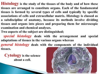
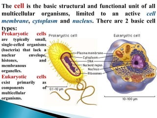
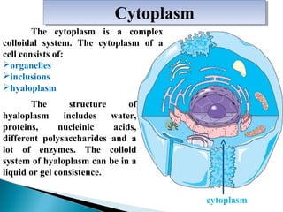
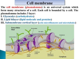
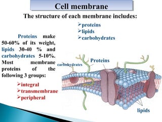
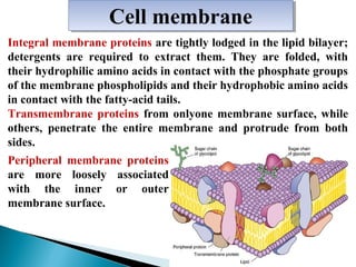
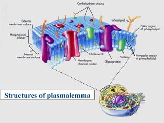
Structure and function of plasma membrane 2ICHHA PURAK
The presentation consists of 72 slides,describes following heads
DEFINITION : STRUCTURE OF PLASMA MEMBRANE
COMPONENTS OF PLASMA MEMBRANE ( (BIOCHEMICAL PROPERTIES)
LIPID BILAYER
PROTEINS
CARBOHYDRATES
CHOLESTEROL
MODELS EXPLAINING STRUCTURE OF BIO MEMBRANE
FLUID MOSAIC MODEL
MOBILITY OF MEMBRANE
GLYCOCALYX : GLYCOPROTEINS AND GLYCOLIPIDS
TRANSPORT OF IONS AND MOLECULES ACROSS PLASMA MEMBRANE
FUNCTIONS OF PLASMA MEMBRANE
DIVERSITY OF CELL MEMBRANES
SITE OF ATPASE ION CARRIER CHANNELS AND PUMPS-RECEPTORS
For More Medicine Free PPT - http://playnever.blogspot.com/
For Health benefits and medicine videos Subscribe youtube channel - https://www.youtube.com/playlist?list=PLKg-H-sMh9G01zEg4YpndngXODW2bq92w
the presentation gives the structure, function, and electron microscopic image of the various cytoplasmic organelles. it also includes the clinical significance of various organelle damage.
Introduction to the histology. Basics of cytology. The structural and molecular basis of cell function / Вступ до предмету гістології. Основи цитології. Структурні та молекулярні основи функціонування клітин.
Lection about Histology of the Respiratory System / Лекція на тему "Гістологія дихальної системи" (Author / Автор - Khodorovska Alla / Ходоровська Алла)
Cell: The cell is the ultimate structural and functional unit of the body.
The three principal constituents of the cell are:
1. Cell membrane
2. Cytoplasm and its organelles
3. Nucleus
power point presentation on the topic cellular level of organization from unit first of subject human anatomy and physiology I for first year B.PHARM it is useful for the student to study easily and find out the material easily for their study it is also useful for techers
this presentation providing about the cell .Cell is the basic living, structural, and functional unit of the body.
Cells are grouped together to form tissues, each of which has a specialized function, e.g.- Bone and blood tissue.
Different tissues are grouped together to form a organs, e.g. liver, stomach, and kidney etc.
Organs are grouped together to form a system, each of which performs a particular function responsible for maintaining homeostasis .
e.g. Urinary system, Respiratory system etc.
This presentation was a hard work of investigation and searching time by veterinary students of ULPGC Gran Canaria university. we learned to much about cytology in the process of recompilation of information. We share this knowledge with the desire that it may be useful and instructive for some one interested in cience or maybe other student arround the world. enjoy it!
Cells and its components(Anatomy) Easy explanationSwatilekha Das
Cells and its components,discussion on cell membrane, cytoplasm, nucleus with pictures....
easy explanation of anatomy topic for 1 st year GNM & B.Sc nursing students...
Comment to get explanation on your required topics.....
please like and share and follow.....
Презентація лекції на тему "Гістологія органів серцево-судинної системи" (Лектор - доцент Ходоровська А.А., Буковинський державний медичний університет)
June 3, 2024 Anti-Semitism Letter Sent to MIT President Kornbluth and MIT Cor...Levi Shapiro
Letter from the Congress of the United States regarding Anti-Semitism sent June 3rd to MIT President Sally Kornbluth, MIT Corp Chair, Mark Gorenberg
Dear Dr. Kornbluth and Mr. Gorenberg,
The US House of Representatives is deeply concerned by ongoing and pervasive acts of antisemitic
harassment and intimidation at the Massachusetts Institute of Technology (MIT). Failing to act decisively to ensure a safe learning environment for all students would be a grave dereliction of your responsibilities as President of MIT and Chair of the MIT Corporation.
This Congress will not stand idly by and allow an environment hostile to Jewish students to persist. The House believes that your institution is in violation of Title VI of the Civil Rights Act, and the inability or
unwillingness to rectify this violation through action requires accountability.
Postsecondary education is a unique opportunity for students to learn and have their ideas and beliefs challenged. However, universities receiving hundreds of millions of federal funds annually have denied
students that opportunity and have been hijacked to become venues for the promotion of terrorism, antisemitic harassment and intimidation, unlawful encampments, and in some cases, assaults and riots.
The House of Representatives will not countenance the use of federal funds to indoctrinate students into hateful, antisemitic, anti-American supporters of terrorism. Investigations into campus antisemitism by the Committee on Education and the Workforce and the Committee on Ways and Means have been expanded into a Congress-wide probe across all relevant jurisdictions to address this national crisis. The undersigned Committees will conduct oversight into the use of federal funds at MIT and its learning environment under authorities granted to each Committee.
• The Committee on Education and the Workforce has been investigating your institution since December 7, 2023. The Committee has broad jurisdiction over postsecondary education, including its compliance with Title VI of the Civil Rights Act, campus safety concerns over disruptions to the learning environment, and the awarding of federal student aid under the Higher Education Act.
• The Committee on Oversight and Accountability is investigating the sources of funding and other support flowing to groups espousing pro-Hamas propaganda and engaged in antisemitic harassment and intimidation of students. The Committee on Oversight and Accountability is the principal oversight committee of the US House of Representatives and has broad authority to investigate “any matter” at “any time” under House Rule X.
• The Committee on Ways and Means has been investigating several universities since November 15, 2023, when the Committee held a hearing entitled From Ivory Towers to Dark Corners: Investigating the Nexus Between Antisemitism, Tax-Exempt Universities, and Terror Financing. The Committee followed the hearing with letters to those institutions on January 10, 202
Unit 8 - Information and Communication Technology (Paper I).pdfThiyagu K
This slides describes the basic concepts of ICT, basics of Email, Emerging Technology and Digital Initiatives in Education. This presentations aligns with the UGC Paper I syllabus.
The French Revolution, which began in 1789, was a period of radical social and political upheaval in France. It marked the decline of absolute monarchies, the rise of secular and democratic republics, and the eventual rise of Napoleon Bonaparte. This revolutionary period is crucial in understanding the transition from feudalism to modernity in Europe.
For more information, visit-www.vavaclasses.com
Embracing GenAI - A Strategic ImperativePeter Windle
Artificial Intelligence (AI) technologies such as Generative AI, Image Generators and Large Language Models have had a dramatic impact on teaching, learning and assessment over the past 18 months. The most immediate threat AI posed was to Academic Integrity with Higher Education Institutes (HEIs) focusing their efforts on combating the use of GenAI in assessment. Guidelines were developed for staff and students, policies put in place too. Innovative educators have forged paths in the use of Generative AI for teaching, learning and assessments leading to pockets of transformation springing up across HEIs, often with little or no top-down guidance, support or direction.
This Gasta posits a strategic approach to integrating AI into HEIs to prepare staff, students and the curriculum for an evolving world and workplace. We will highlight the advantages of working with these technologies beyond the realm of teaching, learning and assessment by considering prompt engineering skills, industry impact, curriculum changes, and the need for staff upskilling. In contrast, not engaging strategically with Generative AI poses risks, including falling behind peers, missed opportunities and failing to ensure our graduates remain employable. The rapid evolution of AI technologies necessitates a proactive and strategic approach if we are to remain relevant.
Honest Reviews of Tim Han LMA Course Program.pptxtimhan337
Personal development courses are widely available today, with each one promising life-changing outcomes. Tim Han’s Life Mastery Achievers (LMA) Course has drawn a lot of interest. In addition to offering my frank assessment of Success Insider’s LMA Course, this piece examines the course’s effects via a variety of Tim Han LMA course reviews and Success Insider comments.
Operation “Blue Star” is the only event in the history of Independent India where the state went into war with its own people. Even after about 40 years it is not clear if it was culmination of states anger over people of the region, a political game of power or start of dictatorial chapter in the democratic setup.
The people of Punjab felt alienated from main stream due to denial of their just demands during a long democratic struggle since independence. As it happen all over the word, it led to militant struggle with great loss of lives of military, police and civilian personnel. Killing of Indira Gandhi and massacre of innocent Sikhs in Delhi and other India cities was also associated with this movement.
Welcome to TechSoup New Member Orientation and Q&A (May 2024).pdfTechSoup
In this webinar you will learn how your organization can access TechSoup's wide variety of product discount and donation programs. From hardware to software, we'll give you a tour of the tools available to help your nonprofit with productivity, collaboration, financial management, donor tracking, security, and more.
Francesca Gottschalk - How can education support child empowerment.pptxEduSkills OECD
Francesca Gottschalk from the OECD’s Centre for Educational Research and Innovation presents at the Ask an Expert Webinar: How can education support child empowerment?
Normal Labour/ Stages of Labour/ Mechanism of LabourWasim Ak
Normal labor is also termed spontaneous labor, defined as the natural physiological process through which the fetus, placenta, and membranes are expelled from the uterus through the birth canal at term (37 to 42 weeks
Normal Labour/ Stages of Labour/ Mechanism of Labour
Basic cytology
1. Professor asastent of department histology, cytology and
embryology - KHODOROVSKA ALLA
2. Histology is the study of the tissues of the body and of how these
tissues are arranged to constitute organs. Each of the fundamental
tissues is formed by several types of cells and typically by specific
associations of cells and extracellular matrix. Histology is classed as
a subdiscipline of anatomy, because its methods involve dividing
tissues and organs into pieces and preparing them for microscopic
examination and chemical analyses.
Two aspects of the subject are distinguished;
special histology deals with the arrangement and special
adaptations of tissues in the various organs whereas
general histology deals with the components of the individual
tissues.
Cytology is the science
about a cell.
3. The cell is the basic structural and functional unit of all
multicellular organisms, limited to an active cell
membrane, cytoplasm and nucleus. There are 2 basic cell
types:
Prokaryotic
cells
are typically small,
single-celled organisms
(bacteria) that lack a
nuclear
envelope,
histones,
and
membranous
organelles.
Eukaryotic
exist
primarily
components
multicellular
organisms.
cells
as
of
4. Сytoplasm
Сytoplasm
The cytoplasm is a complex
colloidal system. The cytoplasm of a
cell consists of:
organelles
inclusions
hyaloplasm
The
structure
of
hyaloplasm
includes
water,
proteins,
nucleinic
acids,
different polysaccharides and a
lot of enzymes. The colloid
system of hyaloplasm can be in a
liquid or gel consistence.
cytoplasm
5. Сell membrane
Сell membrane
The cell membrane (plasmalemma) is an universal system which
form many structures of a cell. Each cell is bounded by a cell. The
plasmalemma includes 3 layer:
I. Glycocalyx (carbohydrates)
II. Lipid bilayer (lipid molecule and proteins)
III. Submembrane cortical layer (actin microfilaments and microtubules)
6. Сell membrane
Сell membrane
The structure of each membrane includes:
Proteins make
50-60% of its weight,
lipids 30-40 % and
carbohydrates 5-10%.
Most
membrane
proteins
of
the
following 3 groups:
proteins
lipids
carbohydrates
carbohydrates
Proteins
іntegral
transmembrane
рeripheral
lipids
7. Сell membrane
Сell membrane
Integral membrane proteins are tightly lodged in the lipid bilayer;
detergents are required to extract them. They are folded, with
their hydrophilic amino acids in contact with the phosphate groups
of the membrane phospholipids and their hydrophobic amino acids
in contact with the fatty-acid tails.
Тransmembrane proteins from onlyone membrane surface, while
others, penetrate the entire membrane and protrude from both
sides.
Peripheral membrane proteins
are more loosely associated
with the inner or outer
membrane surface.
8. Сell membrane
Сell membrane
The inner and the outer dense line composed of a single layer of
phospholipids, between them settle some proteins. Carbohydrates
occur on plasma membranes mainly as oligosaccharide moieties of
membrane glycoproteins and glycolipids.
Membrane oligosaccharides
have
a
characteristic
branching structure and
project from the cell's outer
surface,
forming
a
superficial coat called the
glycocalyx that participates
in cell adhesion and
recognition.
Structures of plasmalemma
9. The plasmalemma carry out some of the important functions:
The plasmalemma carry out some of the important functions:
•Barrier the plasmalemma separates a cell from an environment
and other cells; the nucleus is separated from cytoplasm,
membranes organelles from hyaloplasm.
•Receptor on a surface of plasmalemma. There are special
structures-receptors due to which the cell finds out different
chemical substances, physical factors, other cells, hormones and
antigens.
special structures-receptors
10. •Transport-through a membrane of cell freely passes water, salts and
substances with low molecular weight. Such transport is called
passive.
•The transport against a gradient of concentration is called active.
Passive diffusion
Passive diffusion
Certain
substances
(e.g.,
water) can cross the
membrane in either
direction, following
a
concentration
gradient.
Passive diffusion
does not require
energy expenditure.
11. Facilitated diffusion
Facilitated diffusion
Certain molecules (e.g.,
glucose) cannot freely
diffuse across membranes
but must be helped across
by
a
membrane
component.
This
facilitated diffusion is
often unidirectional, but it
follows a concentration
gradient and requires no
energy.
12. Some nondiffusible molecules can move into or out
of cell either along or against a concentration gradient.
Such movement requires energy, usually as ATP. An
example of this active transport is the sodium pump
(Na+/K+-ATPase), which can expel sodium ions from a cell
even when the external sodium concentration is higher
than the internal one.
Active
Active
transport
transport
13. Endocytosis сells engulf extracellular substances and bring
them into the cytoplasm in membrane-limited vesicles by
mechanisms described collectively a endocytosis.
Exocytosis removes substances from the cell. Cells use this
process both for secretion and for excretion of undigested material.
A membrane-limited vesicle or secretory granule fuses with the
plasma membrane and releases its contents into the extracellular
space, without disrupting the plasma membrane.
Endocytosis
Exocytosis
14. •In phagocytosis (“cell eating”), the cell engulfs insoluble
extracellular substances, such as large macromolecules or
entire bacteria. The vesicles formed are termed
phagosomes.
•In pinocytosis (“cell drinking”), the cell engulfs small
amounts of intercellular fluid, which may contain a variety
of solutes. Pinocytotic vesicles are usually smaller than
phagosomes.
Pinocytosis
a
f
Phagocytosis
f
16. Intercellular connections
Simple contact – membranes of two cells are on distance of 10-12
nm in such manner that glycocalyx one cell adjoins with glycocalyx
of another cell. The basic function is metabolism and information
interchange between cells.
Zonulae occludentes — also
called tight junctions. Zonula
occludens are located between
adjacent plasma membranes most
typically near the apices of
epithelial cells. They form a "beltlike" junction that encircles the
entire circumference of the cell.
These junctions act as barriers
that prevent the movement of
molecules into the intercellular
spaces.
18. Intercellular connections
Zonular adherentes
are band-like
adhesion. This device surrounds the cell and
joins it to its neighbors.
Desmosomes (Maculae adherens).
This is the most common type of tight junction
between adjoining cells. A desmosome is a small
circumscribed area of attachment – attachment
plaques. At the side of a desmosome the plasma
membrane (of each cell) is thickened because of
the presence of dense layer of protein on inner
surface.
Desmosomes are serving
to attach the basal cell
membrane to the basal
lamina.
19. Gap junctions,
Intercellular connections
also called communicating junctions, are regions of
intercellular communication.
They are widespread in epithelial tissues, in
cardiac muscle smooth muscle cells and neurons. Gap junctions are built by six
closely packed transmembrane proteins connexins that assemble to from
structures called connexons. The two connexons fuse, forming the functional
intercellular communication channel. The hydrophilic channel permits the
passage of ions, small molecules and hormones.
20. Intercellular connections
Plasma membrane enfoldings of the
basal plasma
membrane increase the surface area available for transport. The
basal surface of some epithelia, especially those involved in ion
transport, possesses multiple enfoldings of the basal plasma
membrane. These enfoldings partition the basal cytoplasm and
many mitochondria into the finger-like enfoldings.
Synapse
- type of contact
between two nervous cells or
between a nervous cell and a
muscle. Through synapses pass
nervous impulses.
21. Cytoplasm structures can be divided into 3 groups:
•Organelles
are
membrane-bound,
enzymecontaining, permanent subcellular compatrments.
•Cytoplasmic inclusions are structures, membranebound or not, that are generally more transient than
organelles and less actively involved in cell metabolism.
•Cytoplasmic matrix the cytoskeleton is composed of
proteinaceous elements that form a supporting network
within the cytoplasm; some of these elements
(microtubules) also form discrete cytoplasmic structures
such as centrioles.
22. Organelles constantly present in a cell are described as
membranous and nonmembranous.
The membranous organelles include:
endoplasmic reticulum (rER and sER)
mitochondria
Golgi apparatus
lysosomes
peroxisomes
The nonmembranous organelles include:
microtubules
filaments (different varieties)
centrioles
ribosomes
Membranous organelles are the most part of organelles
of a cell.
23. The largest of the cytoplasmic organelles, mitochondria are the
energy providers of the cell. The size to bacteria (usually 2-6 mm
in lenght and 0.2 mm in diameter but quite variable) and have
varios shapes: spheric, ovoid, filamentous. Each mitochondrion is
bounded by 2 unit membranes.
The outer mitochondrial membrane has a smooth contour and
forms a continuous but relatively porous covering. It is freely
permeable to various small molecules.
The
inner
mitochondrial
membrane is less porous and is
therefore semipermeable. It has
numerous infoldings, or cristae, that
project into the mitochondrion’s
interior. The mitochondrial cristae
of most cells are shelflike, but those
in
steroid-secreting
cells
are
typically more tubular.
24. The mitochondrial membranes create 2 membrane-limited spaces. The
intermembrane space is located between the inner and outer membranes and is
continuous with the interacristal space that extends into the cristae. The
intercristal space, or matrix space, is enclosed by the inner membrane and
contains the mitochondrial matrix.
The mitochondrial matrix contains
water, solutes, and large matrix
granules, believed to be concerned
with
mitochondrial
calcium-ion
concentrations. It also contains
circular DNA and mitochondrial
ribosomes similar to those of bacteria.
The matrix contains numerous
soluble enzymes involved in such
specialized mitochondrial functions as
(citric acid
the Krebs cycle cycle, tricarboxylic acid cycle),
b-oxidation of lipids, and
mitochondrial
DNA
DNA synthesis.
25.
26. Function the provide of cell with the energy for chemical and
mechanical work by storing energy generated from cellular
metabolites in the high-energy bonds of ATP.
Mitochondria are found in nearly all eukaryotic cells, and in most
they are dispersed throughout the cytoplasm.
However, they accumulate in the
highest concentrations in cell types and
intracellular regions with the highest
energy requirements.
Cardiac muscle cells are notable for
the abundance of their mitochondria.
Epithelial cells lining the kidney
tubules have abundant mitochodria
interdigitated between basal plasma
membrane infoldings where active
transport of ions and water occurs.
DNA
27. The ribosomes are protein-synthesizing organelles. Each type of
ribosome has 2 unequal ribosomal subunits. Cytoplasmic ribosomes
are composed of ribosomal RNA (rRNA) synthesized in the nucleos
and associated proteins synthesized in the cytoplasm.
ribosomes
28. They are intensely basophilic. Light microscopy reveals cytoplasmic
accumulations of ribosomes as basophilic patches, formerly termed
ergastoplasm in grandular cells and Nissl bodies in neurons. In
electron micrographs, ribosomes appear as small, electron-dense
cytoplasmic granules.
Nissl bodies
29. Cytoplasmic ribosomes occur in 2 forms.
Free ribosomes are individual ribosomes dispersed in cytoplasm.
Polysomes are groups of ribosomes evenly distributed along a
single strand of messenger RNA (mRNA), an arrangement that
permits synthesis of multiple copies of a protein from the same
message. Polysomal ribosomes read (translate) the mRNA code and
thus play a critical role in assembling amino acids into specific
proteins, are found free in the cytoplasm (free polysomes) and
attached to membranes of the rER.
Free polysomes are involved in the
synthesis of structural proteins and
enzymes for intracellular use.
Polysomes of the rER are involved
in synthesizing proteins that are
secreted or
isolated.
30. Endoplasmic Reticulum
The endoplasmic reticulum (ER) is a complex organelle involved in the
synthesis, packaging, and processing of various cell substances. It is a
freely anastomosing network (reticulum) of membranes that form vesicles,
or cisternae; these may be elongated, flattened, rounded, or tubular.
Transfer vesicles (transitional
vesicles) are small, membranelimited vesicles that bud from
the ER and cross the
intervening cytoplasm to reach
the Golgi complex for further
processing or packaging of
their contents.
ER occurs in 2 forms:
rough
smooth
31. Rough endoplasmic reticulum (rER)
The also called granular endoplasmic reticulum, is studded with
ribosomes, many of them in polysomal clusters.
rER
cisternae
are
typically
parallel,
flattened, and elongated,
especially
in
cells
specialized for protein
secretion
(pancreatic
acinar cells, plasma cells).
The ribosomes give rER
basophilic
staining
properties.
The fine structure of
rER (membranes and
individual ribosomes) is visible only with the
electron microscope.
32. Rough endoplasmic reticulum (RER)
rER is mainly concerned with the synhtesis of proteins for
sequestration from the rest of the cytoplasm, ie, secretory proteins
such as collagen, proteins for incorporation into cell membranes,
and lysosomal enzymes (separated from the rest of the cytoplasm to
prevent autolysis).
RER in protein-secreting
epithelial cells often lies in
the
basal
cytoplasm,
between
the
plasma
membrane and the nucleus.
33. Smooth endoplasmic reticulum (SER)
The smooth endoplasmic reticulum (SER) lacks ribosomes
and thus appears smooth in electron micrographs.
SER cisternae are more
tubular or vesicular than those
of RER.
SER stains poorly, if at all, so
with the light microscope it is
indistinguishable from the rest
of the cytoplasm.
Because it lacks ribosomes, the
SER cannot synthesize proteins.
It has many enzymes, important
in lipid metabolism, steroid
hormone synthesis, glycogen
synthesis
(glucose-6phosphatase), and detoxification.
34. Smooth endoplasmic reticulum (SER)
The sER is suspended in the
cytoplasm of many cells and
is especially abundant in cells
that
synthesize
steroid
hormones (cells of the adrenal
cortex, gonads in liver cells
“hepatocytes”, where it is
involved in glycogen synthesis
and drug detoxification).
Specialized
SER
termed
sarcoplasmic reticulum is
found in striated muscle cells,
where it helps to regulate
muscle
contraction
by
sequestering and releasing
calcium ions.
35. The Golgi complex (Golgi apparatus) participates in many activities,
particularly those associated with secretion. It has an essential role
in coordinating membrane flow and vesicle traffic among organelles.
flattened cisternae;
The composed of 3 major
compartments:
conspicious stack of 3-10
discrete, slightly curved,
flattened cisternae;
numerous small vesicles
peripheral to the stack;
a few large vacuoles,
sometimes
called
condensing vacuoles, at
the concave surface of the
stack.
flattened cisternae;
36. The cis face (convex face,
forming face) of the stack is
usually closest to adjacent
dilated ER cisternae and is
surrounded by transfer
vesicles. Its cisternae stain
more darkly with osmium.
The trans face (concave
face, maturing face) often
harbors several condensing
vacuoles and generally faces
away from the nucleus.
flattened cisternae;
37. Lysosomes are spheric, membrane-limited vesicles that
may contain more than 50 enzymes each and function as
the cellular digestive system. Their characteristic enzyme
activities distinguish them from other cellular granules.
The enzyme most widely exploited for their identification
is acid phosphate, because it occurs almost exclusively in
lysosomes. Other enzymes common in lysosomes are
ribonucleases, deoxyribonucleases, cathepsins, sulfatases,
b-glucoronidase, and phospholipases and other proteases,
glucosidases, and lipases.
Lysosomal enzymes usually occur as glycoproteins
and are most active at an acidic pH. Lysosomes
occur in various sizes and electron densities,
depending on their level of activity.
38. Primary lysosomes are small (5-8 nm), with electron-dense
contents; they appear as black circles in electron micrographs. They
are the storage form of lysosomes, and their enzymes are mostly
inactive. Lysosomes enzymes synthesized and coreglycosylated in the
RER are transferred to the Golgi complex for further glycosylation;
it is uncertain whether their final packaging as primary lysosomes
occurs in the Golgi complex. The primary lysosomes disperse
through the cytoplasm.
They are found in most cells
but are most abundant in
phagocytic cells (macrophages,
neutrophils).
lysosomes
40. Secondary lysosomes are larger and less electron-dense and have a
mottled appearance in electron micrographs. They are formed by
the fusion of one or more primary lysosomes with a phagosome.
Their primary function is the digestion of products of heterophagy
and autophagy; when the lysosomal enzymes mix with the
phagosome contents, they become active.
Lysosomal enzymes also catabolize certain products of cell synthesis,
thus regulating the quality and quantity of secretory material.
Secondary lysosomes occur
throughout the cytoplasm in
many cells, in numbers that
reflect the cell’s lysosomal
and phagocytic activity.
41. Residual bodies
are
membranelimited inclusion of
varying size and
electron
density
associated with the
terminal phases of
lysosome function.
They
contain
undigestible
materials such as
pigments, crystals,
and certain lipids.
43. Peroxisomes are membrane-limited, enzyme-containing vesicles
somewhat larger than primary lysosomes. Peroxisomes function in
hydrogen peroxide metabolism. They contain urate oxidase,
hydroxyacid oxidase, and d-amino acid oxidase, which produce
hydrogen peroxide capable of killing bacteria; they also contain
catalase, which oxidizes various substrates and uses the hydrogen
removed in the process to convert the toxic hydrogen peroxide to
water.
Peroxisomes
also
participate
in
gluconeogenesis by assisting in the boxidation of fatty acids. They are found
dispersed in the cytoplasm or in
association with the SER.
with
44. Microtubules are the thickest components of the cytoskeleton, with
diameters of 24 nm. They are fine tubular structures of variable
lenght, with dense walls (5 nm thick) and a clear internal space (14
nm in internal diameter). The walls are composed of subunits called
tubulin heterodimers, each of which consists of one a-tubulin and
one b-tubulin protein molecule. The tubulin heterodimers are
arranged in protofilaments.
Thirteen of these threadlike b-tubulin
polymers of a- and b-tubulin
align parallel to one another to
form the wall of each
microtubule.
Microtubules
increase in length by adding
new heterodimers to one end,
called the nucleation site.
a-tubulin
protofilaments
45. Microfilaments are the thinnest cytoskeletal components (5-7 nm
wide). They are usually composed of one of several types of actin
protein. In striated muscle cells, actin filaments form a stable
paracrystalline array in association with filaments of myosin. Actin
filaments in other cells are less stable and can dissociate and
reassemble. These changes are regulated in part by calcium ions and
cyclic AMP and by actin-binding proteins in the cytoplasm.
Microfilaments are contractile, but to contract, they usually interact
with myosin. In muscle cells, myosin forms thick filaments. In
nonmuscle cells, it exists in soluble form.In nonmuscle cells,
microfilaments are generally distributed as an irregular meshwork
throughout the cytoplasm.
tropomyosin
myosin
actin
46. A centriole is a cylindric group of microtubules, 150 nm in overall
diameter and 350-500 nm long, containing 9 microtubule triplets in a
pinwheel array. Each microtubule in a triplet shares a portion of the
wall of the neighboring microtubule.
An interphase (nondividing)
cell has a pair of adjacent
centrioles with perpendicular
long axes, each surrounded by
several
electron-dense
satellites, or pericentriolar
bodies.
Other
cytoplasmic
microtubules
originate
from the pericenriolar
bodies and radiate into the
cytoplasm.
47. Centrioles are the structural organizers of the cell. Centriole
duplication is a prerequisite for cell division, and during mitosis
the centrioles organize the microtubules of the mitotic spindle.
Location. Between cell divisions,
centrioles are near the nucleus, often
surrounded by Golgi complexes. The
centrioles and associated Golgi
complexes
constitute
the
cell
cytocenter, which appears as a clear
zone near the nucleus. During the S
phase of interphase, each centriole
duplicates by giving rise to a
procentriole that grows at right
angels to the original. During mitosis,
the new centriole pairs migrate to
opposite cell poles to organize the
spindle.
48. Prominent among inclusions serving as storage depots are spheric
lipid droplets, which differ in appearance depending upon the type
of histologic preparation.
Glycogen granules are inclusions that are PAS-positive in light
microscopy and appear in electron micrographs as rosettes of
electron-dense particles. Both lipid droplets and glycogen granules
lack a limiting membrane.
Melanin is a brownish pigment widely distributed in vertebrates,
often found in electron-dense, membrane-limited granules termed
melanosomes. It is particularly abundant in epidermal cells and in
the pigment layer of the retina.
49. NUCLEUS
Nuclei vary in appearance from tissue to tissue and cell to cell, but
they generally have a:
nuclear envelope
nucleoplasm
chromatin
one to several nucleoli
generally
50. NUCLEUS
The nuclear contents are set apart from the cytoplasm by a double
membrane called the nuclear envelope and a narrow (40-70 nm)
intermembrane space called the perinuclear space.
The nuclear envelope is often considered an exctension of the
RER, because its outer surface is often peppered with ribosomes and
shows occasional continuities with the RER.
The inside of the inner
membrane is lined with the
fibrous lamina, a layer
consisting of proteins called
nuclear laminа.
generally
inside
51. NUCLEUS
The envelope is perforated by many nuclear pores,
each of which has a diameter of about 70 nm and is
bounded by:
8 globular subunits
called annular proteins,
which
present
an
octagonal appearance in
some preparations.
Each pore is covered
by
a
proteinaceous
diaphragm
that
is
thinner
than
the
envelope.
globular subunits
generally
52. PORES
The pores provide a channel
for
the
movement
of
important molecules between
the nucleus and cytoplasm.
These
molecules include
nucleic acids synthesized in the
nucleus and used in the
cytoplasm (mRNA, rRNA,
tRNA)
and
proteins
synthesized in the cytoplasm
and used in the nucleus
(histones, polymerases).
nucleus
generally
54. Nucleoplasm
The nucleoplasm is the matrix in which the other
intranuclear components are embedded. It is composed of
enzymatic and nonenzymatic proteins, metabolites, ions,
and water.
heterochromatin
It includes the nuclear
matrix – a fibrillar
“nucleoskeletal” structure
that appears to bind
certain
hormone
receptors-and
newly
synthesized DNA.
55. Nuclear chromatin
Nuclear chromatin is an intensely basophilic substance
consisting of DNA and associated histone and nonhistone
proteins.
Nuclei containing highly coiled chromatin, termed
heterochromatin, stain darkly with basic dyes.
heterochromatin
coiled
56. Nuclear chromatin
Chromatin is a complex of DNA and proteins. It
is responsible for the characteristic basophilia of
the nucleus.
The
densely
staining
material
is
highly
condensed chromatin called
heterochromatin and the
lightly staining material is a
dispersed
form
called
euchromatin.
57. Chromosomes
The chromosomes, the most highly condensed form of
chromatin, are visible during mitosis. In females, only one
X chromosome (either of the 2) is used by each cell;
The inactive X chromosome is often visible as a clump of
heterochromatin termed sex chromatin, or the Barr body.
In most cells, the Barr
body is attached to the
inner surface of the
nuclear envelope. In a
neutrophilic leukocyte,
it may appear as a
drumstick-shaped
appendage
of
the
lobulated nucleus.
58. Nucleolus
During interphase (between mitoses), each nucleus
usually has at least one (or2) intensely basophilic body
called a nucleolus. Nucleoli are the synthesis sites for
most ribosomal RNA (rRNA).
The
nucleolus
disappears
in
preparation
for
mitosis and reappears
after
mitosis
is
completed.
59. Nucleolus
The term nucleolonema is used by light microscopists to refer to a
threadlike basophilic substructure of the nucleolus. The nucleolonema
contains 2 rRNA-rich components distinguishable by electron microscopy.
The pars fibrosa consists of densely packed ribonucleoprotein fibers, 510 nm in diameter. These fibers consist of the newly synthesized primary
transcripts of the rRNA genes and associated proteins.
The pars granulosa
contains dense granules,
15-20 nm in diameter,
that represent maturing
ribosomal
subunits
during
assembly
for
export to the cytoplasm.
Nucleolus
pars fibrosa
pars granulosa