Downloaded 113 times


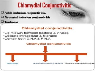

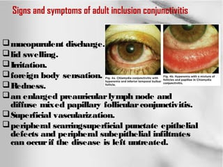


This document discusses Chlamydia trachomatis, the bacteria that causes chlamydial conjunctivitis and trachoma. It describes the signs and symptoms of adult and neonatal inclusion conjunctivitis caused by C. trachomatis infection, including mucopurulent discharge, redness, lid swelling, and irritation. Treatment involves topical and systemic antibiotics like erythromycin or tetracycline. The document also discusses trachoma, a chronic inflammatory disease caused by repeated C. trachomatis infection. Trachoma is endemic in Egypt and clinical features include trichiasis, entropion, and corneal opacities. Prevention strategies like the SAFE approach (sur


