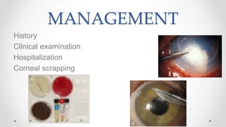
Infectious Corneal
Ulcers
Corneal Ulcers
Def: Corneal ulcers are defects in the corneal epithelium
with or without stromal infiltration.
Types:
A) Infectious ulcerative keratitis
B) Non infectious ulcerative keratitis
Bacteria and
Fungi Viruses Acanthamoeba
Systemic
Autoimmune/
Inflammatory
Local Toxic
InfectiousNon infectious
Etiology
Non Infectious Ulcerative Keratitis
Causes:
Local causes:
Punctate marginal keratitis: Staphylococci, Streptococci, hypersensitivity to
medications
Peripheral keratitis associated with blepharitis:
Systemic causes:
Generally manifestation of systemic, immune-mediated disease
Most common: Rheumatoid arthritis, Wegener’s granulomatosis and
polyarteritis nodosa
Non INFECTIOUSINFECTIOUS
No painPain
No dischargeDischarge
AC reaction: absentAC reaction: present
PeripheralCentral
Trauma: -------Trauma : ++++
Infectious keratitis
Important Facts
 A corneal ulcer is an ocular emergency that
raises high stakes of questions about diagnosis
and management.
 When a large corneal ulcer is staring you in the
face time isn't in your side.
 Despite varying etiologies and presentations,
as well as different treatment approaches ,
corneal ulcers have one thing in common : the
potential to cause devastating loss of vision.
PRINCIPLES OF MANAGEMENT OF CORNEAL
DISEASE
1- Control of infection
2- Control of inflammation
3- Promotion of re-epithelialization
– lubrication
– lid closure
– bandage soft contact lens
4- Prevention of perforation
Tissue adhesive glue
Conjunctival flap
Systemic immunosuppressive agents
Corneal grafting
MICROBIAL KERATITIS ( Bacterial)
Predisposing factors:
Ocular surface disease: Trauma,
post-herpetic
corneal disease,
Bullous Keratopathy,
Corneal exposure,
Dry eye
Diminished corneal sensation
Contact lens wear
Causative Organisms
80 % of cases >>>>Staphylococcus aurous, Streptococcus
pneumonia and Pseudomonas species
Pseudomonas aeruginosa
is the most frequent and the most pathogenic ocular
pathogen which can cause corneal perforation in just 72
hours
MICROBIAL KERATITIS ( Bacterial)
Pathogens which can produce corneal infection in intact
epithelium.
1.Neisseria gonorrhoeae
2.Corynebacterium diphtheriae
3.Listeria
4.Haemophilus
Staph. aureus and strep. pneumoniae
Oval, yellow-white, densely opaque stromal suppuration
surrounded by relatively clear cornea
MANAGEMENT
History
Clinical examination
Hospitalization
Corneal scrapping
Differentiators
 Acute painful injected eye.
 Profuse tearing and discharge.
 Decrease visual acuity.
 Large F.B
 Stromal invasion with epithelial
excavating edge.
Treatment
1. Fluroquinolones 
 Every 5 mins / hour
 Hour / 24 hs
 2 hour / 24 hs
2. Fortified eye drops  ulcer < 2 ws ,
improvement not obvious.
(N.B)Don’t miss resistant bacteria.
3. Steriods????
FUNGAL KERATITIS
Filamentous fungal keratitis
–Aspergillus - Fusarium
Yeasts candida
Clinically
History of vegetable matter injury
Greyish-white ulcer with indistinct margins
Surrounded by feathery infiltrates
Ring infiltrate
Endothelial plaque
Hypopyon
Differentiators
 Dull grey infiltrate.
 Satellite lesions.
 Awareness of those ulcers resembling
bacterial keratitis
 Awareness of those caused by yeast  better
defined borders
 Real flags
Candida keratitis
Usually develops in pre-existing corneal disease or
immunocompromised patient
Yellow-white ulcer
Dense suppuration
Filamentous fungal keratitis
Firm elevated necrotic slough, “hyphate” lines that extend beyond the
edge of the ulcer into the normal cornea,
multifocal granular (or feathery) gray-white “satellite” infiltrates in the
corneal stroma
MANAGEMENT
Culture
Biopsy
Antifungal therapy
– Initially broad-spectrum econazole 1%, voriconazole 1% topically
– Then depending upon sensitivity Natamycin or imidazole for 6 weeks
Systemic ketoconazole
Therapeutic penetrating keratoplasty
ACANTHAMOEBA KERATITIS
Protozoan
–Active (trophozoite)
–Dormant (cystic)
Common in swimmers
and CL wearers
Clinically
Blurred vision and disproportionate pain
Patchy anterior stromal infilterates
Perineural infilterates (radial keratoneuritis)
Infilterates coalesce –ring abcess, ulceration and
hypopyon
White satellite lesions
Acanthamoeba Keratitis
MANAGEMENT
Corneal scrappings stained with calcoflour white
Corneal biopsy
Treatment with chlorhexidine,
polyhexamethylenebiguanide drops,
dipropamidine and propamidine.
Therapeutic penetrating keratoplasty
VIRAL KERATITIS
HERPES SIMPLEX VIRUS
Basically it is epitheliotropic and
may become neurotropic
Two types-HSV-1 & HSV-2
Primary HSV-1 infection occurs
most commonly in the mucocutaneous
distribution of the trigeminal nerve
Classification
1. Epithelial keratitis
2. Disciform keratitis (localized endotheliitis)
4.Metaherpitic ulcer
Necrotizing keratitis
Keratouveitis
Differentiators
 Dentritic ulcer.
 Loss of corneal sensation.
 Photophobia.
Types of HSV keratitis:
 Primary
 Recurrent
 Dentritic , Geographic , Metaherptica
 Diabetic foot in the eye  Neurotrophic
TREATMENT
Epithelial keratitis is treated with topical antivirals
Aciclovir ophthalmic ointment and Trifluridine eye drops
Herpetic stromal keratitis is treated initially with prednisolone
drops every 2 hours accompanied by a prophylactic antiviral drug
Metaherpetic ulcer treated by artificial tears and eye lubricants,
stopping toxic medications, performing punctal occlusion, bandage
contact lens and amniotic membrane transplant
Herpes Zoster Keratitis
Caused by Varicella zoster virus
Causes-varicella(chickenpox) & herpes zoster(shingles)
RISK FACTORS:
• Increasing risk(>70 years)
• Neoplastic diseases
• Immunosuppressive drugs
• Organ transplant recipients
• HIV
Herpes Zoster Ophthalmicus
(HZO)
Unilateral painful skin rash in one or more dermatome distributions of
the fifth cranial nerve (trigeminal nerve)
HUTCHINSON’S sign:
Vesicles at the side or
tip of
the nose are potential
indicators of
ocular diseases
ACUTE EYE DISEASES
Acute epithelial keratitis:
Dendritic lesions with tapered ends without terminal bulbs
Nummular keratitis:
Fine granular subepithelial deposits
Episcleritis:
Scleritis and sclerokeratitis:
Stromal (interstitial) keratitis:
Anterior uveitis
Posterior uveitis
Neurotrophic keratopathy
Scleritis
Eyelid scarring
POST HERPETIC NEURALGIA
Neurological complications
Treatment
Oral acyclovir 800 mg five times daily for 7 to 10 days is the
standard treatment
Topical antiviral ttt
Topical steroids in uveitis
Pain should be treated with narcotics
Topical skin ointment
Thank you

Infectious corneal ulcers

  • 1.
  • 2.
    Corneal Ulcers Def: Cornealulcers are defects in the corneal epithelium with or without stromal infiltration. Types: A) Infectious ulcerative keratitis B) Non infectious ulcerative keratitis
  • 3.
    Bacteria and Fungi VirusesAcanthamoeba Systemic Autoimmune/ Inflammatory Local Toxic InfectiousNon infectious Etiology
  • 4.
    Non Infectious UlcerativeKeratitis Causes: Local causes: Punctate marginal keratitis: Staphylococci, Streptococci, hypersensitivity to medications Peripheral keratitis associated with blepharitis: Systemic causes: Generally manifestation of systemic, immune-mediated disease Most common: Rheumatoid arthritis, Wegener’s granulomatosis and polyarteritis nodosa
  • 5.
    Non INFECTIOUSINFECTIOUS No painPain NodischargeDischarge AC reaction: absentAC reaction: present PeripheralCentral Trauma: -------Trauma : ++++
  • 8.
  • 9.
    Important Facts  Acorneal ulcer is an ocular emergency that raises high stakes of questions about diagnosis and management.  When a large corneal ulcer is staring you in the face time isn't in your side.  Despite varying etiologies and presentations, as well as different treatment approaches , corneal ulcers have one thing in common : the potential to cause devastating loss of vision.
  • 10.
    PRINCIPLES OF MANAGEMENTOF CORNEAL DISEASE 1- Control of infection 2- Control of inflammation 3- Promotion of re-epithelialization – lubrication – lid closure – bandage soft contact lens 4- Prevention of perforation Tissue adhesive glue Conjunctival flap Systemic immunosuppressive agents Corneal grafting
  • 11.
    MICROBIAL KERATITIS (Bacterial) Predisposing factors: Ocular surface disease: Trauma, post-herpetic corneal disease, Bullous Keratopathy, Corneal exposure, Dry eye Diminished corneal sensation Contact lens wear
  • 12.
    Causative Organisms 80 %of cases >>>>Staphylococcus aurous, Streptococcus pneumonia and Pseudomonas species Pseudomonas aeruginosa is the most frequent and the most pathogenic ocular pathogen which can cause corneal perforation in just 72 hours
  • 13.
    MICROBIAL KERATITIS (Bacterial) Pathogens which can produce corneal infection in intact epithelium. 1.Neisseria gonorrhoeae 2.Corynebacterium diphtheriae 3.Listeria 4.Haemophilus
  • 14.
    Staph. aureus andstrep. pneumoniae Oval, yellow-white, densely opaque stromal suppuration surrounded by relatively clear cornea
  • 15.
  • 16.
    Differentiators  Acute painfulinjected eye.  Profuse tearing and discharge.  Decrease visual acuity.  Large F.B  Stromal invasion with epithelial excavating edge.
  • 17.
    Treatment 1. Fluroquinolones  Every 5 mins / hour  Hour / 24 hs  2 hour / 24 hs 2. Fortified eye drops  ulcer < 2 ws , improvement not obvious. (N.B)Don’t miss resistant bacteria. 3. Steriods????
  • 18.
    FUNGAL KERATITIS Filamentous fungalkeratitis –Aspergillus - Fusarium Yeasts candida
  • 19.
    Clinically History of vegetablematter injury Greyish-white ulcer with indistinct margins Surrounded by feathery infiltrates Ring infiltrate Endothelial plaque Hypopyon
  • 20.
    Differentiators  Dull greyinfiltrate.  Satellite lesions.  Awareness of those ulcers resembling bacterial keratitis  Awareness of those caused by yeast  better defined borders  Real flags
  • 21.
    Candida keratitis Usually developsin pre-existing corneal disease or immunocompromised patient Yellow-white ulcer Dense suppuration
  • 22.
    Filamentous fungal keratitis Firmelevated necrotic slough, “hyphate” lines that extend beyond the edge of the ulcer into the normal cornea, multifocal granular (or feathery) gray-white “satellite” infiltrates in the corneal stroma
  • 23.
    MANAGEMENT Culture Biopsy Antifungal therapy – Initiallybroad-spectrum econazole 1%, voriconazole 1% topically – Then depending upon sensitivity Natamycin or imidazole for 6 weeks Systemic ketoconazole Therapeutic penetrating keratoplasty
  • 24.
  • 25.
    Clinically Blurred vision anddisproportionate pain Patchy anterior stromal infilterates Perineural infilterates (radial keratoneuritis) Infilterates coalesce –ring abcess, ulceration and hypopyon White satellite lesions
  • 26.
  • 27.
    MANAGEMENT Corneal scrappings stainedwith calcoflour white Corneal biopsy Treatment with chlorhexidine, polyhexamethylenebiguanide drops, dipropamidine and propamidine. Therapeutic penetrating keratoplasty
  • 28.
    VIRAL KERATITIS HERPES SIMPLEXVIRUS Basically it is epitheliotropic and may become neurotropic Two types-HSV-1 & HSV-2 Primary HSV-1 infection occurs most commonly in the mucocutaneous distribution of the trigeminal nerve
  • 29.
    Classification 1. Epithelial keratitis 2.Disciform keratitis (localized endotheliitis)
  • 30.
  • 31.
    Differentiators  Dentritic ulcer. Loss of corneal sensation.  Photophobia. Types of HSV keratitis:  Primary  Recurrent  Dentritic , Geographic , Metaherptica  Diabetic foot in the eye  Neurotrophic
  • 32.
    TREATMENT Epithelial keratitis istreated with topical antivirals Aciclovir ophthalmic ointment and Trifluridine eye drops Herpetic stromal keratitis is treated initially with prednisolone drops every 2 hours accompanied by a prophylactic antiviral drug Metaherpetic ulcer treated by artificial tears and eye lubricants, stopping toxic medications, performing punctal occlusion, bandage contact lens and amniotic membrane transplant
  • 33.
    Herpes Zoster Keratitis Causedby Varicella zoster virus Causes-varicella(chickenpox) & herpes zoster(shingles) RISK FACTORS: • Increasing risk(>70 years) • Neoplastic diseases • Immunosuppressive drugs • Organ transplant recipients • HIV
  • 34.
    Herpes Zoster Ophthalmicus (HZO) Unilateralpainful skin rash in one or more dermatome distributions of the fifth cranial nerve (trigeminal nerve) HUTCHINSON’S sign: Vesicles at the side or tip of the nose are potential indicators of ocular diseases
  • 35.
    ACUTE EYE DISEASES Acuteepithelial keratitis: Dendritic lesions with tapered ends without terminal bulbs
  • 36.
    Nummular keratitis: Fine granularsubepithelial deposits Episcleritis: Scleritis and sclerokeratitis: Stromal (interstitial) keratitis: Anterior uveitis
  • 37.
  • 38.
    Eyelid scarring POST HERPETICNEURALGIA Neurological complications
  • 39.
    Treatment Oral acyclovir 800mg five times daily for 7 to 10 days is the standard treatment Topical antiviral ttt Topical steroids in uveitis Pain should be treated with narcotics Topical skin ointment
  • 40.