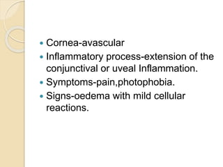
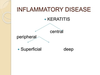
PATHOLOGY OF CORNEA
OPTOM FASLU MUHAMMED
 Cornea-avascular
 Inflammatory process-extension of the
conjunctival or uveal Inflammation.
 Symptoms-pain,photophobia.
 Signs-oedema with mild cellular
reactions.
INFLAMMATORY DISEASE
 KERATITIS
central
peripheral
 Superficial deep
CONGENITAL ABNORMALITIES
 INCLUDES:size,curvature,altrations
and congenital oppacity.
 Micro cornea:
 Megalo cornea:
 Corneal plana:
 Keratoconus
 CONGENITAL CORNEAL
OPPACITIES
CENTRAL
PERIPHERAL
 Peter’s anomaly juvanile
arcus senillis
 Posterior keratoconus
Ulcerative keratitis
 Commonest form of keratitis
 Bacterial , fungal or viral in aetiology
 Described in three stages
 Progressive stage
 Regressive stage
 Healing stage
 Progressive stage-oedeme with
migration of polymorph to the area of
site
 Regressive stage – demolition with the
development of a disticnt line of
demarcation
 Healing stage- keratocytes and
fibroblast , migrating from the limbus
along with blood vessels , lay down
collagen and produce the groud
COMPLICATIONS
Hypopyon-in bacterial ulcer , it results
from irritants and toxins that cross the
descemet’s membrane
Descemetocoele - there is a significant
stromal loss due to necrosis , the thin
descemet;s membrane cannot withstand
iop and protrudes forewards
Retrocorneal abscess – extention of
reactions
 Neutrophillic aggregrates
 Oedema-encounters in all disease that
effects endithelium
 Accomponies both infective and non
infective
 Varies with severity of the process
DEGENERATION
 Superficial deep
SUPERFICIAL
 Mainly in epithelial layers
 XEROPHTHALMIA – Dryness of
conjuctivaand cornea due to
excessive keratinisation
DEEP- Corneal complicaton of retinal
deficiency
 Dry eye- watering eye is
nuicense,while dry eye is a potential
threat to vision
DYSTROPHIES
 Bilateral primary corneal disorder
 Epithelial
 Bowman’s
 Stromal
 endothelial
EPITHELIAL –epithelial cysts may be
present
ANTERIOR MEMBRANE
DYSTROPHY –Known as reis-
buckler’s dystrophy is na autosomal
domonant disorder
STROMAL
GRANULAR DYSTROPHY-
EOSINOPHILIC DEPOSITS
OCCUPYING DIFFERENT ZONES
 MACULAR DYSTROPHY-This stromal
dystrophy is an autosomal recessive
lession characterised by diffuse
corneal oedema with whitish cloudy
deposits in the stroma
 Lattice dystrophy – is primary
amylodosis of cornea
 Endothelial- fuch’s dystrophy fuch’s
endothelial dystrophy-thickning of
descemets membrane with variable
loss of endothelial cells
 Congenital heriditary endothelial
dystrophy
 keratoconus
THANK YOU….

Pathology of cornea