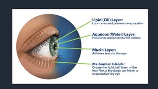
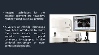
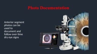
- Imaging techniquesfor the
anterior segment are nowadays
routinely used in clinical practice.
- A variety of imaging techniques
have been introduced to study
the ocular surface, such as
anterior segment optical
coherence tomography, in vivo
confocal microscopy, or non-
contact meibography.
9.
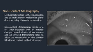
Non-Contact Meibography
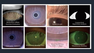
- Meibographyrefers to the visualization
and quantification of Meibomian gland
drop-out using photo documentation.
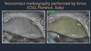
- Non contact Meibography consist of a
‐
slit lamp equipped with an infrared
charge coupled device video camera
‐
and an infrared transmitting filter to
allow the observation of the everted
lid without contact to the instrument.
10.
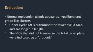
Evaluation:
- Normal meibomianglands appear as hypoilluminant
grape-like clusters.
- Upper eyelid MGs outnumber the lower eyelid MGs
and are longer in length.
- The MGs that did not transverse the total tarsal plate
were indicated as a “dropout.”
11.
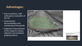
Advantages:
• Easy toperform, with
entire gross evaluation of
the lid.
• Good tool for
documentation and
monitoring of the glands.
• Subjective interpretation
of the image by various
objective and analytical
grading systems.
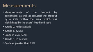
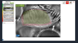
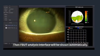
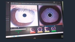
Measurements:
• Measurements ofthe dropout by
percentage, as well as grouped the dropout
by a scale within the area, which was
highlighted by the users’ free-hand tool:
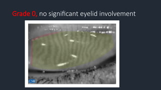
• Grade 0, no loss at all;
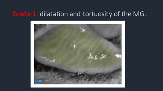
• Grade 1, ≤25%;
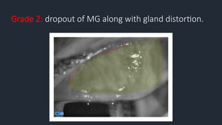
• Grade 2, 26%–50%;
• Grade 3, 51%–75%;
• Grade 4, greater than 75%
15.
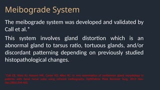
Meibograde System
The meibogradesystem was developed and validated by
Call et al.*
This system involves gland distortion which is an
abnormal gland to tarsus ratio, tortuous glands, and/or
discordant patterning depending on previously studied
histopathological changes.
*Call CB, Wise RJ, Hansen MR, Carter KD, Allen RC. In vivo examination of meibomian gland morphology in
patients with facial nerve palsy using infrared meibography. Ophthalmic Plast Reconstr Surg. 2012 Nov-
Dec;28(6):396-400.
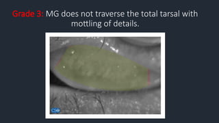
Grade 3: MGdoes not traverse the total tarsal with
mottling of details.
20.
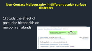
Non-Contact Meibography indifferent ocular surface
disorders
1) Study the effect of
posterior blepharitis on
meibomian glands
21.
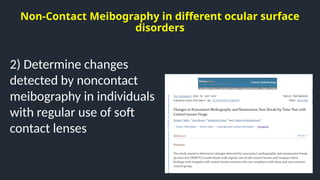
Non-Contact Meibography indifferent ocular surface
disorders
2) Determine changes
detected by noncontact
meibography in individuals
with regular use of soft
contact lenses
22.
Non-Contact Meibography indifferent ocular surface
disorders
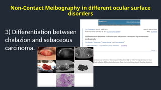
3) Differentiation between
chalazion and sebaceous
carcinoma.
23.
Non-Contact Meibography indifferent ocular surface
disorders
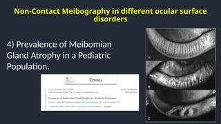
4) Prevalence of Meibomian
Gland Atrophy in a Pediatric
Population.
24.
Non-Contact Meibography indifferent ocular surface
disorders
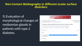
5) Evaluation of
morphological changes of
meibomian glands in
patients with type 2
diabetes.
25.
Non-Contact Meibography indifferent ocular surface
disorders
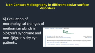
6) Evaluation of
morphological changes of
meibomian glands in
Sjögren’s syndrome and
non-Sjögren’s dry eye
patients.
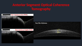
Anterior Segment OpticalCoherence
Tomography
Tear film thickness
Tear meniscus area (TMA).
Tear meniscus height (TMH)
32.
In vivo confocalmicroscopy
• A noninvasive technology that is
useful as a supplementary diagnostic
tool for the in vivo assessment of the
histopathology of many ocular surface
diseases and anterior-segment
disorders at cellular level.
• In the dry eye field, in vivo confocal
microscopy has been applied to the
examination of the cornea, bulbar and
palpebral conjunctiva, Meibomian
gland, and lacrimal gland.


![Dry_Eye_Presentation_Final[1].pptx......](https://cdn.slidesharecdn.com/ss_thumbnails/dryeyepresentationfinal1-250516163834-f963ff70-thumbnail.jpg?width=640&height=640&fit=bounds)


![CASE_PRESENTATION_ON_subdural_hematoma(SDH)[1 FINAL PPT]-1.pptx](https://cdn.slidesharecdn.com/ss_thumbnails/casepresentationonsubduralhematomasdh1finalppt-1-260129172522-d405d375-thumbnail.jpg?width=640&height=640&fit=bounds)


