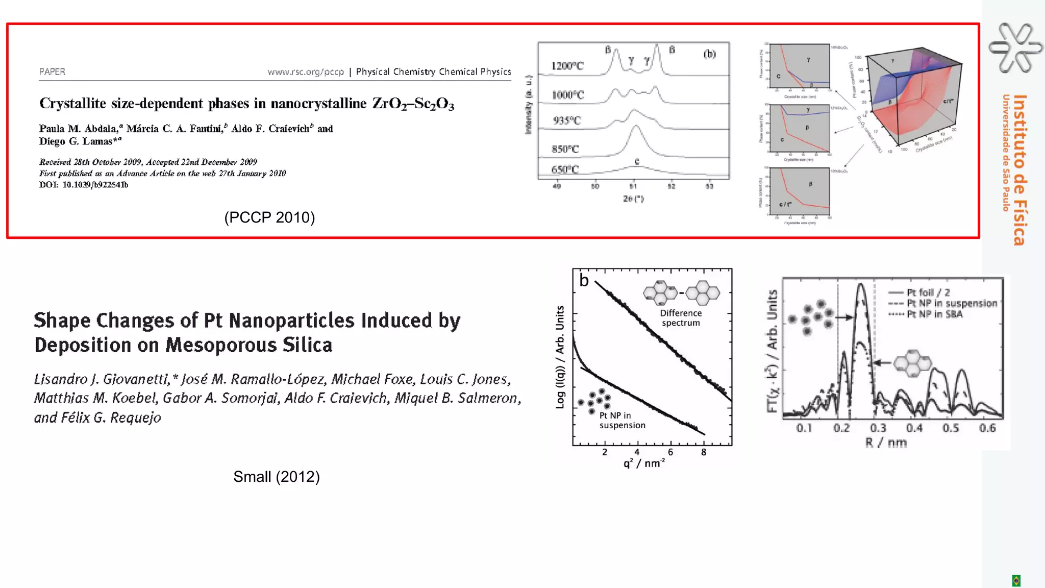
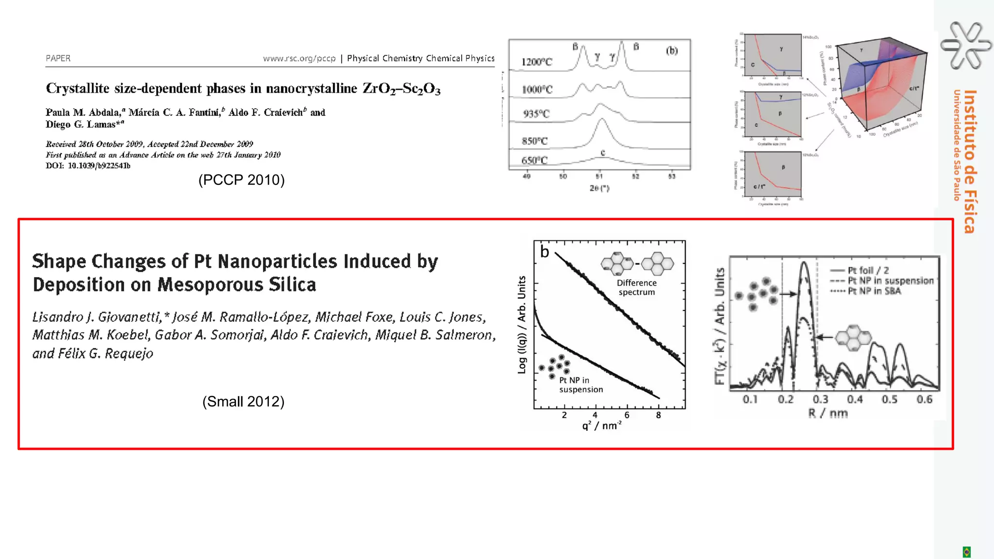
Download as PDF, PPTX
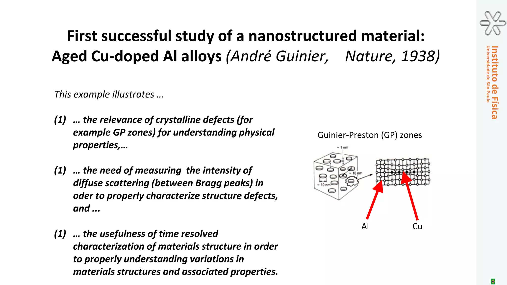
![This example illustrates …
(1) … the relevance of crystalline defects (for
example GP zones) for understanding physical
properties,…
(1) … the need of measuring the intensity of
diffuse scattering (between Bragg peaks) in
oder to properly characterize structure defects,
and ...
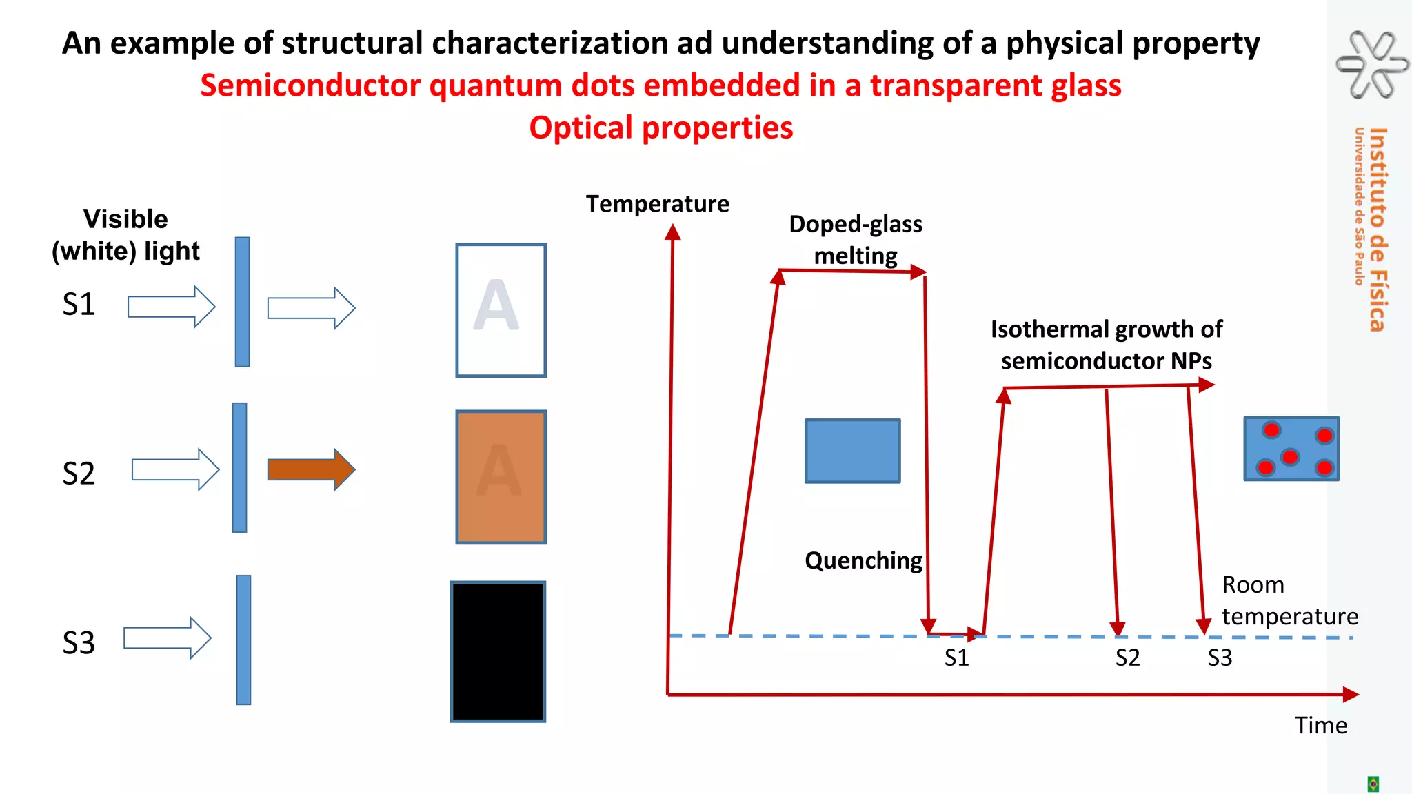
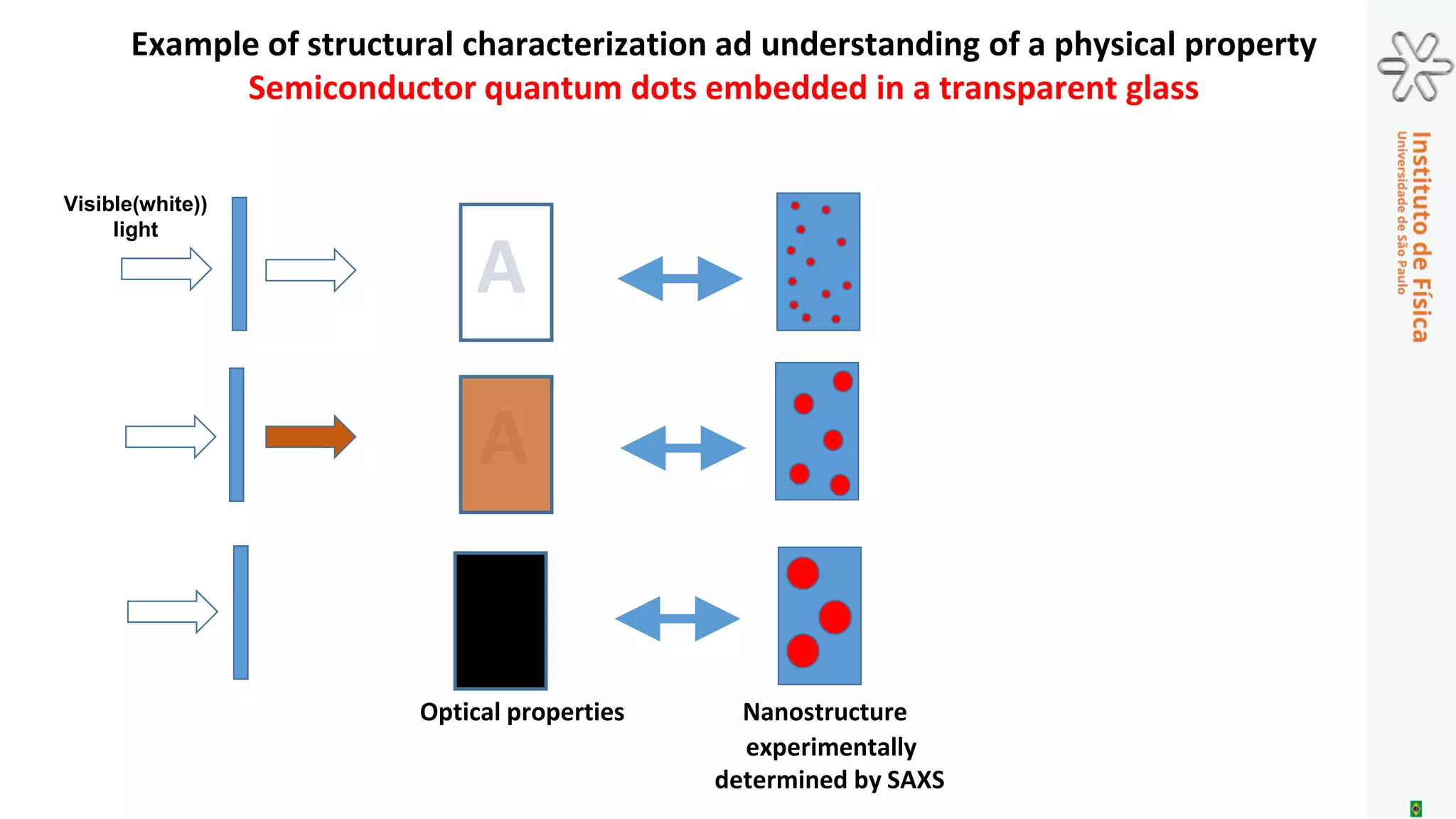
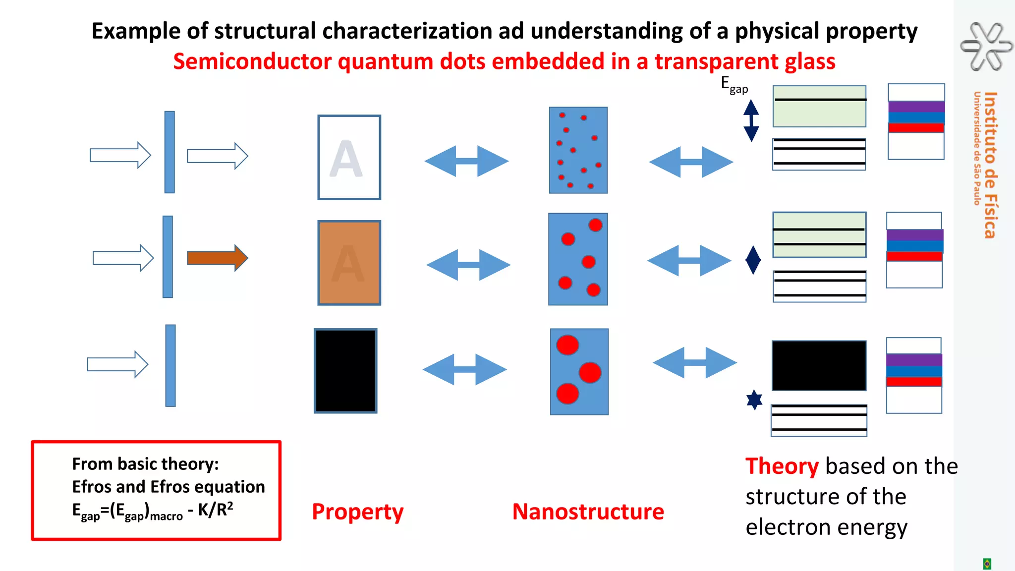
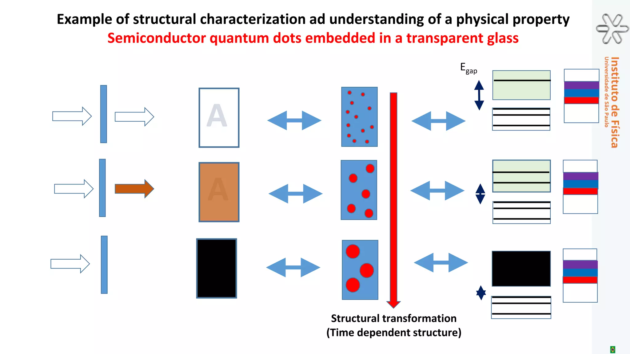
(1) … the usefulness of time resolved
characterization of materials structure in order
to properly understanding variations in
materials structures and associated properties.
]
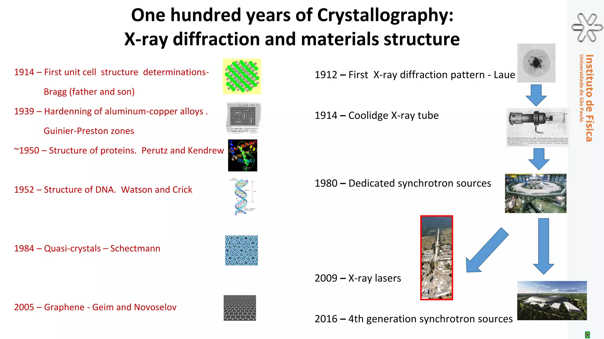
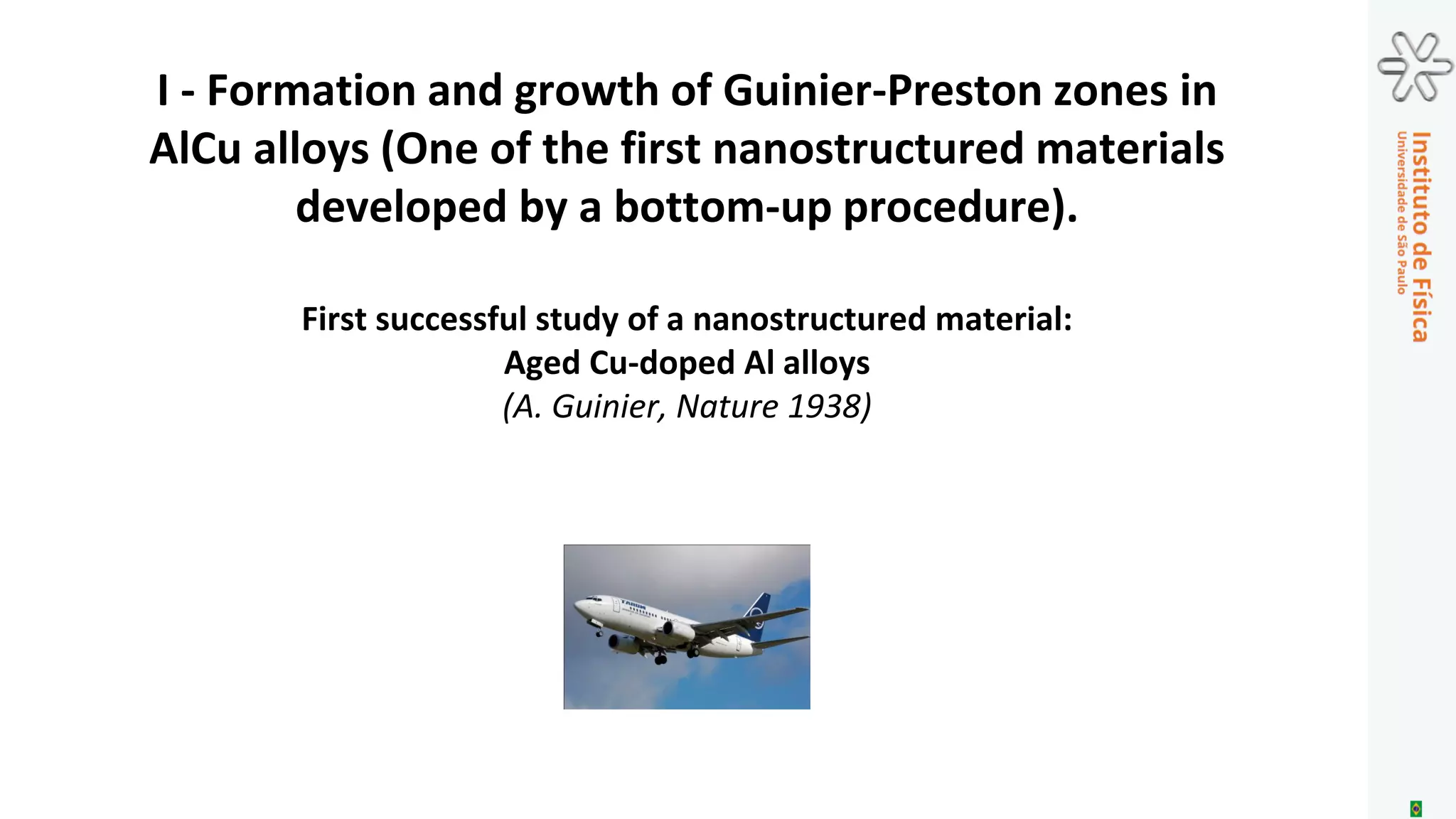
Guinier-Preston (GP) zones
Al Cu
First successful study of a nanostructured material:
Aged Cu-doped Al alloys (André Guinier, Nature, 1938)](https://image.slidesharecdn.com/craievichmemorial2016-161010021942/75/Advanced-Characterization-of-Materials-Relevance-and-Challenges-21-2048.jpg)
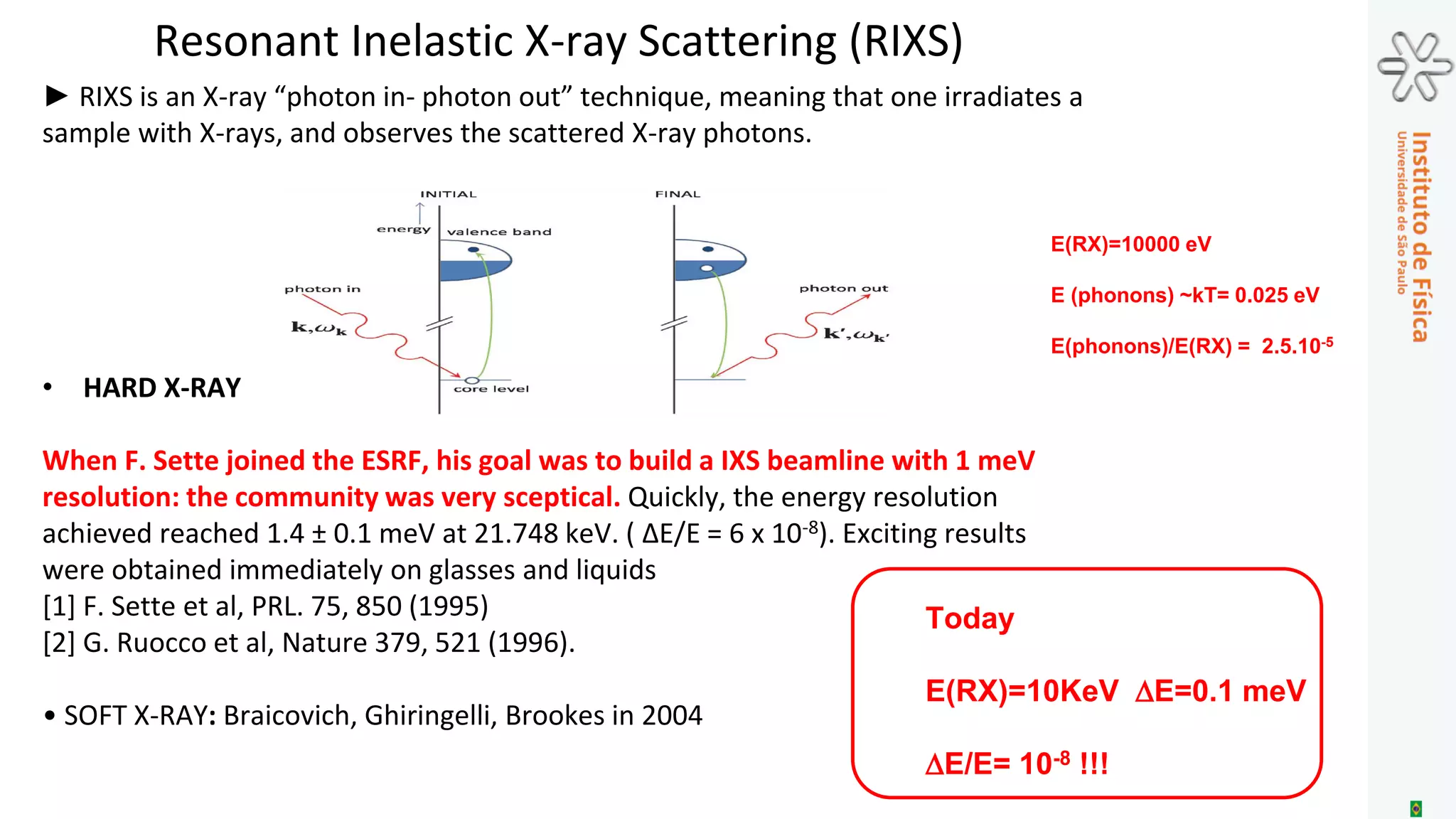
![Resonant Inelastic X-ray Scattering (RIXS)
► RIXS is an X-ray “photon in- photon out” technique, meaning that one irradiates a
sample with X-rays, and observes the scattered X-ray photons.
• HARD X-RAY
When F. Sette joined the ESRF, his goal was to build a IXS beamline with 1 meV
resolution: the community was very sceptical. Quickly, the energy resolution
achieved reached 1.4 ± 0.1 meV at 21.748 keV. ( ΔE/E = 6 x 10⁻⁸). Exciting results
were obtained immediately on glasses and liquids
[1] F. Sette et al, PRL. 75, 850 (1995)
[2] G. Ruocco et al, Nature 379, 521 (1996).
• SOFT X-RAY: Braicovich, Ghiringelli, Brookes in 2004
Today
E(RX)=10KeV DE=0.1 meV
DE/E= 10-8 !!!
E(RX)=10000 eV
E (phonons) ~kT= 0.025 eV
E(phonons)/E(RX) = 2.5.10-5](https://image.slidesharecdn.com/craievichmemorial2016-161010021942/75/Advanced-Characterization-of-Materials-Relevance-and-Challenges-45-2048.jpg)
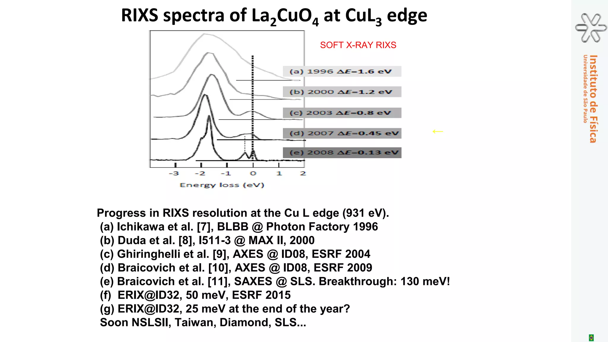
![Progress in RIXS resolution at the Cu L edge (931 eV).
(a) Ichikawa et al. [7], BLBB @ Photon Factory 1996
(b) Duda et al. [8], I511-3 @ MAX II, 2000
(c) Ghiringhelli et al. [9], AXES @ ID08, ESRF 2004
(d) Braicovich et al. [10], AXES @ ID08, ESRF 2009
(e) Braicovich et al. [11], SAXES @ SLS. Breakthrough: 130 meV!
(f) ERIX@ID32, 50 meV, ESRF 2015
(g) ERIX@ID32, 25 meV at the end of the year?
Soon NSLSII, Taiwan, Diamond, SLS...
SOFT X-RAY RIXS
←
RIXS spectra of La2CuO4 at CuL3 edge](https://image.slidesharecdn.com/craievichmemorial2016-161010021942/75/Advanced-Characterization-of-Materials-Relevance-and-Challenges-46-2048.jpg)
O documento discute a caracterização avançada de materiais, destacando a importância da cristalografia e as técnicas de radiação de sincrotron, incluindo suas gerações e desafios futuros. Abordando a evolução histórica das técnicas de difração de raios-X, o texto enfatiza como novas fontes de luz síncrotron e lasers de elétrons livres podem melhorar a compreensão das propriedades dos materiais em diferentes escalas. Além disso, apresenta exemplos de como as transformações estruturais e defeitos em materiais impactam suas propriedades físicas.