Downloaded 266 times
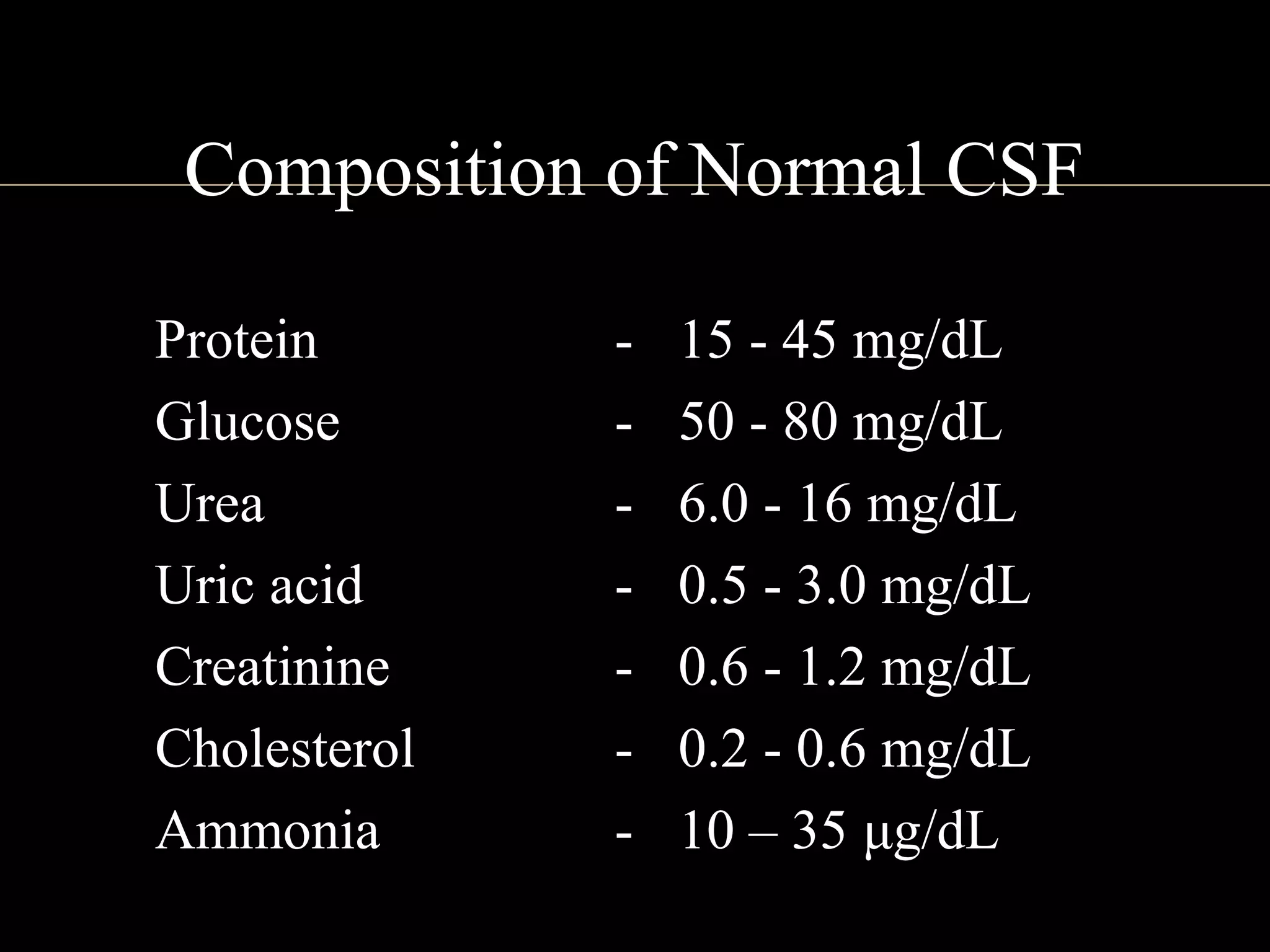

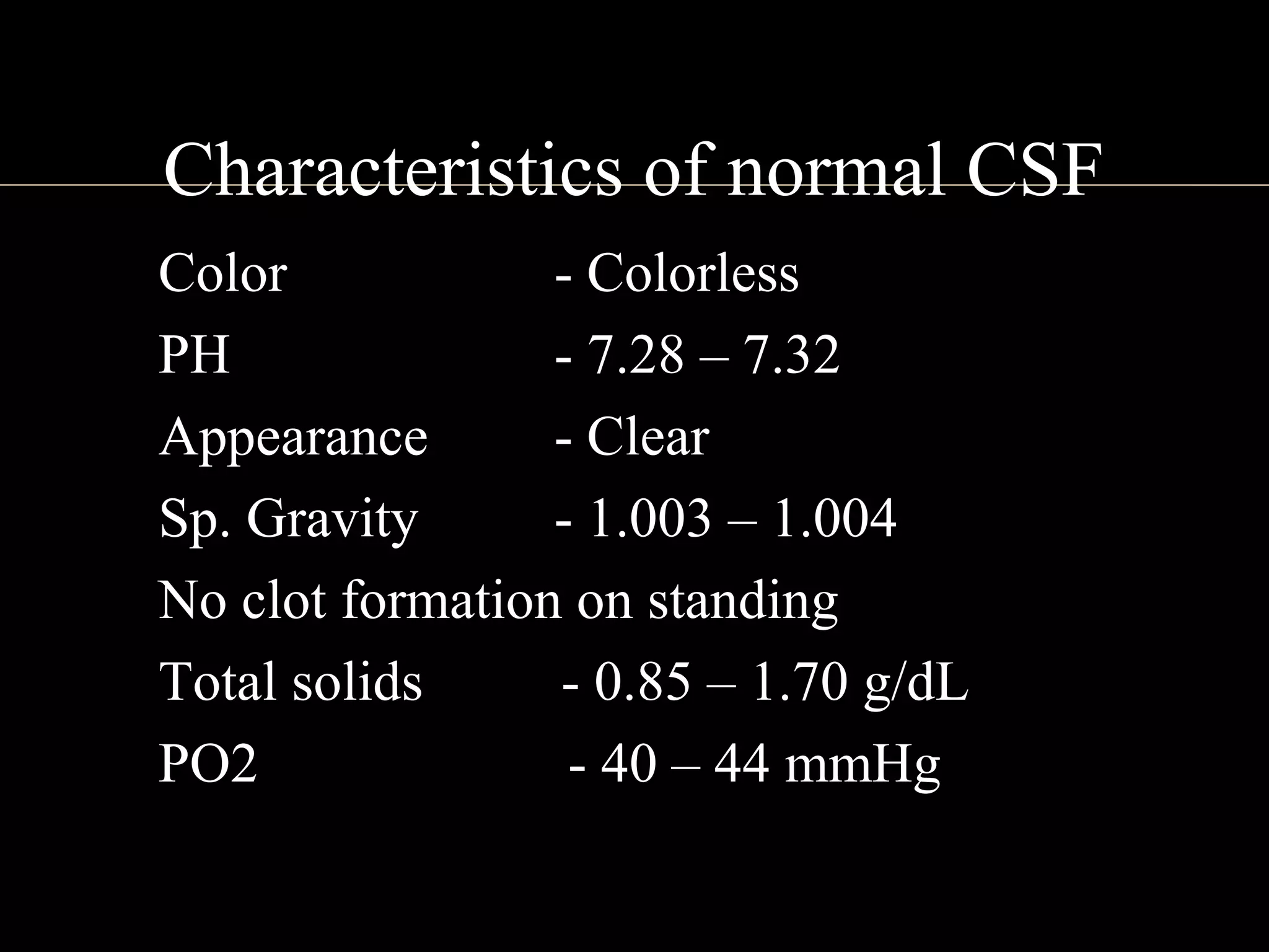
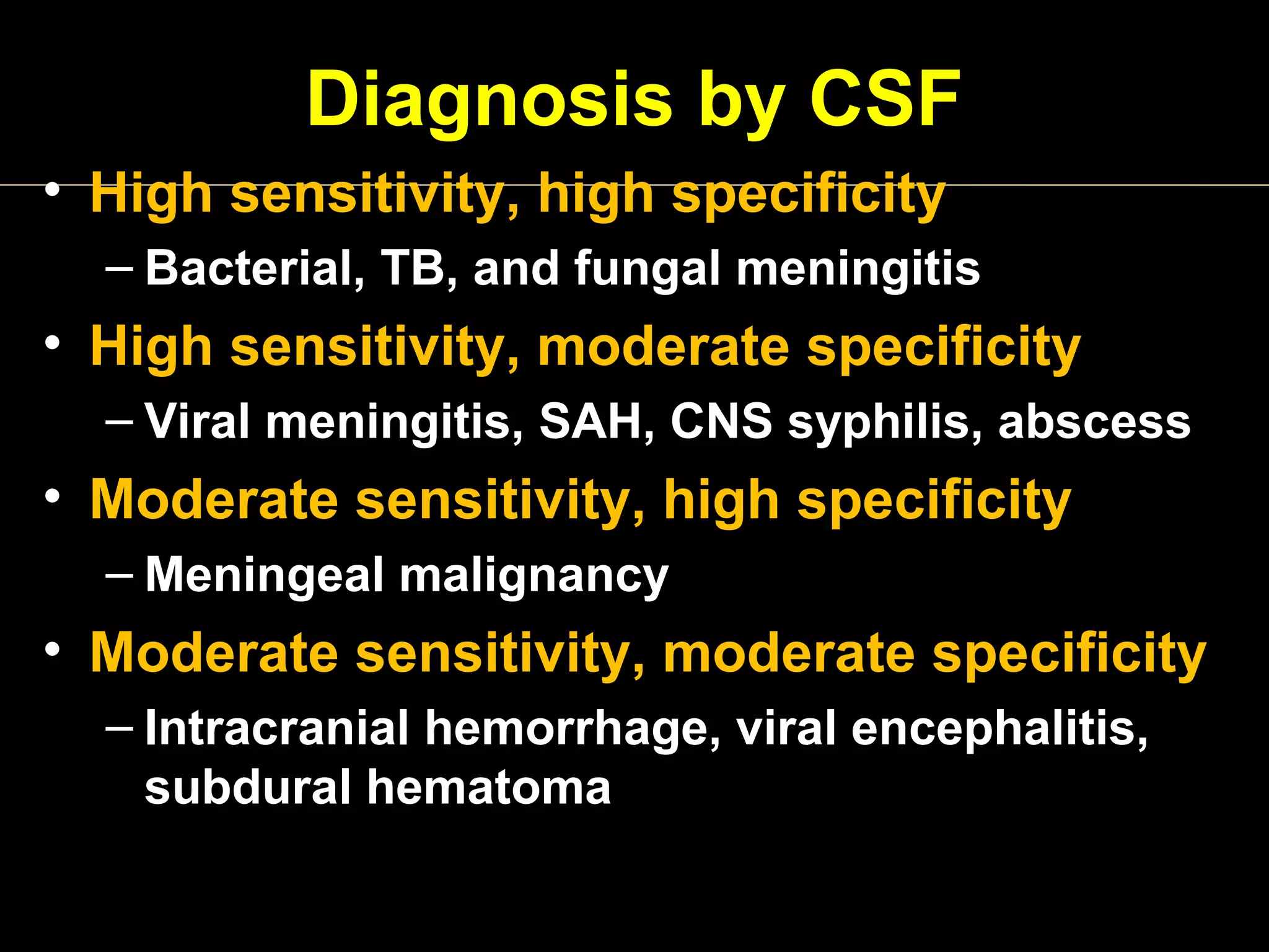
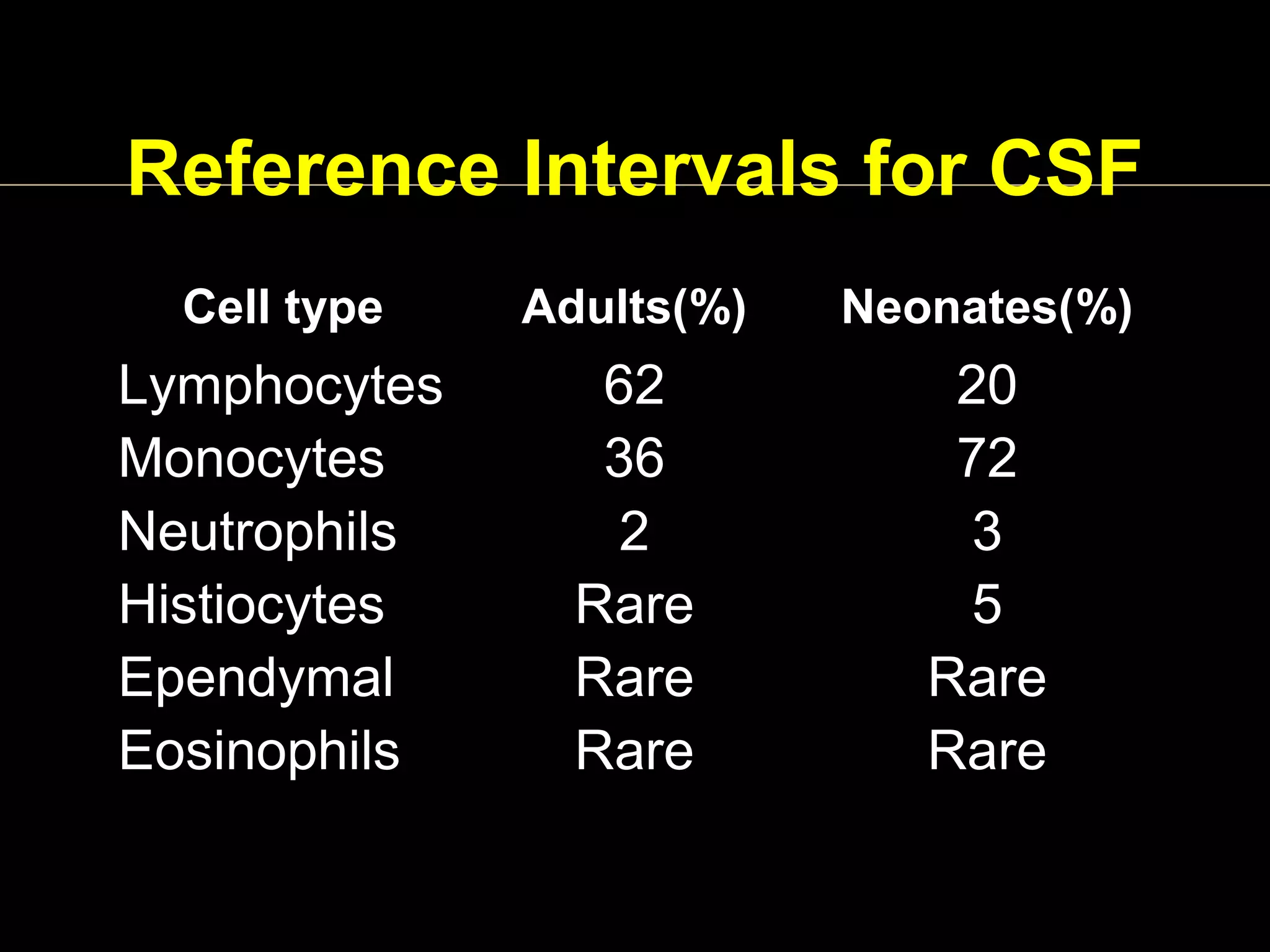

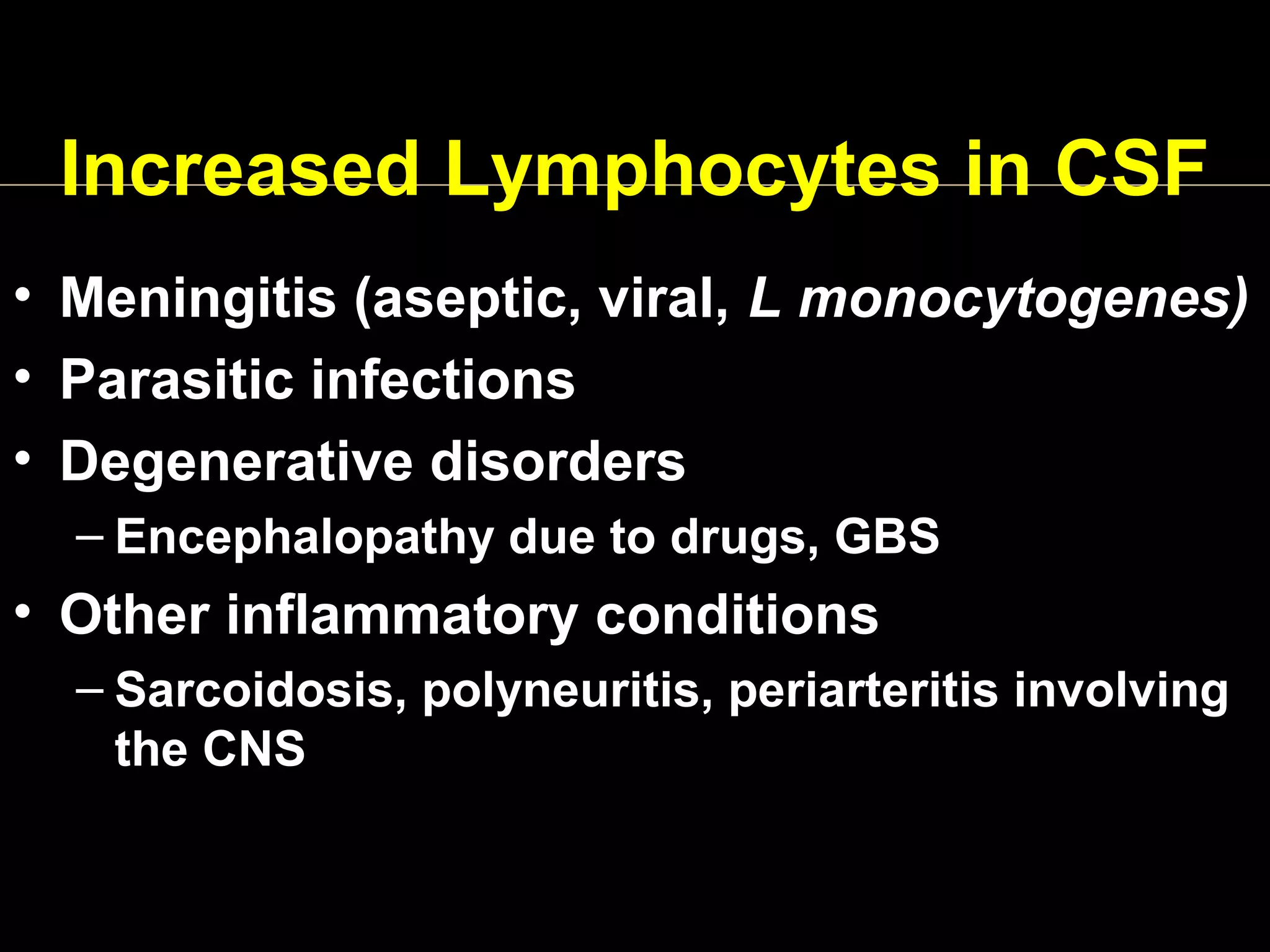
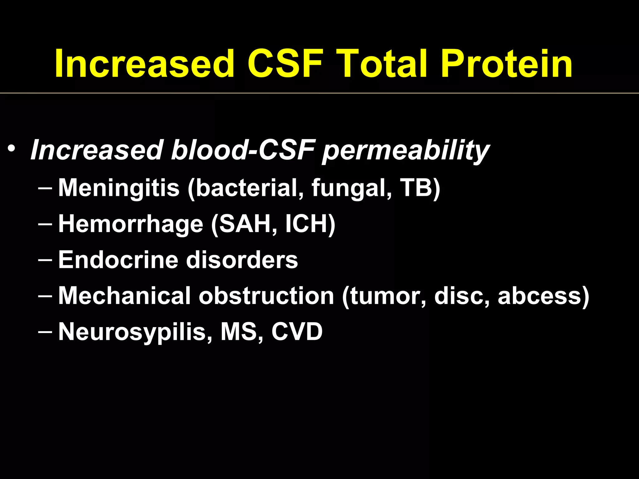


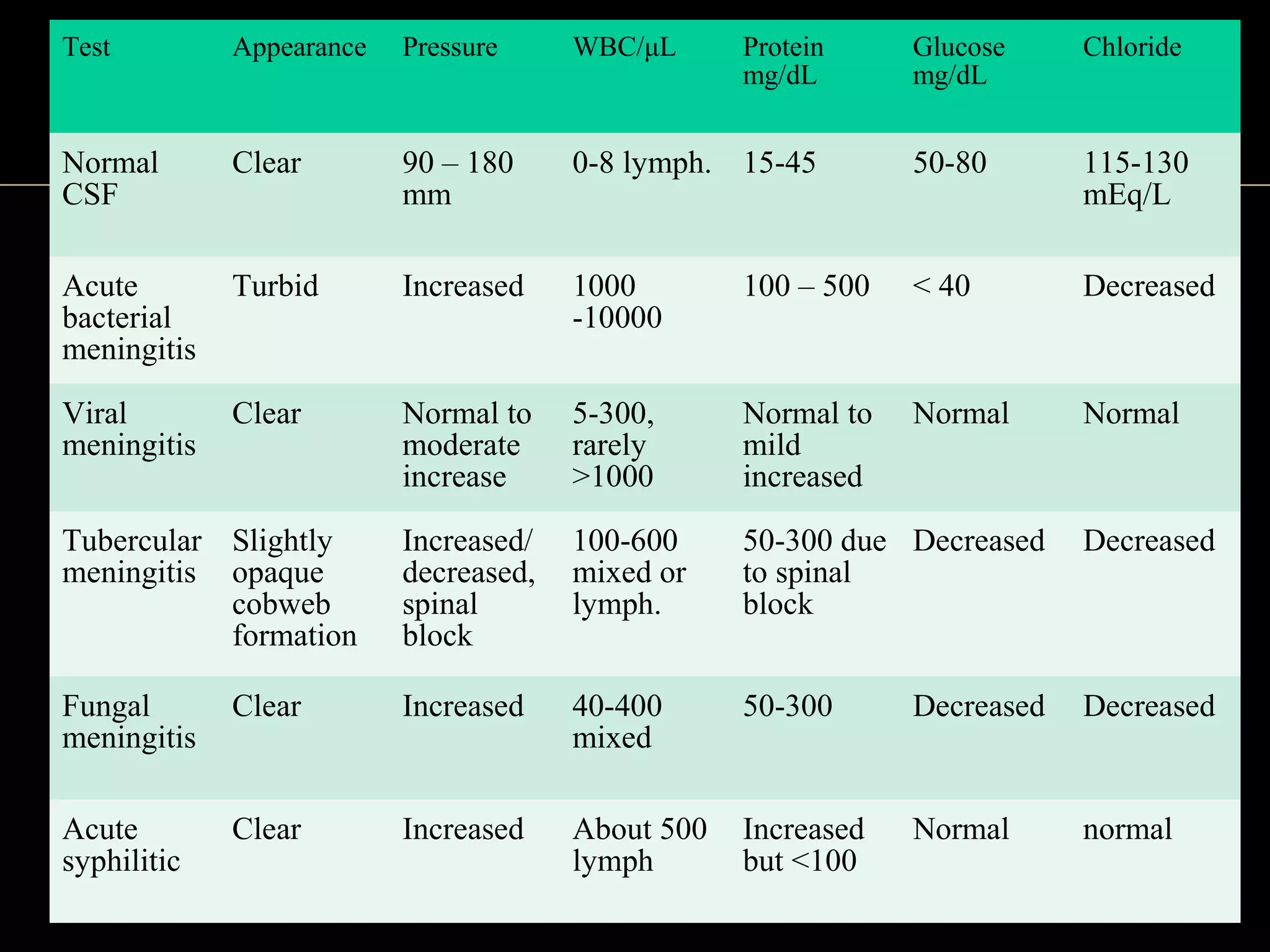

The document discusses the normal composition and characteristics of cerebrospinal fluid (CSF) as well as abnormalities that can indicate various medical conditions: - CSF normally contains low levels of proteins, glucose, and cells while maintaining slightly acidic pH, clarity and specific gravity. - Bacterial, fungal and tuberculous meningitis can be indicated by turbid CSF, high white blood cell count often with neutrophils, low glucose, and elevated proteins. - Viral meningitis presents with clear CSF, mild increase in cells and proteins, and normal glucose.


![Cytopathology Of Cerebrospinal Fluid[1]Power Point](https://cdn.slidesharecdn.com/ss_thumbnails/cytopathologyofcerebrospinalfluid1power-point-1230479978520994-2-thumbnail.jpg?width=640&height=640&fit=bounds)


