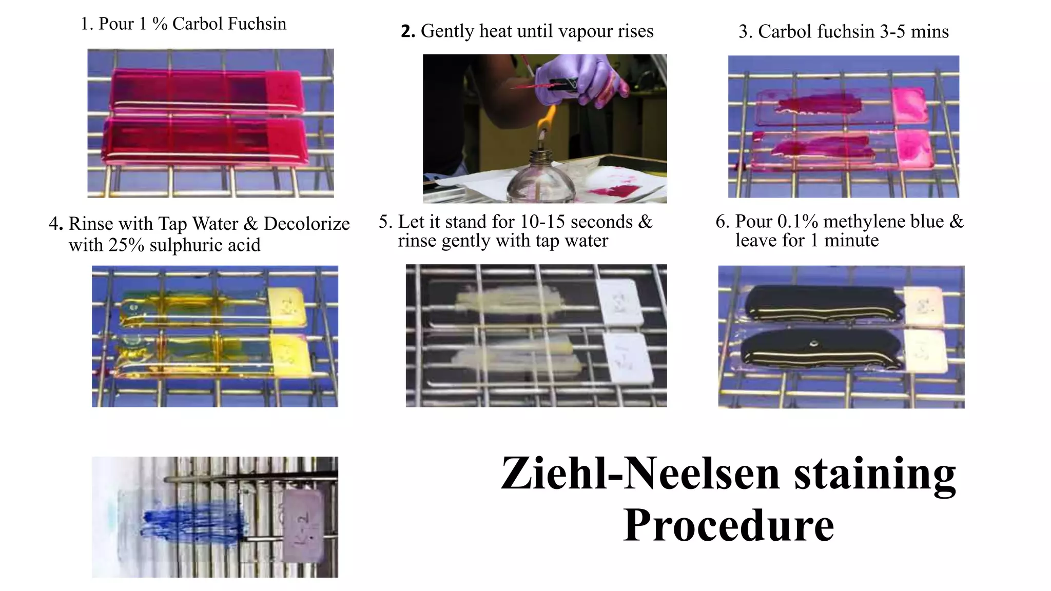
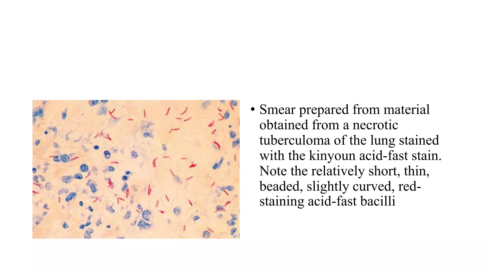
The document summarizes the Ziehl-Neelsen staining technique used to identify acid-fast bacteria like Mycobacterium tuberculosis. The technique involves staining a smear with carbol fuchsin, heating to allow the stain to penetrate the waxy cell walls, washing with acid to decolorize non-acid fast cells, and counterstaining with methylene blue. Acid fast bacteria will appear bright pink against a blue background under the microscope. The document also describes the Kinyoun cold staining method and provides grading scales used to report acid fast bacilli observations.
The PPT is mainly all about Mycobacterium Tuberculosis. Agents causing the disease Tuberculosis, pathogenesis, laboratory diagnosis, treatment and prophylaxis. It was made for both BSc and MSc students.
Acid fast staining is differential staining technique which differentiate bacteria into two group- acid fast bacteria and non acid bacteria. It used to identify acid-fast organisms such as members of the genus Mycobacterium .
Hereby you can get all about bacterial staining.
MICROBIAL STAINING
introduction
Microbial Staining - giving color to microbes. Because microbes are colorless and highly transparent structures.
Staining process in which microbes are getting color.
STAINES / DYES
Staines / dyes - organic compounds which carries either positive charges or negative charges or both .
Based on the charges
Basic stain / dyes :- stain with + ve charge .
Acidic stain / dyes - stain with -ve charge .
2. Based on function of stain :
Neutral stain / dyes - stain with both charges .
Simple staining only one dye is used differentiation among bacteria is impossible Eg . Simple Staining.
Differential staining- more than one dye is used- Differentiation among bacteria is possible- Eg. Gram's staining, Acid - fast staining.
Special staining - more than one dye used - Special structures are seen. Eg. Capsule staining , Spore staining .
Smear preparation:
A drop of water is placed in the centre of a slide
One loopfuls of organisms is transferred to the centre of slide
Spread the organisms over the slide
The smear is allowed to dry
Slide is passed through flame several times to heat-kill and fix organisms
A bacterial stain is stained with crystal violet (fuchsin, methylene blue) 1 min
Observe under microscope
Basic Dyes
Methylene Blue
Crystal Violet
Carbol Fuchsin
Safranin
Malachite Green
Acidic Dyes
Picric Acid
Nigrosin
India Ink
Eosin
differrential statining
Two or more reagents Distinguish
Bacterial groups
Specific Structures
Example
Gram stain
Acid Fast Stain
Pulmonary Thromboembolism - etilogy, types, medical- Surgical and nursing man...VarunMahajani
Disruption of blood supply to lung alveoli due to blockage of one or more pulmonary blood vessels is called as Pulmonary thromboembolism. In this presentation we will discuss its causes, types and its management in depth.
- Video recording of this lecture in English language: https://youtu.be/lK81BzxMqdo
- Video recording of this lecture in Arabic language: https://youtu.be/Ve4P0COk9OI
- Link to download the book free: https://nephrotube.blogspot.com/p/nephrotube-nephrology-books.html
- Link to NephroTube website: www.NephroTube.com
- Link to NephroTube social media accounts: https://nephrotube.blogspot.com/p/join-nephrotube-on-social-media.html
Anti ulcer drugs and their Advance pharmacology ||
Anti-ulcer drugs are medications used to prevent and treat ulcers in the stomach and upper part of the small intestine (duodenal ulcers). These ulcers are often caused by an imbalance between stomach acid and the mucosal lining, which protects the stomach lining.
||Scope: Overview of various classes of anti-ulcer drugs, their mechanisms of action, indications, side effects, and clinical considerations.
Factory Supply Best Quality Pmk Oil CAS 28578–16–7 PMK Powder in Stockrebeccabio
Factory Supply Best Quality Pmk Oil CAS 28578–16–7 PMK Powder in Stock
Telegram: bmksupplier
signal: +85264872720
threema: TUD4A6YC
You can contact me on Telegram or Threema
Communicate promptly and reply
Free of customs clearance, Double Clearance 100% pass delivery to USA, Canada, Spain, Germany, Netherland, Poland, Italy, Sweden, UK, Czech Republic, Australia, Mexico, Russia, Ukraine, Kazakhstan.Door to door service
Hot Selling Organic intermediates
Report Back from SGO 2024: What’s the Latest in Cervical Cancer?bkling
Are you curious about what’s new in cervical cancer research or unsure what the findings mean? Join Dr. Emily Ko, a gynecologic oncologist at Penn Medicine, to learn about the latest updates from the Society of Gynecologic Oncology (SGO) 2024 Annual Meeting on Women’s Cancer. Dr. Ko will discuss what the research presented at the conference means for you and answer your questions about the new developments.
Ozempic: Preoperative Management of Patients on GLP-1 Receptor Agonists Saeid Safari
Preoperative Management of Patients on GLP-1 Receptor Agonists like Ozempic and Semiglutide
ASA GUIDELINE
NYSORA Guideline
2 Case Reports of Gastric Ultrasound
These simplified slides by Dr. Sidra Arshad present an overview of the non-respiratory functions of the respiratory tract.
Learning objectives:
1. Enlist the non-respiratory functions of the respiratory tract
2. Briefly explain how these functions are carried out
3. Discuss the significance of dead space
4. Differentiate between minute ventilation and alveolar ventilation
5. Describe the cough and sneeze reflexes
Study Resources:
1. Chapter 39, Guyton and Hall Textbook of Medical Physiology, 14th edition
2. Chapter 34, Ganong’s Review of Medical Physiology, 26th edition
3. Chapter 17, Human Physiology by Lauralee Sherwood, 9th edition
4. Non-respiratory functions of the lungs https://academic.oup.com/bjaed/article/13/3/98/278874
2. ZIEHL-NEELSEN STAINING
PRINCIPLE
• Acid-fast mycobacteria contain mycolic acid in their outer
membrane, making the cells waxy and resistant to staining with
aqueous based stains such as the Gram stain.
• The primary stain, carbol fuchsin is applied to the cells, and heat
and phenol are used to allow the stain to penetrate into the waxy
surface of acid-fast microorganisms.
• The excess stain is removed with treatment by acid alcohol (ethanol
and hydrochloric acid).
• A secondary stain, methylene blue, is then applied to the cells.
• NOTE: Colour Blind workers are advised to use picric acid
solution (7g/l in water) which yields a yellow background.
3. METHOD OF MAKING SMEAR
1. Vortex concentrated sediment, un-concentrated sputum, other purulent
material, or stool. Place 2 to 3 drops on the slide from the end of the
broom-stick parallel to the slide and slowly spread the liquid uniformly to
make a thin smear.
2. For cerebrospinal fluid (CSF) sediment, vortex thoroughly and apply to
the slide in heaped drops. A heaped drop is allowed to air dry, and a
second application of sediment is placed on the same spot and allowed to
dry. A minimum of three layers, applied to the same 1-cm diameter circle,
should facilitate detection of small numbers of bacilli. (Note: Some
laboratories have stopped performing acid-fast stains on CSF because
positive stains are extremely rare.)
3. Fix the smear at 80° C for 15 minutes or for 2 hours at 65° to 70° C on an
electric hot plate. (Note: Survival of mycobacteria at this temperature has
been reported; handle all specimens with proper precautions.)
4. Ziehl-Neelsen staining
Procedure
4. Rinse with Tap Water & Decolorize
with 25% sulphuric acid
1. Pour 1 % Carbol Fuchsin
5. Let it stand for 10-15 seconds &
rinse gently with tap water
6. Pour 0.1% methylene blue &
leave for 1 minute
3. Carbol fuchsin 3-5 mins2. Gently heat until vapour rises
5. Acid fast bacilli will appear stained pink,
straight curved rods with blue background due
to methylene blue
6. Reporting
• Read at least for 5 minutes & 100 high power field before reporting a
negative result.
7. REVISED RNTCP GUIDELINES
AFB observation Grade
More than 10 AFB per field in atleast 20 fields 3+
1-10 AFB per field in atleast 50 fields 2+
10-99 AFB per 100 fields 1+
1-9 AFB per 100 fields Scanty
(Actual no.)
No AFB seen in atleast 100 field Negative
8. Acid-fast organisms/structures Sulfuric acid (%) needed
for Decolorlzation
Mycobacterium tubercuJosis 25%
Mycobacterium leprae 5%
Nocardia 1%
Acid·fast parasites such as Cryptosporidium,
Cyclospora, Isosopra, Microsporidia, Taenia
saginata (segments and eggs), hooklets of hydatid
cyst and eggs of Schistosoma mansoni
1%
Bacterial spore 0.25-0.5%
Sperm head 0.5-1%
Legionella micdadei 0.5-1%
9. Kinyoun modification (Cold Method)
Identification of acid-fast Mycobacterium spp. and parasites such as
Cryptosporidium and Isopora spp.
PRINCIPLE
• Acid-fast mycobacteria contain mycolic acid in their outer membrane,
making the cells waxy and resistant to staining with aqueous based stains
such as the Gram stain.
• The primary stain, carbol fuchsin, is applied to the cells and phenol is used
to allow the stain to penetrate into the waxy surface of acid-fast
microorganisms.
• The excess stain is removed with treatment by 1% sulfuric acid.
• A secondary stain, methylene blue, is then applied to the cells.
10. RESULTS
• Acid-fast organisms, Mycobacterium spp., will appear pink.
• Nonacid-fast organisms will appear blue.
• In addition, background material should stain blue.
LIMITATIONS
• May be less sensitive than the Ziehl-Neelsen method.
• Smears that are too thick may not properly stain.
11. • Smear prepared from material
obtained from a necrotic
tuberculoma of the lung stained
with the kinyoun acid-fast stain.
Note the relatively short, thin,
beaded, slightly curved, red-
staining acid-fast bacilli
12. REFRENCES
• Bailey & Scott’s Diagnostic Microbiology
• Koneman’s color atlas and textbook 7th Edition
• Mackie & McCartney Practical Medical Microbiology 14th Edition
Two types of acid-fast stains are commonly used (Table 19-3):
1. Fluorochrome stain: auramine O, with or without a second fluorochrome, rhodamine
2. Carbolfuchsin stains: a mixture of fuchsin with phenol (carbolic acid)
a. Ziehl–Neelsen (hot stain)
b. Kinyoun (cold stain
P 497.e1 567 Bailey Scott
P 98 koneman
The Kinyoun modification of the acid-fast stain is called the “cold method” because a surface-active detergent, such as Tergitol, is used rather than heat treatment.
the Fite–Ferraco acid-fast stain (i.e., also a partial acid-fast stain) is recommended for paraffin sections
Ziehl–Neelsen Procedure Carbolfuchsin: Dissolve 3 g of basic fuchsin in 10 mL of 90%–95% ethanol. Add 90 mL of 5% aqueous solution of phenol.
Kinyoun Cold Procedure Carbolfuchsin: Dissolve 4 g of basic fuchsin in 20 mL of 90%–95% ethanol and then add 100 mL of a 9% aqueous solution of phenol (9 g of phenol dissolved in 100 mL of distilled water)