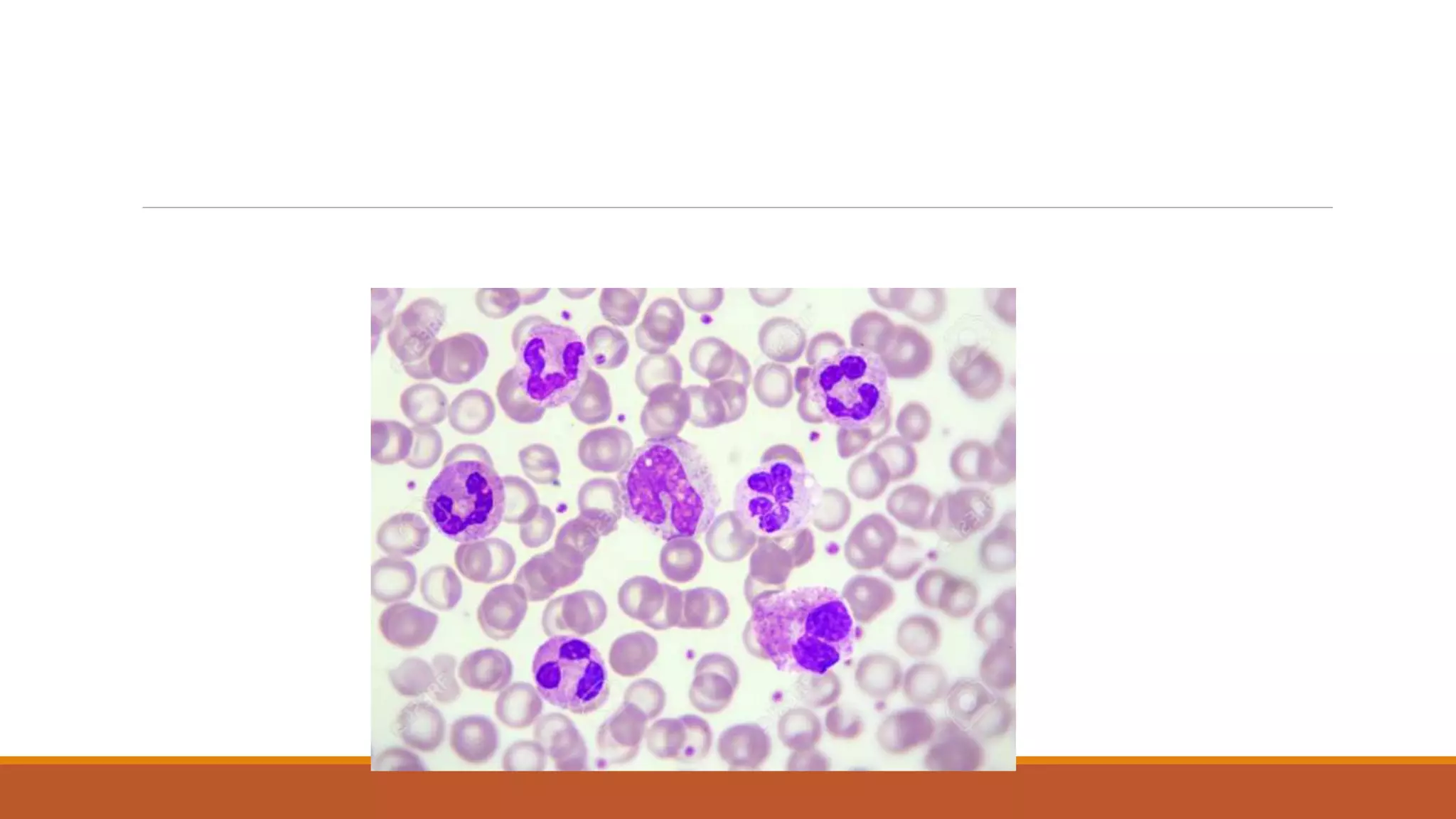
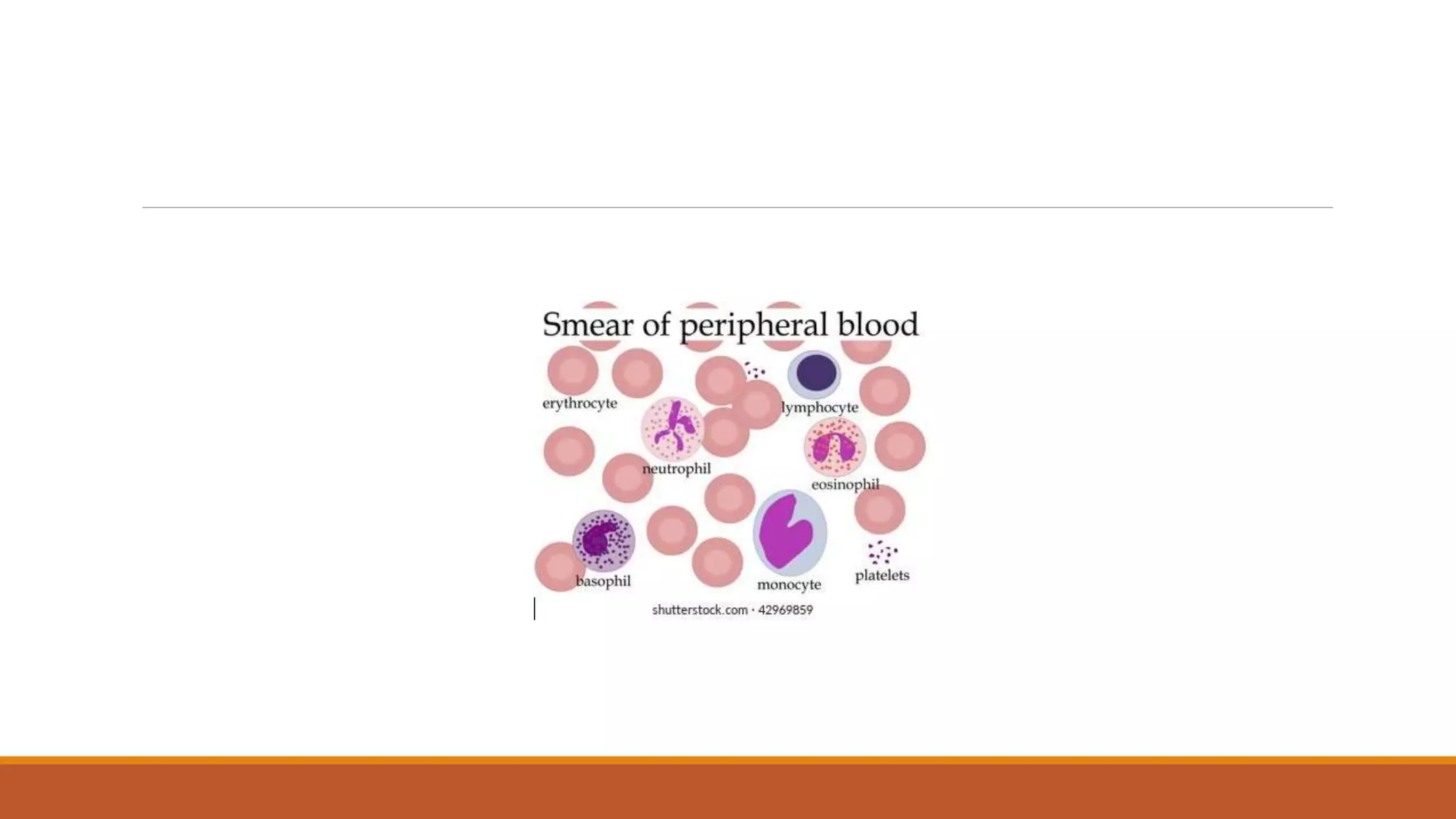
This document provides information on staining blood films and smears. It discusses the different types of stains used including Romanowsky stains like Leishman stain, Giemsa stain, Wright stain, and Field stain. Specimens should be collected in EDTA and smears prepared within an hour then fixed in methanol or ethanol to preserve cell morphology before staining. Romanowsky stains use methylene blue and eosin dyes to reveal subtle differences in cell structures and components.
2. Collection of specimen
Ideally, blood can be collected by finger prick
ā¦ If other tests being performed, can use venipuncture
ā¦ EDTA is preferred as the anticoagulant as heparin may lead to morphological distortion
Smears should be prepared and stained within an hour of drawing the specimen.
ā¦ Alterations in morphology may occur if delayed.
3.
4. THICK FILM THIN FILM
-lysed RBCs -fixed RBCs single layer
-larger volume -smaller volume
-o.25microlt blood/100 fields -0.005microlt blood/100
feilds
-blood elements are more
concentrated
-good screening test -good species differentiation
-difficult to diagnose
species
-used in mass surveys -requires more time to read
-For quick diagnosis
5. Fixing of blood film
ā¢The blood film need to be fixed with acetone free methyl alcohol for 1 minute.
ā¢Alcohol denature the protein and harden the cell contents.
ā¢For wright and leishman no pre fixation is required.
ā¢The stains most commonly used for staining of blood films are romanowsky stains.
6. To preserve the morphology of the cells, films must be fixed as soon as possible after they have
dried.
It is important to prevent contact with water before fixation is complete.
Methyl alcohol (methanol) is the choice, although ethyl alcohol ("absolute alcohol") can be used
Methylated spirit (95% ethanol) must not be used as it contains water.
To fix the films, place them in a covered staining jar or tray containing the alcohol for 2-3
minutes.
In humid climates it might be necessary to replace the methanol 2-3 times per day; the old
portions can be used for storing clean slides.
7. Romanowsky stain
ā¢Romanowsky stain :it is named after dmitri leonovich romanowsky who invented it in 1891.
ā¢ Definition: a stain made from water soluble eosin, methylene blue and acetone free methanol.
ā¢Romanowsky stains are universally employed for staining of blood films. All romanowsky
combinations have two essential ingredients i.E. Methylene blue and eosin
9. 1) Anionic dye
2) Acid dye
3)Pink red-colour Structures
that stain with eosin are termed Eosinophilic
10. Methylene blue combines with anionic components of the cell .eg: DNA ,and stain these blue.
Eosin combines with cationic components of the cell . eg: cytoplasm and stain them red .
Then there occurs stain āstain interaction.
This composition and mode of action allows romanowsky stains to reveal the subtle differences
in shades of staining.
11. A well stained āsmear with any of romanowsky stains shows following features :
1)Red blood cells: Pink red (or)Deep red colour.
2)Polychromatic cells(reticulocytes):Gray blue colour.
3) Neutrophils : Pale,Pink cytoplasm,Purple Granules .
4)Eosinophils: Pale,Pink cytoplasm, orange red granules
5) Basophils: Blue cytoplasm, Dark blue-Violet Granules .
6) Monocytes: Gray āBlue cytoplasm.
7)Lymphocytes: Dark blue cytoplasm.
8) Platelets: Purple Colour.
12. VARIOUS STAIN
VARIOUS STAINS INCLUDED UNDER ROMANOWSKY STAINS ARE AS FOLLOWS: 1)Leishman stain
2)Giemsa stain
3)Wright stain
4)Field stain
5)Jenner stain
6)JSB stain
Among the above mentioned stains ,Jenner is the simplest and Giemsa is the most complex.
Leishman stain it occupies intermediate position and it is still widely used in the routine staining
of blood films.
13. LEISHMAN STAIN:
PRINCIPLE: It is used in microscopy for staining blood smears. It provides excellent stain quality .
COMPONENTS: It contains methylene blue and eosin in methanol(acetone free)in the ratio of
1.5gm/1 litre.
It is generally used to differentiate and identify leucocytes, malaria parasites etc
Preparation Dissolve 0.2 g of powdered Leishmanās dye in 100 ml of acetone-free methyl alcohol
in a conical flask. Warm it to 50Ā°C for half an hour with occasional shaking.
14. Procedure for staining
1)Pour Leishmanās stain dropwise (counting the drops) on the slide and wait for 2 minutes. This
allows fixation of the PBF in methyl alcohol.
2)Add double the quantity of buffered water dropwise over the slide (i.e. double the number of
drops).
3) Mix for 8 minutes
4)Wash in water for 1 to 2 minutes.
5) Dry in air and examine under oil immersion lens of the microscope.
16. field stain A
Methylene blue 0.8gm
Azure I 0.5 gm
Disodium hydrogen phosphate 5 gm
Potassium dihydrogen anhydrous 6.25gm
Distilled water 500ml
17. Field stain B
Eosin 1gm
Disodium hydrogen phosphate 5 gm
Potassium dihydrogen phosphate 6.25 gm
Distilled water 500ml
18. Procedure:
A. Fix thin film with methanol for 1 min.
B. Dry microscopic slide on filter paper
C. Immerse slide in Fieldās stain B (Eosin) for 5 seconds
D. Immediately wash with water
E. Immerse slide in Fieldās stain A (Methylene blue) for 10 seconds