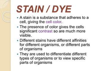
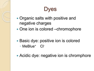
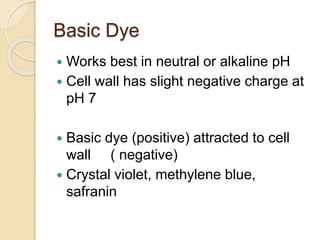
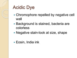
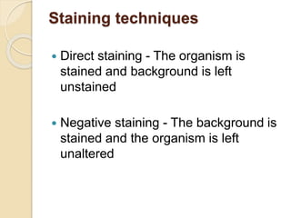
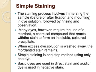
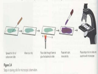
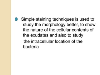
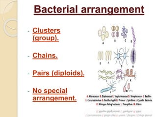
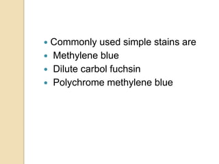
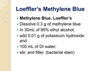
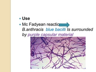
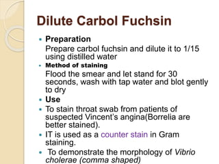
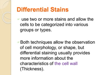
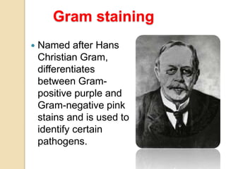
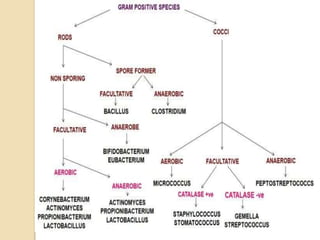
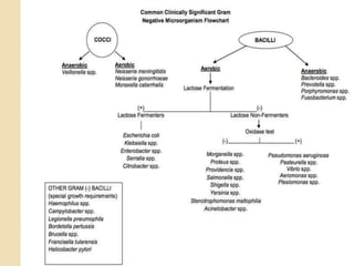
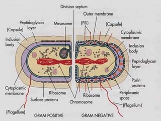
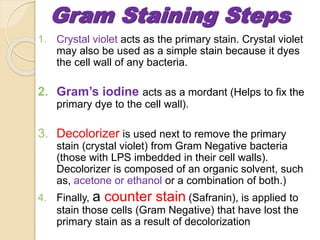
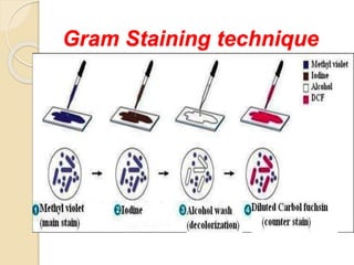
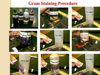
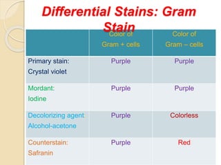
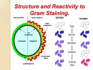
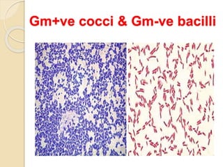
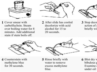
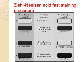
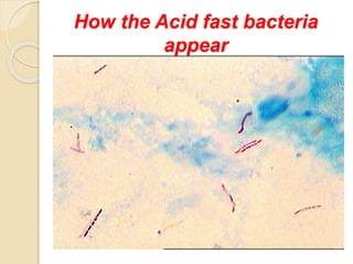
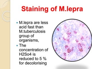
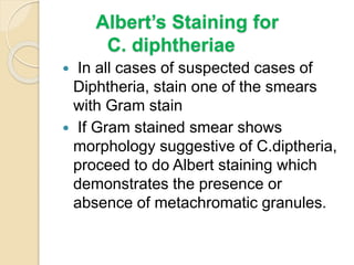
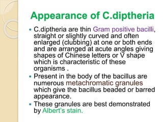
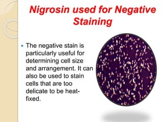
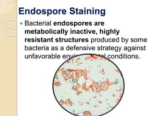
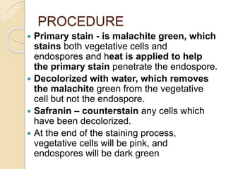
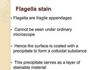
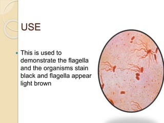
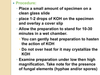
The document discusses various staining techniques used to visualize bacteria under a microscope. It describes the principles of simple staining using single dyes like methylene blue or carbol fuchsin. Differential staining techniques like Gram staining and acid-fast staining are also covered, which use multiple dyes to categorize bacteria based on cell wall characteristics. Special stains used to highlight specific bacterial structures such as capsules, flagella or spores are mentioned. Detailed procedures for common staining methods like Gram stain, acid-fast stain and Albert stain are provided. The document aims to explain the use of staining to differentiate bacterial types and visualize their morphology.


![CTEV [ clubfoot] DR ARUN LAL ,DR MOHAMED ASHRAF travancore medical college k...](https://cdn.slidesharecdn.com/ss_thumbnails/ctevclubfootdrarunlaldrmohamedashraftravancoremedicalcollegekollamkeralaindia-260208063247-18fc466c-thumbnail.jpg?width=640&height=640&fit=bounds)


