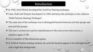
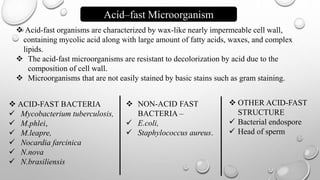
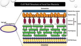
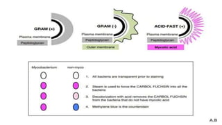
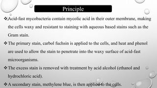
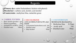
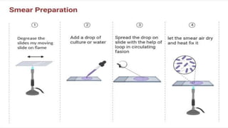
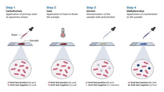
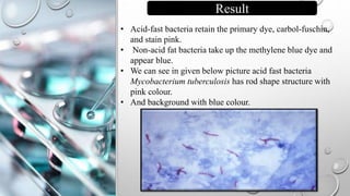
M. J. P. ROHILKAND
UNIVERSITY,
BAREILLY
Introduction
Acid fast
microorganism
Cell wall structure
Principle
Reagent
Procedure
Result
Modification
Applications
Content
Introduction
 In 1882, Paul Ehrlich developed the Acid Fast Staining technique.
 Later, Ziehl and Neelsen developed it in 1883 and hence the technique is also called as
"Ziehl-Neelsen Staining Technique".
 The main aim of this technique was to distinguish bacteria between acid fast groups and
non-acid fast groups.
 This test is exclusively used for identification of Mycobacterium tuberculosis, a
causative agent of TB.
 It is a member of Mycobacterium genus.
 In Ziehl & Neelsen staining method, the acid-fast bacteria appear to be red bright bacilli
with a light blue background.
 Acid-fast organisms are characterized by wax-like nearly impermeable cell wall,
containing mycolic acid along with large amount of fatty acids, waxes, and complex
lipids.
 The acid-fast microorganisms are resistant to decolorization by acid due to the
composition of cell wall.
 Microorganisms that are not easily stained by basic stains such as gram staining.
Acid–fast Microorganism
 ACID-FAST BACTERIA
 Mycobacterium tuberculosis,
 M.phlei,
 M.leapre,
 Nocardia farcinica
 N.nova
 N.brasiliensis
 NON-ACID FAST
BACTERIA –
 E.coli,
 Staphylococcus aureus.
 OTHER ACID-FAST
STRUCTURE
 Bacterial endospore
 Head of sperm
Cell Wall Structure of Acid fast Bacteria
Principle
Acid-fast mycobacteria contain mycolic acid in their outer membrane, making
the cells waxy and resistant to staining with aqueous based stains such as the
Gram stain.
The primary stain, carbol fuchsin is applied to the cells, and heat and phenol
are used to allow the stain to penetrate into the waxy surface of acid-fast
microorganisms.
The excess stain is removed with treatment by acid alcohol (ethanol and
hydrochloric acid).
A secondary stain, methylene blue, is then applied to the cells.
Regents
Primary dye: carbol fuchsin(basic fuchsin with phenol)
Decolorizer : sulfuric acid, alcohol, acid alcohol
Counterstain : methylene blue, malachite green
 CARBOL FUCHSIN
• Basic fuchsin (powder) 1gm
• Phenol (crystalline) 5ml
• Alcohol(95% or100%) 10ml
• Distilled water 100ml
DECOLORIZER
ACID ALCOHOL(3%HCl in 95% ethyl
alcohol)
• Concentrated hydrochloric acid 3ml
• Distilled water 2ml
• Ethyl alcohol 95ml
 COUNTERSTAIN
METHYLENE BLUE(0.25%)
• Methylene blue 0.25gm
• Acetic acid 1ml
• Distilled water 99ml
Procedure
 STEP 1.
• Prepare smear.
 STEP 2.
• Add pour 1% Carbol Fuchsin - the primary stain.
• Heat the slides gently until you see vapors, but do not boil. (for3 to 5 min)
• Rinse slides gently with tap water.
 STEP 3.
• Add pour 20% Sulphuric acid- the decolorizing solution. ( for 15 sec)
• Rinse the slides with tap water.
• Tilt to drain excess water.
 STEP 4.
• Pour 0.1% Methylene Blue - the counter stain. (for 30 sec)
• Rinse the slides gently with tap water.
• Tilt the slides to drain excess water.
• Finally, place slides on rack to air-dry.
• STEP 5.
• Examine under microscope.
• Acid-fast bacteria retain the primary dye, carbol-fuschin,
and stain pink.
• Non-acid fat bacteria take up the methylene blue dye and
appear blue.
• We can see in given below picture acid fast bacteria
Mycobacterium tuberculosis has rod shape structure with
pink colour.
• And background with blue colour.
Result
1.Mycobacterium leprae
Same method is used with 5% sulphuric acid due to less mycolic acid in cell wall.
2.Nocardia and Legionella
1% sulphuric acid is used
2.Stool Specimen
We use 3% acid alcohol instead of sulphuric acid so as to stain Cryptosporidium parvum.
Modifications
Applications
 Used for examination and identification of Mycobacterium species.
 Used to differentiate between acid- fast and non-acid fast bacilli
 Used for the identification of some fungal species such as
Cryptosporidium.
THANK
YOU

ZN Staining

  • 1.
    M. J. P.ROHILKAND UNIVERSITY, BAREILLY
  • 2.
    Introduction Acid fast microorganism Cell wallstructure Principle Reagent Procedure Result Modification Applications Content
  • 3.
    Introduction  In 1882,Paul Ehrlich developed the Acid Fast Staining technique.  Later, Ziehl and Neelsen developed it in 1883 and hence the technique is also called as "Ziehl-Neelsen Staining Technique".  The main aim of this technique was to distinguish bacteria between acid fast groups and non-acid fast groups.  This test is exclusively used for identification of Mycobacterium tuberculosis, a causative agent of TB.  It is a member of Mycobacterium genus.  In Ziehl & Neelsen staining method, the acid-fast bacteria appear to be red bright bacilli with a light blue background.
  • 4.
     Acid-fast organismsare characterized by wax-like nearly impermeable cell wall, containing mycolic acid along with large amount of fatty acids, waxes, and complex lipids.  The acid-fast microorganisms are resistant to decolorization by acid due to the composition of cell wall.  Microorganisms that are not easily stained by basic stains such as gram staining. Acid–fast Microorganism  ACID-FAST BACTERIA  Mycobacterium tuberculosis,  M.phlei,  M.leapre,  Nocardia farcinica  N.nova  N.brasiliensis  NON-ACID FAST BACTERIA –  E.coli,  Staphylococcus aureus.  OTHER ACID-FAST STRUCTURE  Bacterial endospore  Head of sperm
  • 5.
    Cell Wall Structureof Acid fast Bacteria
  • 7.
    Principle Acid-fast mycobacteria containmycolic acid in their outer membrane, making the cells waxy and resistant to staining with aqueous based stains such as the Gram stain. The primary stain, carbol fuchsin is applied to the cells, and heat and phenol are used to allow the stain to penetrate into the waxy surface of acid-fast microorganisms. The excess stain is removed with treatment by acid alcohol (ethanol and hydrochloric acid). A secondary stain, methylene blue, is then applied to the cells.
  • 8.
    Regents Primary dye: carbolfuchsin(basic fuchsin with phenol) Decolorizer : sulfuric acid, alcohol, acid alcohol Counterstain : methylene blue, malachite green  CARBOL FUCHSIN • Basic fuchsin (powder) 1gm • Phenol (crystalline) 5ml • Alcohol(95% or100%) 10ml • Distilled water 100ml DECOLORIZER ACID ALCOHOL(3%HCl in 95% ethyl alcohol) • Concentrated hydrochloric acid 3ml • Distilled water 2ml • Ethyl alcohol 95ml  COUNTERSTAIN METHYLENE BLUE(0.25%) • Methylene blue 0.25gm • Acetic acid 1ml • Distilled water 99ml
  • 9.
    Procedure  STEP 1. •Prepare smear.  STEP 2. • Add pour 1% Carbol Fuchsin - the primary stain. • Heat the slides gently until you see vapors, but do not boil. (for3 to 5 min) • Rinse slides gently with tap water.  STEP 3. • Add pour 20% Sulphuric acid- the decolorizing solution. ( for 15 sec) • Rinse the slides with tap water. • Tilt to drain excess water.  STEP 4. • Pour 0.1% Methylene Blue - the counter stain. (for 30 sec) • Rinse the slides gently with tap water. • Tilt the slides to drain excess water. • Finally, place slides on rack to air-dry. • STEP 5. • Examine under microscope.
  • 12.
    • Acid-fast bacteriaretain the primary dye, carbol-fuschin, and stain pink. • Non-acid fat bacteria take up the methylene blue dye and appear blue. • We can see in given below picture acid fast bacteria Mycobacterium tuberculosis has rod shape structure with pink colour. • And background with blue colour. Result
  • 13.
    1.Mycobacterium leprae Same methodis used with 5% sulphuric acid due to less mycolic acid in cell wall. 2.Nocardia and Legionella 1% sulphuric acid is used 2.Stool Specimen We use 3% acid alcohol instead of sulphuric acid so as to stain Cryptosporidium parvum. Modifications
  • 14.
    Applications  Used forexamination and identification of Mycobacterium species.  Used to differentiate between acid- fast and non-acid fast bacilli  Used for the identification of some fungal species such as Cryptosporidium.
  • 15.