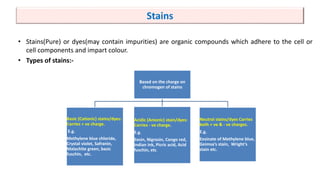
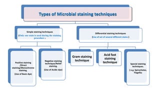
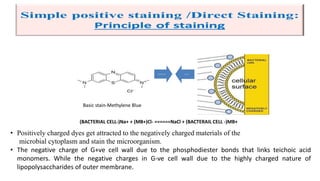
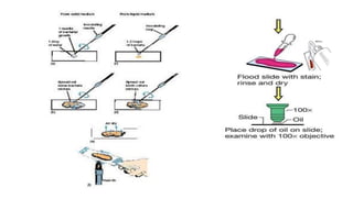
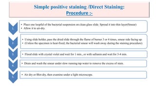
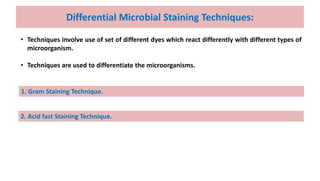
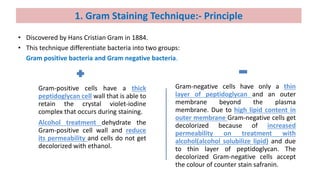
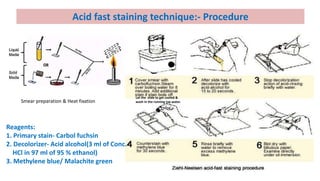
SHITAL TRIVEDI
ASSISTANT PROFESSOR,
Department of pharmaceutical technology,
L.J.INSTITUTE OF PHARMACY
Subject: Pharmaceutical Microbiology
SEMESTER 5
MICROBIAL STAINING
• Most of the microorganisms are colourless and transparent in the light microscope. So, they cannot
be seen easily.
• It is necessary to create contrast between the field and the microorganisms.
 Microbial Staining: Before observation under the light microscope, the process to create contrast
between the background field and the microorganisms by coloration of the microbial specimen
using staining reagent, is called microbial staining.
 Purposes of Microbial Staining :
• Development of contrast enable better visualization of transparent objects.
• Study of size, shapes and arrangement of microorganisms.
• Morphological differentiation of different types of organisms.
• Visualization of external and internal structures.
• Study of distribution and chemical nature of cellular constituents.
• Differentiation between the living and the dead cells.
Stains
• Stains(Pure) or dyes(may contain impurities) are organic compounds which adhere to the cell or
cell components and impart colour.
• Types of stains:-
Based on the charge on
chromogen of stains
Basic (Cationic) stains/dyes:
Carries + ve charge.
E.g.
Methylene blue chloride,
Crystal violet, Safranin,
Malachite green, basic
fuschin, etc.
Acidic (Anionic) stain/dyes:
Carries - ve charge.
E.g.
Eosin, Nigrosin, Congo red,
Indian ink, Picric acid, Acid
fuschin, etc.
Neutral stains/dyes Carries
both + ve & - ve charges.
E.g.
Eosinate of Methylene blue,
Geimsa’s stain, Wright’s
stain etc.
Types of Microbial staining techniques
Simple staining techniques
(Only one stain is used during the staining
procedure )
Positive staining
/Direct
staining/Monochrome
staining.
(Use of Basic dye)
Negative staining
technique/Relief
staining.
(Use of Acidic dye)
Differential staining techniques
(Use of set of several different stains )
Gram staining
technique
Acid fast
staining
technique Special staining
techniques.
( e.g. Spirochetes,
Flagella)
Basic stain-Methylene Blue
------ ---
• Positively charged dyes get attracted to the negatively charged materials of the
microbial cytoplasm and stain the microorganism.
• The negative charge of G+ve cell wall due to the phosphodiester bonds that links teichoic acid
monomers. While the negative charges in G-ve cell wall due to the highly charged nature of
lipopolysaccharides of outer membrane.
(BACTERIAL CELL-)Na+ + (MB+)Cl- ======NaCl + (BACTERAIL CELL -)MB+
Procedure:
Basic steps in Simple Positive Microbial Staining
Smear preparation.
Heat fixation.
Reaction with staining
reagent.
Wash out of excess
staining reagent.
Drying of specimen
1.
• Place one loopful of the bacterial suspension on clean glass slide. Spread it into thin layer(Smear)
• Allow it to air-dry.
2.
• Using slide holder, pass the dried slide through the flame of burner 3 or 4 times, smear side facing up.
• (Unless the specimen is heat-fixed, the bacterial smear will wash away during the staining procedure).
3. • Flood slide with crystal violet and wait for 1 min., or with safranin and wait for 3-4 min.
4.
• Drain and wash the smear under slow running tap water to remove the excess of stain.
5.
• Air dry or Blot dry, then examine under a light microscope.
Simple positive staining /Direct Staining:
Procedure :-
With Methylene Blue With safranin
Simple positive staining /Direct Staining:
Observation:-
All bacteria get colored.
Acidic stain-Nigrosin/Acid black 2, water soluble
Negative staining :
Principle :-
Negative staining technique:- Staining performed with acidic (negatively charged) stains such as
Nigrosin or Congo red.
They are repelled by the negatively charged cytoplasm/cell surface and gather around the cells,
leaving the cells clear and unstained.
------
---
1.
• Place a small drop of Nigrosin (Acidic stain) at the end of the clean glass slide.
2.
• Place a loopful of microbial sample and mix with drop of Nigrosin.
3. • Using the edge of another slide, spread the drop across the slide.
4. • Allow it to air dry and examine under a microscope.
• Note: No heat fixation and no wash out of excess stain.
Negative staining : Procedure :-
With Nigrosin
Negative staining
Observation:-
Colorless microorganisms against colored
background..
Applications :
• This method is used to study morphological characters of cell size, shape, and arrangement of
microbial cells.
• This method is easy and useful in observing the microorganisms which are difficult to stain.
Drawback:
• Microorganisms are not killed in this technique, therefore not suitable for studying pathogens.
Negative staining
Applications and drawback:-
Differential Microbial Staining Techniques:
• Techniques involve use of set of different dyes which react differently with different types of
microorganism.
• Techniques are used to differentiate the microorganisms.
1. Gram Staining Technique.
2. Acid fast Staining Technique.
• Discovered by Hans Cristian Gram in 1884.
• This technique differentiate bacteria into two groups:
Gram positive bacteria and Gram negative bacteria.
1. Gram Staining Technique:- Principle
Gram-positive cells have a thick
peptidoglycan cell wall that is able to
retain the crystal violet-iodine
complex that occurs during staining.
Alcohol treatment dehydrate the
Gram-positive cell wall and reduce
its permeability and cells do not get
decolorized with ethanol.
Gram-negative cells have only a thin
layer of peptidoglycan and an outer
membrane beyond the plasma
membrane. Due to high lipid content in
outer membrane Gram-negative cells get
decolorized because of increased
permeability on treatment with
alcohol(alcohol solubilize lipid) and due
to thin layer of peptidoglycan. The
decolorized Gram-negative cells accept
the colour of counter stain safranin.
Difference in Cell wall structure
Gram Staining Technique:- Procedure
Smear preparation & Heat fixation
Note: 1. Use fresh culture. 2. Avoid over decolorization
Basic Fuchsin/
Methylene blue/
1 min. 1 min. 1 min.
10-15 sec.
• Gram-positive bacteria appear blue or purple.
E.g. Staphylococcus aureus, Streptococcus pyogenes, Corynebacterium diphtheria.
• Gram-negative cells will appear pink to red.
E.g. Neisseria meningitidis, Escherichia coli, Pseudomonas aeruginosa)
Gram Staining Technique:- Observation
Acid fast staining technique:-
( Ziehl-Neelsen staining method)
• This technique was initially developed by Scientist Paul Ehrlich and then was modified by two
scientists Ziehl & Neelsen.
• This technique differentiates Acid fast bacteria from other Acid non fast bacteria.
 Bacteria displaying acid fastness include:
• Genus Mycobacterium – M. leprae, M. Tuberculosis, M. smegmatis, M. Avium complex, M.
kansasii.
• Genus Nocardia – N. brasiliensis, N. cyriacigeorgica, N. farcinica, and N. nova.
 Acid fastness can also be attributed to other structures not classified as bacteria. These
include:
• Bacterial endospores, Head of sperm, Cryptosporidium parvum, Isospora belli, Cyclospora
cayetanensis, Taenia saginata eggs, Hydatid cysts, Sarcocystis, Nuclear inclusion bodies in lead
poisoning.
• In acid-fast organisms, the cell wall is resistant to water-based stains and therefore require a
special staining technique.
• Heat or a lipid solvent is used to carry the first stain, carbol fuchsin, into the cells. Then the cells
are washed with a diluted acid alcohol solution.
• The acid fast organisms have the waxy lipid called mycolic acid in their cell walls. Heat acts as
a physical mordant while phenol (carbol of carbol fuchsin) acts as the chemical mordant and it
allows carbol fuchsin permeability to cell wall containing mycolic acid and it stain the acid fast
as well as nonacid fast organisms.
• It is assumed that mycolic acid resist the effect of the acid alcohol and prevents acid-alcohol
from decolorizing protoplasm and retain the carbol fuchsin stain (they appear bright red
under the microscope).
• Whereas nonacid fast organisms get decolorized and loose the primary stain carbol fuchsin and
take on the subsequent methylene blue stain (they appear blue under the microscope).
Acid fast staining technique:- Principle
Acid fast staining technique:- Procedure
Smear preparation & Heat fixation
Reagents:
1. Primary stain- Carbol fuchsin
2. Decolorizer- Acid alcohol(3 ml of Conc.
HCl in 97 ml of 95 % ethanol)
3. Methylene blue/ Malachite green
Let the slide to get cooled &
wash in the running tap water.
Ziehl-Neelsen staining procedure:- Observation
A- Non-Acid fast bacteria: Appear blue. E.g. Escherichia coli, Staphylococcus aureus etc.
B- Acid fast bacteria: Appear bright red. E.g. Mycobacterium tuberculosis, Mycobacterium avium,
Nocardia species.
Acid fast staining-The Kinyoun method, or Kinyoun stain
• The Kinyoun method, or Kinyoun stain (cold method), developed by Joseph Kinyoun. It’s a
modified method developed by Robert Koch (the one who first identified Mycobacterium)in
1882.
• The Kinyoun method can be modified as a weak acid fast stain, which uses 5% sulfuric acid
instead of hydrochloric acid. The weak acid fast stain in addition to staining mycobacteria will
stain organisms that are not able to maintain the carbol fuchsin after decolorizing with HCl.
• Unlike the Ziehl-Neelsen stain (Z-N stain), the Kinyoun method of staining does not require
heating. Since the Kinyoun stain is a cold method (no heat applied), the concentration of carbol
fuchsin used is increased.

Microbial staining

  • 1.
    SHITAL TRIVEDI ASSISTANT PROFESSOR, Departmentof pharmaceutical technology, L.J.INSTITUTE OF PHARMACY Subject: Pharmaceutical Microbiology SEMESTER 5 MICROBIAL STAINING
  • 2.
    • Most ofthe microorganisms are colourless and transparent in the light microscope. So, they cannot be seen easily. • It is necessary to create contrast between the field and the microorganisms.  Microbial Staining: Before observation under the light microscope, the process to create contrast between the background field and the microorganisms by coloration of the microbial specimen using staining reagent, is called microbial staining.  Purposes of Microbial Staining : • Development of contrast enable better visualization of transparent objects. • Study of size, shapes and arrangement of microorganisms. • Morphological differentiation of different types of organisms. • Visualization of external and internal structures. • Study of distribution and chemical nature of cellular constituents. • Differentiation between the living and the dead cells.
  • 3.
    Stains • Stains(Pure) ordyes(may contain impurities) are organic compounds which adhere to the cell or cell components and impart colour. • Types of stains:- Based on the charge on chromogen of stains Basic (Cationic) stains/dyes: Carries + ve charge. E.g. Methylene blue chloride, Crystal violet, Safranin, Malachite green, basic fuschin, etc. Acidic (Anionic) stain/dyes: Carries - ve charge. E.g. Eosin, Nigrosin, Congo red, Indian ink, Picric acid, Acid fuschin, etc. Neutral stains/dyes Carries both + ve & - ve charges. E.g. Eosinate of Methylene blue, Geimsa’s stain, Wright’s stain etc.
  • 4.
    Types of Microbialstaining techniques Simple staining techniques (Only one stain is used during the staining procedure ) Positive staining /Direct staining/Monochrome staining. (Use of Basic dye) Negative staining technique/Relief staining. (Use of Acidic dye) Differential staining techniques (Use of set of several different stains ) Gram staining technique Acid fast staining technique Special staining techniques. ( e.g. Spirochetes, Flagella)
  • 5.
    Basic stain-Methylene Blue --------- • Positively charged dyes get attracted to the negatively charged materials of the microbial cytoplasm and stain the microorganism. • The negative charge of G+ve cell wall due to the phosphodiester bonds that links teichoic acid monomers. While the negative charges in G-ve cell wall due to the highly charged nature of lipopolysaccharides of outer membrane. (BACTERIAL CELL-)Na+ + (MB+)Cl- ======NaCl + (BACTERAIL CELL -)MB+
  • 6.
    Procedure: Basic steps inSimple Positive Microbial Staining Smear preparation. Heat fixation. Reaction with staining reagent. Wash out of excess staining reagent. Drying of specimen
  • 8.
    1. • Place oneloopful of the bacterial suspension on clean glass slide. Spread it into thin layer(Smear) • Allow it to air-dry. 2. • Using slide holder, pass the dried slide through the flame of burner 3 or 4 times, smear side facing up. • (Unless the specimen is heat-fixed, the bacterial smear will wash away during the staining procedure). 3. • Flood slide with crystal violet and wait for 1 min., or with safranin and wait for 3-4 min. 4. • Drain and wash the smear under slow running tap water to remove the excess of stain. 5. • Air dry or Blot dry, then examine under a light microscope. Simple positive staining /Direct Staining: Procedure :-
  • 9.
    With Methylene BlueWith safranin Simple positive staining /Direct Staining: Observation:- All bacteria get colored.
  • 10.
    Acidic stain-Nigrosin/Acid black2, water soluble Negative staining : Principle :- Negative staining technique:- Staining performed with acidic (negatively charged) stains such as Nigrosin or Congo red. They are repelled by the negatively charged cytoplasm/cell surface and gather around the cells, leaving the cells clear and unstained. ------ ---
  • 11.
    1. • Place asmall drop of Nigrosin (Acidic stain) at the end of the clean glass slide. 2. • Place a loopful of microbial sample and mix with drop of Nigrosin. 3. • Using the edge of another slide, spread the drop across the slide. 4. • Allow it to air dry and examine under a microscope. • Note: No heat fixation and no wash out of excess stain. Negative staining : Procedure :-
  • 12.
    With Nigrosin Negative staining Observation:- Colorlessmicroorganisms against colored background..
  • 13.
    Applications : • Thismethod is used to study morphological characters of cell size, shape, and arrangement of microbial cells. • This method is easy and useful in observing the microorganisms which are difficult to stain. Drawback: • Microorganisms are not killed in this technique, therefore not suitable for studying pathogens. Negative staining Applications and drawback:-
  • 14.
    Differential Microbial StainingTechniques: • Techniques involve use of set of different dyes which react differently with different types of microorganism. • Techniques are used to differentiate the microorganisms. 1. Gram Staining Technique. 2. Acid fast Staining Technique.
  • 15.
    • Discovered byHans Cristian Gram in 1884. • This technique differentiate bacteria into two groups: Gram positive bacteria and Gram negative bacteria. 1. Gram Staining Technique:- Principle Gram-positive cells have a thick peptidoglycan cell wall that is able to retain the crystal violet-iodine complex that occurs during staining. Alcohol treatment dehydrate the Gram-positive cell wall and reduce its permeability and cells do not get decolorized with ethanol. Gram-negative cells have only a thin layer of peptidoglycan and an outer membrane beyond the plasma membrane. Due to high lipid content in outer membrane Gram-negative cells get decolorized because of increased permeability on treatment with alcohol(alcohol solubilize lipid) and due to thin layer of peptidoglycan. The decolorized Gram-negative cells accept the colour of counter stain safranin.
  • 16.
    Difference in Cellwall structure
  • 17.
    Gram Staining Technique:-Procedure Smear preparation & Heat fixation Note: 1. Use fresh culture. 2. Avoid over decolorization Basic Fuchsin/ Methylene blue/ 1 min. 1 min. 1 min. 10-15 sec.
  • 18.
    • Gram-positive bacteriaappear blue or purple. E.g. Staphylococcus aureus, Streptococcus pyogenes, Corynebacterium diphtheria. • Gram-negative cells will appear pink to red. E.g. Neisseria meningitidis, Escherichia coli, Pseudomonas aeruginosa) Gram Staining Technique:- Observation
  • 19.
    Acid fast stainingtechnique:- ( Ziehl-Neelsen staining method) • This technique was initially developed by Scientist Paul Ehrlich and then was modified by two scientists Ziehl & Neelsen. • This technique differentiates Acid fast bacteria from other Acid non fast bacteria.  Bacteria displaying acid fastness include: • Genus Mycobacterium – M. leprae, M. Tuberculosis, M. smegmatis, M. Avium complex, M. kansasii. • Genus Nocardia – N. brasiliensis, N. cyriacigeorgica, N. farcinica, and N. nova.  Acid fastness can also be attributed to other structures not classified as bacteria. These include: • Bacterial endospores, Head of sperm, Cryptosporidium parvum, Isospora belli, Cyclospora cayetanensis, Taenia saginata eggs, Hydatid cysts, Sarcocystis, Nuclear inclusion bodies in lead poisoning.
  • 20.
    • In acid-fastorganisms, the cell wall is resistant to water-based stains and therefore require a special staining technique. • Heat or a lipid solvent is used to carry the first stain, carbol fuchsin, into the cells. Then the cells are washed with a diluted acid alcohol solution. • The acid fast organisms have the waxy lipid called mycolic acid in their cell walls. Heat acts as a physical mordant while phenol (carbol of carbol fuchsin) acts as the chemical mordant and it allows carbol fuchsin permeability to cell wall containing mycolic acid and it stain the acid fast as well as nonacid fast organisms. • It is assumed that mycolic acid resist the effect of the acid alcohol and prevents acid-alcohol from decolorizing protoplasm and retain the carbol fuchsin stain (they appear bright red under the microscope). • Whereas nonacid fast organisms get decolorized and loose the primary stain carbol fuchsin and take on the subsequent methylene blue stain (they appear blue under the microscope). Acid fast staining technique:- Principle
  • 21.
    Acid fast stainingtechnique:- Principle
  • 22.
    Acid fast stainingtechnique:- Procedure Smear preparation & Heat fixation Reagents: 1. Primary stain- Carbol fuchsin 2. Decolorizer- Acid alcohol(3 ml of Conc. HCl in 97 ml of 95 % ethanol) 3. Methylene blue/ Malachite green Let the slide to get cooled & wash in the running tap water.
  • 23.
    Ziehl-Neelsen staining procedure:-Observation A- Non-Acid fast bacteria: Appear blue. E.g. Escherichia coli, Staphylococcus aureus etc. B- Acid fast bacteria: Appear bright red. E.g. Mycobacterium tuberculosis, Mycobacterium avium, Nocardia species.
  • 24.
    Acid fast staining-TheKinyoun method, or Kinyoun stain • The Kinyoun method, or Kinyoun stain (cold method), developed by Joseph Kinyoun. It’s a modified method developed by Robert Koch (the one who first identified Mycobacterium)in 1882. • The Kinyoun method can be modified as a weak acid fast stain, which uses 5% sulfuric acid instead of hydrochloric acid. The weak acid fast stain in addition to staining mycobacteria will stain organisms that are not able to maintain the carbol fuchsin after decolorizing with HCl. • Unlike the Ziehl-Neelsen stain (Z-N stain), the Kinyoun method of staining does not require heating. Since the Kinyoun stain is a cold method (no heat applied), the concentration of carbol fuchsin used is increased.