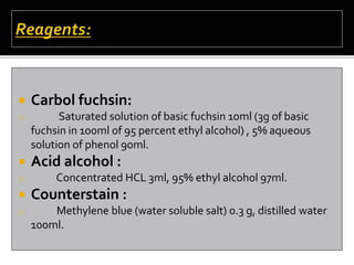
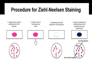
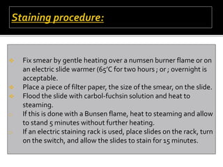
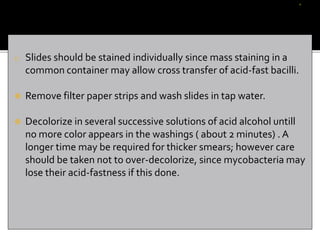
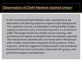
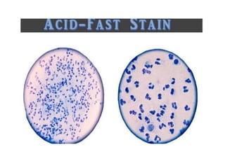
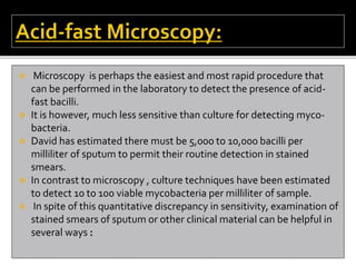
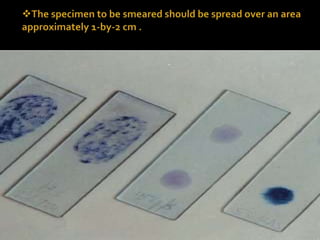
b
Prepared by ‘ M.Phool Badshah ’
 The more widely used Ziehl-Neelsen acid-fast stain still must be
used to confirm the acid-fast nature of bacilli in sputum smears,
in culture, and to standardize the inoculum size for direct drug
susceptibility tests.
 Ziehl–Neelsen staining is a bacteriological stain used to identify
acid-fast organisms, mainly Mycobacteria . It is named for two
German doctors who modified the stain: the bacteriologist
Franz Ziehl (1859–1926) and the pathologist Friedrich Neelsen
(1854–1898).
 Carbol fuchsin:
o Saturated solution of basic fuchsin 10ml (3g of basic
fuchsin in 100ml of 95 percent ethyl alcohol) , 5% aqueous
solution of phenol 90ml.
 Acid alcohol :
o Concentrated HCL 3ml, 95% ethyl alcohol 97ml.
 Counterstain :
o Methylene blue (water soluble salt) 0.3 g, distilled water
100ml.
 Fix smear by gentle heating over a numsen burner flame or on
an electric slide warmer (65’C for two hours ; or ; overnight is
acceptable.
 Place a piece of filter paper, the size of the smear, on the slide.
 Flood the slide with carbol-fuchsin solution and heat to
steaming.
o If this is done with a Bunsen flame, heat to steaming and allow
to stand 5 minutes without further heating.
o If an electric staining rack is used, place slides on the rack, turn
on the switch, and allow the slides to stain for 15 minutes.
o Slides should be stained individually since mass staining in a
common container may allow cross transfer of acid-fast bacilli.
 Remove filter paper strips and wash slides in tap water.
 Decolorize in several successive solutions of acid alcohol untill
no more color appears in the washings ( about 2 minutes) . A
longer time may be required for thicker smears; however care
should be taken not to over-decolorize, since mycobacteria may
lose their acid-fastness if this done.
 Wash with tap water.
 Counter-stain with aqueous methylene blue for 30
seconds.
 Wash with water and dry in the air or over gentle heat.
 In the conventional Ziehl-Neelsen stain, mycobacteria are
observed as red staining organisms against a blue background.
 The organisms may be coccobacillary to long bacillary bodies,
ranging in size from 0.8-5.0 microns long and 0.2-0.6 micron in
width.The longer bacilli may exhibit uneven staining, with
everything from bipolar to beaded stains having been reported.
 The characteristics banded cells commonly seen in M.Kansasii
often enable a presumptive diagnosis of the presence of this
organism, while the suggestion of pleomorphic and sometimes
branched forms more commonly is observed with group 3 non-
photochromogenic mycobacteria.
 In reading stained smears the microscopist should keep in mind
that he must prepare a preliminary report for the clinician
indicating the type of stain used, the presence or absence of
acid-fast bacilli, and also that he must utilize his findings to
enable a standardization of the digested inoculum for use in
direct drug susceptibility testing.
 Clinicians are interested at least in a rough indication of the
numbers of acid-fast bacilli seen on the smear. For most
purposes the old Gaffky scale is much too cumbersome to be
practical.The following method recommended by the national
tuberculosis association is perhaps most widely used.
Number of organisms seen Report
 3-9 per slide Rare
 10 or more per slide Few
 Greater than 1 per oil immersion
field Numerous
 The presence of only 1 or 2 acid-fast bacilli on an entire slide
should not be reported untill confirmed by another smear
prepared from the same or another specimen.
 Many bacteriologists tend to by-pass smear examination
because they feel it either is too time-consuming or they are
using the more sensitive culture techniques.
 The value of microscopy , not only as a means for the rapid
presumptive diagnosis of tuberculosis but also for the purpose
of observing a given patients response
 To chemotherapy, should not be underestimated . It has been
repeatedly demonstrated that patients who are microscopically
smear positive provide the greatest threat to the community.
 These are the “Infectious reservoirs” the source of spread of
tubercle bacilli.
 On the other hand, the smear negative patient, especially one
covered by an adequate chemotherapeutic regimen, is less likely
to be a source for spread of the disease to his contacts.
 In addition to the reasons above, microscopic smear
examination of digested sputum concentrates is valuable for the
preparation of proper “inoculum size” for direct drug
susceptibility testing.
 It is not the infection to imply that the staining methods
described here are the only acceptable ones, Many other
techniques have been described, and in the experience of the
writers , seem wholly acceptable for routine use.
 For example, bacause of individual preference, different
counterstains , such as brilliant green or picric acid, may replace
the methylene blue commonly employed in the Ziehl-Neelsen
stain.
 To speed up the staining procedure, some investigators have
combined the de-colorization and counterstaining into one
solution, while others have advocated various “Cold Staining”
procedure . By the same token.
 Microscopy is perhaps the easiest and most rapid procedure that
can be performed in the laboratory to detect the presence of acid-
fast bacilli.
 It is however, much less sensitive than culture for detecting myco-
bacteria.
 David has estimated there must be 5,000 to 10,000 bacilli per
milliliter of sputum to permit their routine detection in stained
smears.
 In contrast to microscopy , culture techniques have been estimated
to detect 10 to 100 viable mycobacteria per milliliter of sample.
 In spite of this quantitative discrepancy in sensitivity, examination of
stained smears of sputum or other clinical material can be helpful in
several ways :
 (a) It provides a presumptive diagnosis of mycobacterial disease.
 (b) It enables the rapid identification of most infectious cases, i.e., those
smear positive.
 (c) It may be used to follow the progress of tuberculosis patients on
chemotherapy.
 (d) It is of vital importance in regard to the patient’s discharge from the
hospital, or return to gainful employment.
 (e) it can confirm that cultures growing on media are indeed acid-fast.
 (f) It is useful in determining appropriate dilutions of sediments for direct
drug susceptibility tests.
 Precautions:
 Microscope slides used for acid-fast staining should be new,
clean, and unscratched.
 Acid-fast material from previous smears may be retained on old,
washed, and reused slides and, thus, may lead to “false-positive
reports.”
 One end of the slide should be labeled with the patient’s name
and/or hospital identification number and date.
 To smear the specimen over a larger area would be self-defeating. If
micro-scopist examines 100 to 300 fields on a stained smear 1-by-2
cm, only 1/100 t0 4/100 of the entire area of the smear is observed
(i.e., 1% to 4% of the smear area), depending on the magnification
used for scanning.
 A larger smear, examined in the same manner, would only further
reduce examining efficiency.
 Keep the area of the smear fairly small, 1-by-2 cm is satisfactory.
 Use a bacteriological loop, applicator stick, or pipette to make smears,
Loop may be reused after it is dipped into an alcohol sand flask and
sterilized in the flame of a Bunsen burner, but applicator sticks and
pipettes should be discarded after each smear is prepared.
 Let the smear air dry and then heat fix it.
 Use either an electric slide warmer at 65’C to 75’C for at least 2 hours
(overnight is acceptable), or pass the slide 3 to 4 times through the
blue cone of a burner flame as for other bacteriological smears.
 Heat-fixing does not always kill the mycobacteria , so handle
smears carefully.
 Discard stained slides into covered pans containing disinfectant
solution and autoclave them before disposal, or discard them
into 3% amphyl solution overnight.
 Many acid-fast staining pr0cedures have been described,
including hot and cold staining methods and those which
employ fluorescing dyes.
 All are based on the property of mycobacteria to retain the
primary stain, even after exposure to strong mineral acids or
acid alcohol-thus the term “acid-fast bacilli ”.
o Avoiding some problems about “False-Positive and
False-Negative reports” :
 Prepare and stain smears carefully to avoid such problems as poorly
stained preparations and false-positive or false-negative results .The
following suggestions may be helpful in avoiding some of these
problems:
I. Avoid insufficient de-staining with acid-alcohol:
o Most Myco-bacteria are strongly acid-fast and are not easily decolorized.
o Smears that are too thick are difficult to de-stain , and the subsequent
counter stain may obscure acid-fast bacilli.
I. Use a contrasting counter-stain:
o Counter-stain should not be so intense that it “hides” the stained
mycobacteria.
o Color-blind individuals may find fluorochrome-stained smears easier to
examine.
I. Be aware of possible sources (environmental) of
false-positive smears. Common sources of acid-fast
bacilli that contribute to this problem are:
o Tap water and infrequently cleaned distilled water reservoirs. Check these
sources from time to time to see if acid-fast bacilli can be detected.Tap
water may be especially problematic after heavy rainstorms.The distilled
water reservoir that is always left on the shelf and refilled from an outside
supply source is especially suspect.
 Ice-making machines. Ice is often used to facilitate passage of
gastric tubes. If the tap water used to make the ice is
contaminated with mycobacteria, it could lead to false-positive
smears or cultures.
 Transfer of material from slide to slide in bulk-staining tanks.To
avoid this problem, stain slides individually.
 Transfer of “ Positive” flakes from thick slides to other slides via
immersion oil. Do not make thick smears; Carefully heat-fix
smears; wipe oil immersion lens between slides; allow oil to free-
fall from applicator onto the smear instead of touching
applicator to the slide.
---------------------Thank you----------------------

AFB (Acid-Fast Bacilli) smear staining.pptx

  • 1.
    b Prepared by ‘M.Phool Badshah ’
  • 2.
     The morewidely used Ziehl-Neelsen acid-fast stain still must be used to confirm the acid-fast nature of bacilli in sputum smears, in culture, and to standardize the inoculum size for direct drug susceptibility tests.  Ziehl–Neelsen staining is a bacteriological stain used to identify acid-fast organisms, mainly Mycobacteria . It is named for two German doctors who modified the stain: the bacteriologist Franz Ziehl (1859–1926) and the pathologist Friedrich Neelsen (1854–1898).
  • 3.
     Carbol fuchsin: oSaturated solution of basic fuchsin 10ml (3g of basic fuchsin in 100ml of 95 percent ethyl alcohol) , 5% aqueous solution of phenol 90ml.  Acid alcohol : o Concentrated HCL 3ml, 95% ethyl alcohol 97ml.  Counterstain : o Methylene blue (water soluble salt) 0.3 g, distilled water 100ml.
  • 5.
     Fix smearby gentle heating over a numsen burner flame or on an electric slide warmer (65’C for two hours ; or ; overnight is acceptable.  Place a piece of filter paper, the size of the smear, on the slide.  Flood the slide with carbol-fuchsin solution and heat to steaming. o If this is done with a Bunsen flame, heat to steaming and allow to stand 5 minutes without further heating. o If an electric staining rack is used, place slides on the rack, turn on the switch, and allow the slides to stain for 15 minutes.
  • 6.
    o Slides shouldbe stained individually since mass staining in a common container may allow cross transfer of acid-fast bacilli.  Remove filter paper strips and wash slides in tap water.  Decolorize in several successive solutions of acid alcohol untill no more color appears in the washings ( about 2 minutes) . A longer time may be required for thicker smears; however care should be taken not to over-decolorize, since mycobacteria may lose their acid-fastness if this done.
  • 7.
     Wash withtap water.  Counter-stain with aqueous methylene blue for 30 seconds.  Wash with water and dry in the air or over gentle heat.
  • 8.
     In theconventional Ziehl-Neelsen stain, mycobacteria are observed as red staining organisms against a blue background.  The organisms may be coccobacillary to long bacillary bodies, ranging in size from 0.8-5.0 microns long and 0.2-0.6 micron in width.The longer bacilli may exhibit uneven staining, with everything from bipolar to beaded stains having been reported.  The characteristics banded cells commonly seen in M.Kansasii often enable a presumptive diagnosis of the presence of this organism, while the suggestion of pleomorphic and sometimes branched forms more commonly is observed with group 3 non- photochromogenic mycobacteria.
  • 9.
     In readingstained smears the microscopist should keep in mind that he must prepare a preliminary report for the clinician indicating the type of stain used, the presence or absence of acid-fast bacilli, and also that he must utilize his findings to enable a standardization of the digested inoculum for use in direct drug susceptibility testing.  Clinicians are interested at least in a rough indication of the numbers of acid-fast bacilli seen on the smear. For most purposes the old Gaffky scale is much too cumbersome to be practical.The following method recommended by the national tuberculosis association is perhaps most widely used.
  • 10.
    Number of organismsseen Report  3-9 per slide Rare  10 or more per slide Few  Greater than 1 per oil immersion field Numerous  The presence of only 1 or 2 acid-fast bacilli on an entire slide should not be reported untill confirmed by another smear prepared from the same or another specimen.  Many bacteriologists tend to by-pass smear examination because they feel it either is too time-consuming or they are using the more sensitive culture techniques.  The value of microscopy , not only as a means for the rapid presumptive diagnosis of tuberculosis but also for the purpose of observing a given patients response
  • 11.
     To chemotherapy,should not be underestimated . It has been repeatedly demonstrated that patients who are microscopically smear positive provide the greatest threat to the community.  These are the “Infectious reservoirs” the source of spread of tubercle bacilli.  On the other hand, the smear negative patient, especially one covered by an adequate chemotherapeutic regimen, is less likely to be a source for spread of the disease to his contacts.  In addition to the reasons above, microscopic smear examination of digested sputum concentrates is valuable for the preparation of proper “inoculum size” for direct drug susceptibility testing.
  • 12.
     It isnot the infection to imply that the staining methods described here are the only acceptable ones, Many other techniques have been described, and in the experience of the writers , seem wholly acceptable for routine use.  For example, bacause of individual preference, different counterstains , such as brilliant green or picric acid, may replace the methylene blue commonly employed in the Ziehl-Neelsen stain.  To speed up the staining procedure, some investigators have combined the de-colorization and counterstaining into one solution, while others have advocated various “Cold Staining” procedure . By the same token.
  • 14.
     Microscopy isperhaps the easiest and most rapid procedure that can be performed in the laboratory to detect the presence of acid- fast bacilli.  It is however, much less sensitive than culture for detecting myco- bacteria.  David has estimated there must be 5,000 to 10,000 bacilli per milliliter of sputum to permit their routine detection in stained smears.  In contrast to microscopy , culture techniques have been estimated to detect 10 to 100 viable mycobacteria per milliliter of sample.  In spite of this quantitative discrepancy in sensitivity, examination of stained smears of sputum or other clinical material can be helpful in several ways :
  • 15.
     (a) Itprovides a presumptive diagnosis of mycobacterial disease.  (b) It enables the rapid identification of most infectious cases, i.e., those smear positive.  (c) It may be used to follow the progress of tuberculosis patients on chemotherapy.  (d) It is of vital importance in regard to the patient’s discharge from the hospital, or return to gainful employment.  (e) it can confirm that cultures growing on media are indeed acid-fast.  (f) It is useful in determining appropriate dilutions of sediments for direct drug susceptibility tests.  Precautions:  Microscope slides used for acid-fast staining should be new, clean, and unscratched.  Acid-fast material from previous smears may be retained on old, washed, and reused slides and, thus, may lead to “false-positive reports.”  One end of the slide should be labeled with the patient’s name and/or hospital identification number and date.
  • 17.
     To smearthe specimen over a larger area would be self-defeating. If micro-scopist examines 100 to 300 fields on a stained smear 1-by-2 cm, only 1/100 t0 4/100 of the entire area of the smear is observed (i.e., 1% to 4% of the smear area), depending on the magnification used for scanning.  A larger smear, examined in the same manner, would only further reduce examining efficiency.  Keep the area of the smear fairly small, 1-by-2 cm is satisfactory.  Use a bacteriological loop, applicator stick, or pipette to make smears, Loop may be reused after it is dipped into an alcohol sand flask and sterilized in the flame of a Bunsen burner, but applicator sticks and pipettes should be discarded after each smear is prepared.  Let the smear air dry and then heat fix it.  Use either an electric slide warmer at 65’C to 75’C for at least 2 hours (overnight is acceptable), or pass the slide 3 to 4 times through the blue cone of a burner flame as for other bacteriological smears.
  • 18.
     Heat-fixing doesnot always kill the mycobacteria , so handle smears carefully.  Discard stained slides into covered pans containing disinfectant solution and autoclave them before disposal, or discard them into 3% amphyl solution overnight.  Many acid-fast staining pr0cedures have been described, including hot and cold staining methods and those which employ fluorescing dyes.  All are based on the property of mycobacteria to retain the primary stain, even after exposure to strong mineral acids or acid alcohol-thus the term “acid-fast bacilli ”. o Avoiding some problems about “False-Positive and False-Negative reports” :  Prepare and stain smears carefully to avoid such problems as poorly stained preparations and false-positive or false-negative results .The following suggestions may be helpful in avoiding some of these problems:
  • 19.
    I. Avoid insufficientde-staining with acid-alcohol: o Most Myco-bacteria are strongly acid-fast and are not easily decolorized. o Smears that are too thick are difficult to de-stain , and the subsequent counter stain may obscure acid-fast bacilli. I. Use a contrasting counter-stain: o Counter-stain should not be so intense that it “hides” the stained mycobacteria. o Color-blind individuals may find fluorochrome-stained smears easier to examine. I. Be aware of possible sources (environmental) of false-positive smears. Common sources of acid-fast bacilli that contribute to this problem are: o Tap water and infrequently cleaned distilled water reservoirs. Check these sources from time to time to see if acid-fast bacilli can be detected.Tap water may be especially problematic after heavy rainstorms.The distilled water reservoir that is always left on the shelf and refilled from an outside supply source is especially suspect.
  • 20.
     Ice-making machines.Ice is often used to facilitate passage of gastric tubes. If the tap water used to make the ice is contaminated with mycobacteria, it could lead to false-positive smears or cultures.  Transfer of material from slide to slide in bulk-staining tanks.To avoid this problem, stain slides individually.  Transfer of “ Positive” flakes from thick slides to other slides via immersion oil. Do not make thick smears; Carefully heat-fix smears; wipe oil immersion lens between slides; allow oil to free- fall from applicator onto the smear instead of touching applicator to the slide. ---------------------Thank you----------------------